. 2017 Mar 27:8:14091.
doi: 10.1038/ncomms14091.

Macrophages induce AKT/β-catenin-dependent Lgr5+ stem cell activation and hair follicle regeneration through TNF

Affiliations

Macrophages induce AKT/β-catenin-dependent Lgr5+ stem cell activation and hair follicle regeneration through TNF

Xusheng Wang et al. Nat Commun. .

Abstract

Skin stem cells can regenerate epidermal appendages; however, hair follicles (HF) lost as a result of injury are barely regenerated. Here we show that macrophages in wounds activate HF stem cells, leading to telogen-anagen transition (TAT) around the wound and de novo HF regeneration, mostly through TNF signalling. Both TNF knockout and overexpression attenuate HF neogenesis in wounds, suggesting dose-dependent induction of HF neogenesis by TNF, which is consistent with TNF-induced AKT signalling in epidermal stem cells in vitro. TNF-induced β-catenin accumulation is dependent on AKT but not Wnt signalling. Inhibition of PI3K/AKT blocks depilation-induced HF TAT. Notably, Pten loss in Lgr5+ HF stem cells results in HF TAT independent of injury and promotes HF neogenesis after wounding. Thus, our results suggest that macrophage-TNF-induced AKT/β-catenin signalling in Lgr5+ HF stem cells has a crucial role in promoting HF cycling and neogenesis after wounding.


Conflict of interest statement

The authors declare no competing financial interests.

Figures

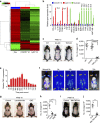
Figure 1
Figure 1. Ly6C+ inflammatory macrophages contribute to WIH-A and WIHN.
(a) Wounding the skin of 8-week-old mice induced a transition of HF from the telogen phase to the anagen phase around wound areas. n=56. (b) The longitudinal section of the wound at the 15th day post-wound (PWD-15) (upper panel) and of the dermal side of the skin showed the pigmented anagen HFs around the wound (lower panel). (c) Flow cytometry analysis of wound tissue single cell suspensions showed the extensive depletion of F4/80+/CD11b+ macrophages after clodronate liposome (Clo) treatment. Mice receiving clodronate, n=7; control mice, n=12. (d) WIH-A analysis in Clo treated (n=8) and the control mice (n=11). (e,f) Clo treatment resulted in complete elimination of WIHN (n=7) (e), and the number of neogenic HFs in the two groups of mice was quantified (f). (g) Immunofluorescence (IF) analysis of wound tissue showed the infiltration of both Ly6C+ inflammatory macrophages and CX3CR1+ resident macrophages at PWD-3. For all IF analyses, representative images from 8 to 16 tissue sections of wounds in 4–6 mice are shown. (h) IF image showed CX3CR1-YFP cells (green) in the dermis of the normal skin of CX3CR1CreER/+:R26iDTR/+ mice. Flow cytometry analysis indicated that TM and DT treatments depleted over 90% of CX3CR1-EYFP cells in the blood of CX3CR1CreER/+:R26iDTR/+ mice (n=9). (i) Flow cytometry analysis showed that the DT treatments depleted over 95% of Ly6C+ cells in the blood of LysM-Cre:R26iDTR/+ mice (n=9). (j) IF staining of Ly6C+ macrophages in the wound tissue (PWD-3) of LysM-Cre:R26iDTR/+ mice with or without DT treatment. (k,l) Treatment of LysM-Cre:R26iDTR/+ mice with DT resulted in the complete inhibition of WIH-A. n=8 for DT- mice; n=10 for DT+ mice. (m,n) Deletion of Ly6C+ macrophages resulted in no neogenic HF in the wound (n=7), but the deletion of CX3CR1+ macrophages showed no obvious effect on the number of neogenic HF (n=8 mice). W, wound; HA, hair follicle anagen analysis; HN, hair follicle neogenesis analysis; TM, tamoxifen; DT, diphtheria toxin; WT, wild type. Scale bars, 50 μm. Data are expressed as the mean±s.e.m. ***P<0.005, unpaired t-test, two-tailed.
Figure 2
Figure 2. TNF is a crucial mediator to induce the HF TAT.
(a) Gene expression profile of Ly6C+/F4/80+ macrophages (M), CX3CR1+/F4/80+ macrophages and Ly6G+/F4/80 neutrophils (Neu), which were sorted from wound tissue at PWD-3. Total RNA was extracted from samples of three mice. (b) Differentially expressed cytokine genes from microarray analysis were further validated by real-time PCR analysis. (For all real-time PCR analyses, gene expression was normalized to GAPDH with 40 cycles, data are represented as the mean±s.d., and n=3.) (c,d) WIH-A analysis in WT and deficient for IL6 (IL6-KO) or TNFA (TNFα-KO) gene mice, and the number of anagen HFs in different mice was quantified (d). IL6-knockout mice, n=5; TNFA-knockout mice, n=6; WT mice, n=6. (e) Real-time PCR analysis showed TNFA expression levels in the tissue surrounding the wounds (2 mm in width) at different times post-wounding. (f) Bioluminescent imaging of Tnf-Luc-eGFP mice at different times after wounding showed changes of the TNF-α level in the wound. Data are representative of 7–9 independent experiments at each time point. (g,h) Lenalidomide and QNZ treatment resulted in decreased anagen HFs as assessed at PWD-15 (g), and the number of anagen HFs in different groups was quantified (h). Control mice, n=9; lenalidomide-treated mice, n=7; QNZ-treated mice, n=8. (i,j) Wound-induced anagen HFs was significantly decreased in Lysmcre/+:TNFflox/flox mice (i), and the number of anagen HFs in two groups was quantified (j). Control mice (Lysmcre/+:TNFflox/+), n=6; Lysmcre/+:TNFflox/flox mice, n=4. Data are expressed as the mean±s.e.m. **P<0.01, ***P<0.005, unpaired t-test, two-tailed.
Figure 3
Figure 3. TNF-α is sufficient to induce HF TAT and is crucial for WIHN.
(a,b) Intracutaneous injection of TNF-α can induce the HF telogen–anagen transition at the injection site in 7-week (W)-old mice (refractory phase) and 9-week-old mice (competent phase) (a), and the number of TNF-α-induced anagen hair follicles in the two different group was quantified (b). Seven-week-old mice, n=6 for each group; 9-week-old mice, n=8 for each group. (c,d) Wounding to the skin induced more anagen HFs in 9-week-old mice than in 7-week-old mice (c), and the number of anagen HFs in the two different groups was quantified (d). n=7 for each age group. (e,f) WIH-A analysis in TNFR1−/− mice, TNFR2−/− mice and WT mice (e), and the number of anagen HFs in different groups was quantified (f). Wild-type (C57BL/6) mice, n =7; TNFR1−/− mice, n=6; TNFR2−/− mice, n=9. (g,h) Wound-induced anagen HFs in mice constitutively expressing TNF-α (Tg-TNF-α) were significantly increased compared with WT mice, and the number of anagen HFs in the two different groups was quantified (h). Control mice (WT), n=6; Tg-TNF-α mice, n=5. (i) Bioluminescent imaging of Tnf-Luc-eGFP mice at different days (D) after wounding showed changes in the TNF-α level in the wound. Areas with high TNF-α levels (green/red) shifted from the wound (W) periphery to the wound centre with the progression of wound healing. n=15 for Tnf-Luc-eGFP mice, and n=10 for wild-type (WT) mice. (j) IF analysis of sections from the PWD-14 wound showed the presence of F4/80+ macrophages in the wound, which largely co-localized with TNF-α. Scale bar, 50 μm. (k,l) WIHN analysis in WT, TNFA−/− and Tg-TNF-α mice at PWD-30 (k), and the number of neogenic HFs in wounds was quantified (l). n=6 for both wild-type and TNFA−/− mice; n=12 for Tg-TNF-α mice. Scale bars, 2 mm. Data are expressed as the mean±s.e.m. *P<0.05, **P<0.01, ***P<0.005, unpaired t-test, two-tailed.
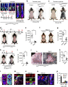
Figure 4
Figure 4. TNF activates the PI3K/AKT pathway in HF stem cells.
(a) Using TMT labelling and affinity enrichment followed by high-resolution LC–MS/MS and quantitative phosphor-proteomics, functional enrichment-based cluster analysis identified up-regulated signals in cultured epidermal stem cells after TNF-α treatment. (b) Western blot analysis of p-AKT (at Serine 473) and total AKT in cultured murine epidermal stem cells in the presence of different concentrations of TNF-α for 0.5 h. For all western blot analysis, data are representative of 3–5 independent experiments. (c) TNF-α treatment (8 h) resulted in no obvious changes in p-GSK-3β(ser9) and p-β-catenin (Ser33/37/Thr45, Ser675) but significantly increased the level of p-β-catenin (Ser552), which also showed in a TNF-dose-dependent manner, quite similar to p-AKT. (d) Perifosine diminished p-β-catenin (Ser552) levels that were elevated by TNF-α in cultured epidermal stem cells. (e) Western blot analysis indicated that TNF-α increased the level of β-catenin, and the effect was attenuated by Perifosine or LY294002. (f) TCF/LEF Dual-luciferase reporter analysis showed the relative transcription activity in differently treated groups. (g) At PWD-3, both p-AKT (Ser473) and p-β-catenin (Ser552) were detected in the wound adjacent to Lgr5+ hair follicle stem cells, and the p-AKT (Ser473) and p-β-catenin (Ser552) were highly co-localized. (h) At PWD-3, accumulated β-catenin was detected in the wound adjacent to Lgr5+ hair follicle stem cells, and the accumulated β-catenin was highly co-localized with p-AKT (Ser473). Scale bars, 30 μm.
Figure 5
Figure 5. Lgr5+ HFSCs are indispensable for WIH-A and important for WIHN.
(a) IF analysis of skin tissue sections indicated that tamoxifen treatment for Lgr5-Cre:Ptenflox/flox mice induced marked p-AKT and the division of Lgr5-EGFP+ cells in the HF. Scale bars, 50 μm. (b) Subcutaneous injection of tamoxifen induced HF TAT at the injection site in Lgr5-Cre:Ptenflox/flox mice (n=6). (c) Intraperitoneal administration of tamoxifen induced widespread HF TAT in back skin. Lgr5-Cre:Ptenflox/flox mice, n=9; WT mice, n=12. (d,e) Deletion of Lgr5+ cells in Lgr5-Cre:R26iDTR/+ mice completely attenuated the WIH-A (d), and the number of anagen HFs was quantified (n=7) (e). (f,g) Wound-induced anagen HFs were markedly decreased in mice that received Lgr5-mTNFR1-ShRNA (ShRNA) compared with mice that received a mock sequence. (h,i) WIH-A analysis in β-catenin knockout and the control mice at PWD-15 (n=7). (j,k) Depletion of Lgr5+ stem cells result in extensive reduction of neogenic HFs. n=5 for TM-treated mice, and n=6 for TM- and DT-treated mice. (l) Lgr5+ cells (mTomato+/mGFP+, mT+/mG+) were mainly located in the ORS of the anagen hair follicle (Anagen V), and the Lgr5+ cell lineage cells (mT/mG+) contributed to all the cellular components of the hair bulb. Lgr5+, Lgr5 expressing cells. Lgr5-L, Lgr5+ cell lineage tracing. Scale bars, 20 μm. (m) Lgr5+ progeny cells (mT/mG+) migrated from the HF toward the wound area at PWD-7. (n) Lgr5+ progeny cells (mT/mG+) formed clones in newly formed epithelium at PWD-30. (o,p) Lgr5+ progeny cells participated in the neogenic HF in wounds (o), and the number of Lgr5+ progeny cells in different neogenic HFs varied greatly as follows: the neogenic HFs without or with less than 3% of Lgr5+ lineage cells comprised 53% of all analysed neogenic HFs (50 in 94), and those with 3–30%, 30–60%, 60–90% and over 90% of Lgr5+ lineage cells comprised 16% (15 in 94), 19% (18 in 94), 8% (7 in 94) and 4% (4 in 94) of all analysed neogenic HFs, respectively (p). All 94 neogenic HFs were analysed from 5 different Lgr5+ cell lineage tracing mice. Scale bars, 50 μm. Data are expressed as the mean±s.e.m. **P<0.01, ***P<0.005, unpaired t-test, two-tailed.
Figure 6
Figure 6. AKT activation in Lgr5+ cells promotes de novo hair regeneration.
(a) New hair follicles in the wound began to form after PWD-14, and marked p-AKT was detected in the newly formed epidermis and in the hair germ (HG) that initiated hair follicle (HF) neogenesis. Similar to the p-AKT signal, abundant β-catenin was also detected. Scale bars, 30 μm. (b,c) p-AKT and β-catenin in the epidermis of re-epithelialized wounds of TNFA/ and WT mice at PWD-20. Scale bars, 50 μm. (d,e) Lgr5+ stem cells (GFP+) were present in the upper portion of the hair follicle at PWD-3 and in the re-epithelialized epidermis at PWD-5 in Pten knockout and the control mice (d). The number of GFP+ cells in the new epidermis of the two different groups was counted (e). n=15 tissue sections from 5 mice in each group were analysed. Scale bars, 50 μm. (f,g) Pten knockout in Lgr5+ cells promoted de novo HF regeneration, and synchronous knockout of β-catenin with Pten completely attenuated the Pten loss-induced hair follicle neogenesis in the wound (f). Then, the number of neogenic HFs in different groups was quantified (g). n=8 for tamoxifen-treated mice; n=9 for control mice. Scale bars, 1 mm. Bu, budge; SG, sebaceous gland; HG, hair germ; DP, dermal papilla. Data are expressed as the mean±s.e.m. **P<0.01. ***P<0.005, unpaired t-test, two-tailed.

References

    1. Oshima H., Rochat A., Kedzia C., Kobayashi K. & Barrandon Y. Morphogenesis and renewal of hair follicles from adult multipotent stem cells. Cell 104, 233–245 (2001). - PubMed
    1. Stenn K. S. & Paus R. Controls of hair follicle cycling. Physiol. Rev. 81, 449–494 (2001). - PubMed
    1. Morris R. J. et al.. Capturing and profiling adult hair follicle stem cells. Nat. Biotechnol. 22, 411–417 (2004). - PubMed
    1. Jaks V. et al.. Lgr5 marks cycling, yet long-lived, hair follicle stem cells. Nat. Genet. 40, 1291–1299 (2008). - PubMed
    1. Snippert H. J. et al.. Lgr6 marks stem cells in the hair follicle that generate all cell lineages of the skin. Science 327, 1385–1389 (2010). - PubMed

Publication types