. 2017 Mar 27:7:45270.
doi: 10.1038/srep45270.

A primary human macrophage-enteroid co-culture model to investigate mucosal gut physiology and host-pathogen interactions

Affiliations

A primary human macrophage-enteroid co-culture model to investigate mucosal gut physiology and host-pathogen interactions

Gaelle Noel et al. Sci Rep. .

Erratum in

Abstract

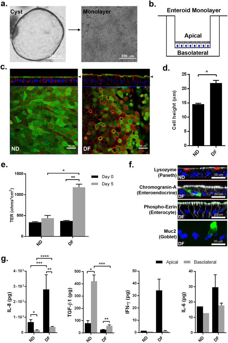
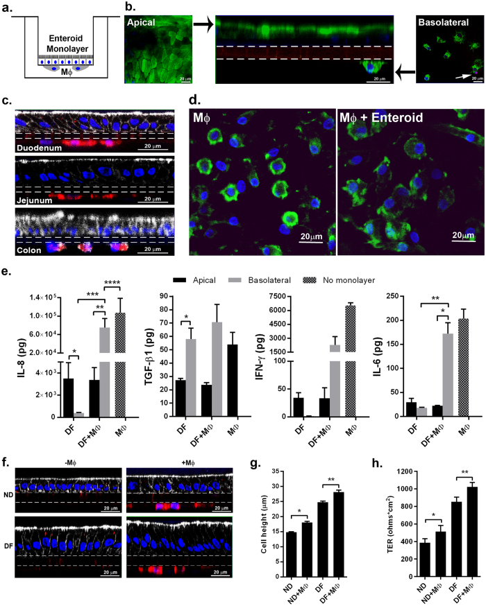
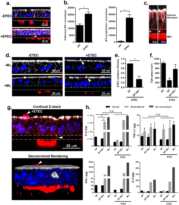
Integration of the intestinal epithelium and the mucosal immune system is critical for gut homeostasis. The intestinal epithelium is a functional barrier that secludes luminal content, senses changes in the gut microenvironment, and releases immune regulators that signal underlying immune cells. However, interactions between epithelial and innate immune cells to maintain barrier integrity and prevent infection are complex and poorly understood. We developed and characterized a primary human macrophage-enteroid co-culture model for in-depth studies of epithelial and macrophage interactions. Human intestinal stem cell-derived enteroid monolayers co-cultured with human monocyte-derived macrophages were used to evaluate barrier function, cytokine secretion, and protein expression under basal conditions and following bacterial infection. Macrophages enhanced barrier function and maturity of enteroid monolayers as indicated by increased transepithelial electrical resistance and cell height. Communication between the epithelium and macrophages was demonstrated through morphological changes and cytokine production. Intraepithelial macrophage projections, efficient phagocytosis, and stabilized enteroid barrier function revealed a coordinated response to enterotoxigenic and enteropathogenic E. coli infections. In summary, we have established the first primary human macrophage-enteroid co-culture system, defined conditions that allow for a practical and reproducible culture model, and demonstrated its suitability to study gut physiology and host responses to enteric pathogens.


Conflict of interest statement

The authors declare no competing financial interests.

Figures

Figure 1
Figure 1. Human enteroid monolayers form mature intestinal epithelium ex vivo.
(a) Human small intestinal enteroids cysts derived from biopsies (left panel) were grown as confluent epithelial monolayers (right panel) on permeable inserts. (b) Schematic representation of polarized enteroid monolayer established on permeable insert. (c) Non-differentiated (ND; left panel) and differentiated (DF; right panel) enteroid monolayers from small intestinal biopsies were visualized by confocal microscopy and showed polarized apical surfaces (wheat germ agglutinin [WGA]), green; indicated with black arrowheads in top panels). Mature microvilli were observed on the apical surface of DF enteroid monolayers (actin, red). XZ projection, top panels; XY projection, bottom panels; nuclei, blue. (d) Cell heights significantly increased in DF enteroid monolayers (*indicates p = 3.21 × 10−21). (e) TER increased upon 5 days of differentiation (*indicates p = 2.13 × 10−6; **indicates p = 4.96 × 10−9). (f) Paneth cells (lysozyme, red; top panel) were found in ND enteroid monolayers. Enteroendocrine cells (chromogranin-A, green), enterocytes (phospho-ezrin, green), and goblet cells (MUC2, green) were observed in DF enteroid monolayers. Nuclei, blue; actin, white. (g) Total amount of secreted cytokines released in the apical and basolateral media were quantified following 24 h of culture for IL-1β, IL-2, IL-4, IL-6, IL-8, IL-10, IL-12p70, IL-13 IFN-γ, and TNF-α, and 72 h of culture for TGF-β1, in ND and DF enteroid monolayer cultures. IL-8: *indicates p = 0.0132; **indicates p = 0.0010; ***indicates p = 0.0133; ****indicates p = 0.0009. TGF-β1: *indicates p = 0.0067; **indicates p = 0.0448; ***indicates p = 0.0018. Values are presented as Mean + SEM of at least three independent experiments involving enteroids generated from at least three individual donors with the exception of IFN-γ and IL-6 production in ND cultures that represent Mean +© SEM of two independent experiments with enteroids generated from two different donors (Fig. 1g).
Figure 2
Figure 2. Human macrophages display immune phenotype and bacterial sensing.
(a) Freshly isolated human monocytes (Mo; upper panels) and the derivative differentiated macrophages (MΦ; lower panels) were assessed by flow cytometry for surface expression of immune cell markers including CD14 and CD16 (shown as dot plots) and CD64, CX3CR1, CD89 and HLA-DR (shown as histograms). For Mo, the three main populations were reported as Q1, Q2 and Q3. (b) ETEC incubated with MΦ (schematic representation, left panel). Following overnight infection, bacteria were phagocytosed by MΦ. DNA, blue; ETEC, green; CD14 (MΦ), red. (c,d) MΦ were seeded underneath the filter with EPEC added onto the insert (schematic representation, left panel). MΦ extended projections across the 1.0 μm pore insert filter to reach EPEC, added on the opposite surface of the insert membrane. Dashed lines indicate the position of the 1.0 μm filter. (c) DNA, blue; actin, green; CD14, red; EPEC DNA, arrowheads. (d) EPEC flagella, green; CD14, red.
Figure 3
Figure 3. Human enteroid monolayers and monocyte-derived macrophages communicate in co-culture.
(a) Schematic representation of human enteroid co-cultured with macrophages (MΦ). (b) Macrophage-enteroid co-culture showed polarized DF duodenal enteroid monolayer indicated by apical WGA labeling (green; left and middle panels) and MΦ facing the basolateral side of the epithelium (middle and right panels), both separated by a permeable membrane shown by dashed lines (middle panel). Propidium iodide (red, white arrow in right panel) did not label substantial number of cells. Nuclei, blue; XZ projection, middle panel; XY projection, left and right panels. (c) Macrophage-enteroid co-cultures were established with duodenal-, jejunal-, and colon-derived enteroid monolayers. Nuclei, blue; actin, white; CD14 (MΦ), red; filter, dashed lines. (d) MΦ cultured 24 h with DF enteroid monolayer (right panel) showed morphological changes when compared to MΦ cultured alone (left panel). Nuclei, blue; WGA, green. (e) IL-8, IFN-γ, and IL-6 secretion was increased in basolateral media of DF enteroid cultured 24 h with MΦ. TGF-β1 secreted in apical and basolateral media of 72 h macrophage-enteroid co-cultures was not significantly modulated by the addition of MΦ. IL-8: *indicates p = 0.0156; **indicates p = 0.0156; ***indicates p = 0.0156; ****indicates p = 0.0469. TGF-β1: *indicates p = 0.0448. IL-6: *indicates p = 0.0226; **indicates p = 0.0184. (f) Both ND and DF enteroid monolayers (left panels) could be cultured with MΦ (right panels). Filter, dashed lines; actin, white; nuclei, blue; CD14 (MΦ), red. (g) The presence of MΦ increased cell height in both ND and DF enteroid monolayers. *Indicates p = 1.06 × 10−12; **indicates p = 4.39 × 10−5. (h) Differentiation of the enteroid monolayers and the addition of MΦ increased TER. *Indicates p = 0.0391; **indicates p = 0.0105. Data are presented as Mean + SEM of at least three independent experiments involving enteroids generated from at least three individual donors.
Figure 4
Figure 4. Bacterial infection of macrophage-enteroid monolayer induces morphological and physiological cell changes.
(a) MΦ projections extended through Transwell filters into DF enteroid monolayers following overnight EPEC infection. Actin, white; nuclei, blue; CD14 (MΦ), red; filter, dashed lines. (b) Adherent MΦ (left panel; *indicates p = 4.42 × 10−5) and number of MΦ projections (right panel; *indicates p = 2.33 × 10−9) increased upon apical overnight EPEC infection of co-cultures. UN, uninfected. (c) High resolution immunofluorescence confocal microscopy showed MΦ projections (arrowheads) going through permeable support and along lateral epithelial membranes upon overnight apical EPEC infection. Actin, white; CD14 (MΦ), red; filter, dashed lines. (d) ETEC adhered to DF enteroid monolayers alone (upper right panel) and co-cultured with MΦ (bottom right panel) following 3 h of infection. Uninfected cultures are represented in left panels. Actin, white; nuclei, blue; ETEC, green; CD14 (MΦ), red; filter, dashed lines. (e) Relative number of viable ETEC collected from the enteroid monolayer following 16 h of infection decreased in the presence of MΦ. *Indicates p = 0.0332. (f) DF enteroid monolayers infected with ETEC for 16 h displayed reduced TER values that were partially restored by the presence of MΦ in the co-culture. (g) Confocal Z-stack image (upper panel) and derivative 3D representation (lower panel) of MΦ extending projections through the membrane to reach the apical surface of DF epithelium infected 3 h with ETEC (arrowheads). Actin, white; CD14 (MΦ), red; nuclei, blue; filter, dashed lines. (h) Overnight ETEC infection did not significantly change the levels of IL-8, TGF-β1, IFN-γ, and IL-6 secreted by the cultures. IL8: *indicates p = 0.0447; **indicates p = 0.0455. Data are presented as the Mean + SEM of three independent experiments involving enteroids generated from three different donors with the exception of IFN-γ and IL-6 production that represent Mean + SEM of two independent experiments with enteroids generated from two different donors.

References

    1. Peterson L. W. & Artis D. Intestinal epithelial cells: regulators of barrier function and immune homeostasis. Nat Rev Immunol 14, 141–153, doi: 10.1038/nri3608 (2014). - DOI - PubMed
    1. Duell B. L., Cripps A. W., Schembri M. A. & Ulett G. C. Epithelial Cell Coculture Models for Studying Infectious Diseases: Benefits and Limitations. Journal of Biomedicine and Biotechnology 2011, 852419, doi: 10.1155/2011/852419 (2011). - DOI - PMC - PubMed
    1. Gibbons D. L. & Spencer J. Mouse and human intestinal immunity: same ballpark, different players; different rules, same score. Mucosal Immunol 4, 148–157 (2011). - PubMed
    1. Tsilingiri K. et al.. Probiotic and postbiotic activity in health and disease: comparison on a novel polarised ex-vivo organ culture model. Gut 61, 1007–1015, doi: 10.1136/gutjnl-2011-300971 (2012). - DOI - PubMed
    1. Bimczok D. et al.. Human gastric epithelial cells contribute to gastric immune regulation by providing retinoic acid to dendritic cells. Mucosal Immunol 8, 533–544, doi: 10.1038/mi.2014.86 (2015). - DOI - PMC - PubMed

Publication types