. 2017 Mar 27:8:14754.
doi: 10.1038/ncomms14754.

Rational combination of oncolytic vaccinia virus and PD-L1 blockade works synergistically to enhance therapeutic efficacy

Affiliations

Rational combination of oncolytic vaccinia virus and PD-L1 blockade works synergistically to enhance therapeutic efficacy

Zuqiang Liu et al. Nat Commun. .

Abstract

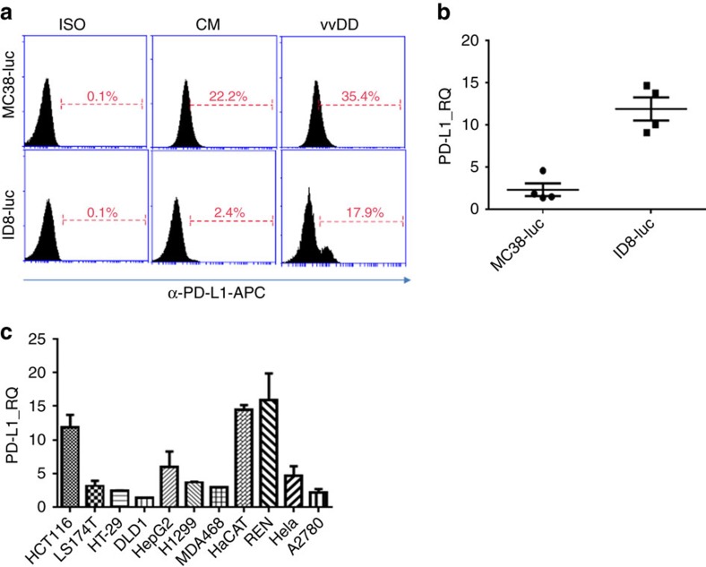
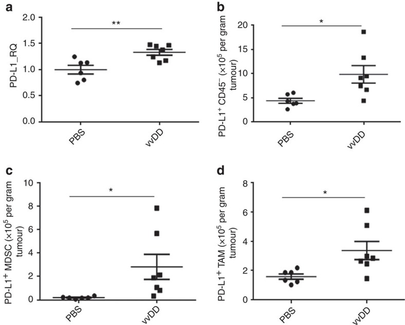
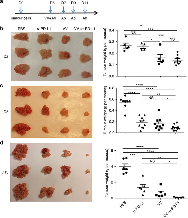
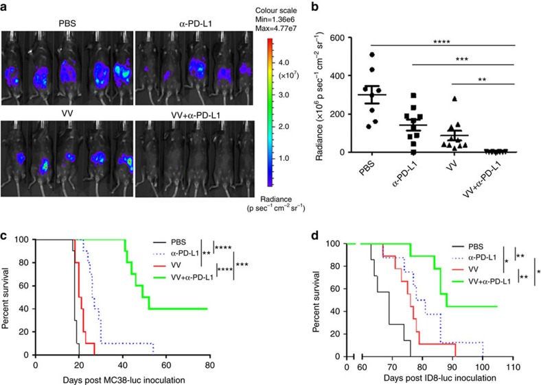
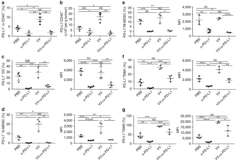
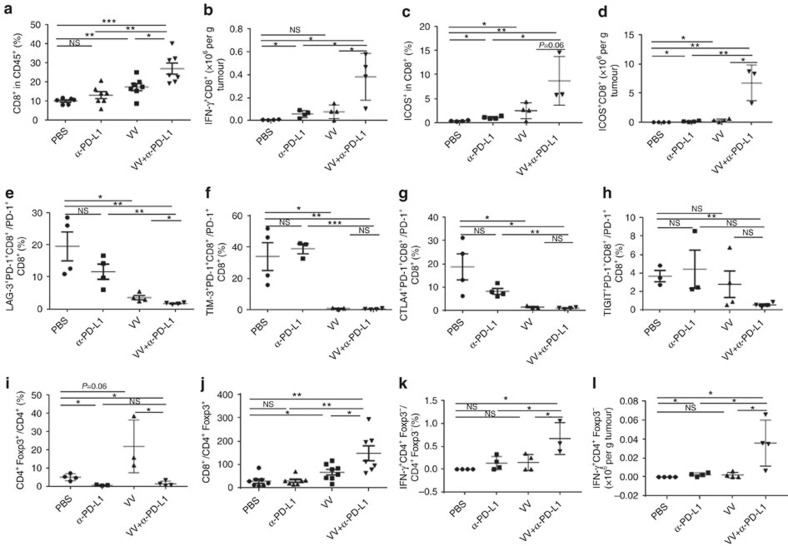
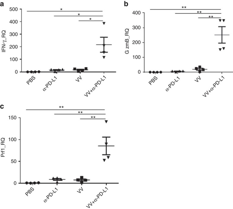
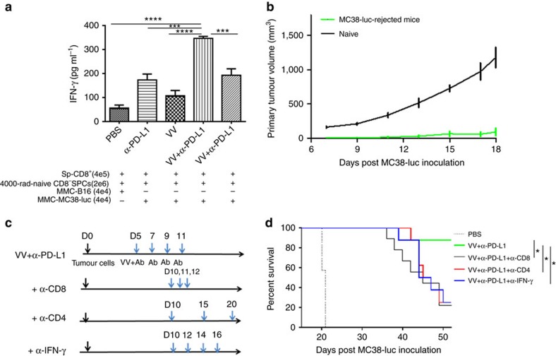
Both anti-PD1/PD-L1 therapy and oncolytic virotherapy have demonstrated promise, yet have exhibited efficacy in only a small fraction of cancer patients. Here we hypothesized that an oncolytic poxvirus would attract T cells into the tumour, and induce PD-L1 expression in cancer and immune cells, leading to more susceptible targets for anti-PD-L1 immunotherapy. Our results demonstrate in colon and ovarian cancer models that an oncolytic vaccinia virus attracts effector T cells and induces PD-L1 expression on both cancer and immune cells in the tumour. The dual therapy reduces PD-L1+ cells and facilitates non-redundant tumour infiltration of effector CD8+, CD4+ T cells, with increased IFN-γ, ICOS, granzyme B and perforin expression. Furthermore, the treatment reduces the virus-induced PD-L1+ DC, MDSC, TAM and Treg, as well as co-inhibitory molecules-double-positive, severely exhausted PD-1+CD8+ T cells, leading to reduced tumour burden and improved survival. This combinatorial therapy may be applicable to a much wider population of cancer patients.


Conflict of interest statement

D.L.B. is a shareholder of Sillajen Biotherapeutics, a company developing oncolytic viruses. The remaining authors declare no competing financial interests.

Figures

Figure 1
Figure 1. PD-L1 is elevated post vvDD infection in vitro.
(a) MC38-luc colon cancer or ID8-luc ovarian cancer cells (4 × 10⁵ cells each) were mock-infected or infected with vvDD. These cells were harvested 24 h later, blocked with α-CD16/32 Ab and then stained with α-PD-L1 for flow cytometry. (b) Total RNA was extracted from the harvested tumour cells and used in RT–qPCR to determine PD-L1 expression. In a, ISO: isotype IgG control used for staining; CM: condition medium. Data are presented as individuals, mean +/− s.d. (c) Cells from a panel of human cancer cell lines representing colorectal, ovarian, lung, cervical cancer and mesothelioma were infected or mock-infected with vvDD for 24 h and total RNA was prepared and subjected to RT–qPCR to determine the relative expression of PD-L1. The values on the y axis indicate the ratio of PD-L1 expression between infected versus mock-infected cancer cells. Data are presented as mean +/− s.d.
Figure 2
Figure 2. PD-L1 is elevated in cancer tissue post vvDD treatment in vivo.
B6 mice were subcutaneously inoculated with MC38-luc cells (4 × 10⁵ per mouse). PBS or vvDD was intratumorally injected at 2 × 10⁸ pfu per tumour when the s.c. tumour area reached 5 × 5 mm2. Tumour tissues were collected from PBS or virus-treated mice 4 days post treatment. Collected tumour tissues were weighed and incubated in RPMI 1640 medium containing 2% FBS, 1 mg ml−1 collagenase, 0.1 mg hyaluronidase, and 200 U DNase I at 37 °C for 1–2 h to make single cell preparations. The single cells were used for extraction of total RNA for RT–qPCR assay (a), or they were blocked with α-CD16/32 Ab and then stained with antibodies against CD45, CD11b, Gr1, PD-L1, F4/80 to determine PD-L1 expression on tumour cell (CD45) (b), MDSC (CD45+CD11b+Gr1+) (c) and TAM (CD45+CD11b+F4/80+) (d). In this experiment, the anti-PD-L1 Ab clone 10F.9G2 was used for analysis. Significant differences are indicated by *(P<0.05) or ** (P<0.01) determined by t-test. In this and other figures, the standard symbols for P values are *P<0.05; **P<0.01.
Figure 3
Figure 3. Primary tumour growth is dynamically monitored post treatments.
B6 mice were intraperitoneally inoculated with 5 × 10⁵ MC38-luc cancer cells and treated as described. Tumour-bearing mice were killed on days 2, 5 and 13 after first treatment, and primary tumour tissues were collected. Shown are schematic representations of the experiment (a) or weight of primary tumours on day 2 (b), day 5 (c) and day 13 (d) post first treatment. Numbers of mice per group are 5–11 (n=5∼11). In photographs, only representative three tumours from each group are shown. In the figures, VV=vvDD-CXCL11. Data were presented as individuals and means. Student's t-test was used to analyse the statistical significance (*P<0.05; **P<0.01; ***P<0.001; ****P<0.0001; and NS: not significant).
Figure 4
Figure 4. VV and PD-L1 blockade synergistically elicit anti-tumour effect.
B6 mice were intraperitoneally inoculated with 5 × 10⁵ MC38-luc or 3.5 × 10⁶ ID8-luc cells, respectively, and divided into required groups according to tumour growth condition based on live animal IVIS imaging 5 days post tumour cell injection. Grouped mice were intraperitoneally injected with VV (2 × 10⁸ pfu per 100 μl), α-PD-L1 Ab, VV plus α-PD-L1 Ab, or PBS (100 μl) per mouse, respectively. α-PD-L1 Ab (200 μg per 100 μl) was injected every 2 days for a total of four times, according to the scheme in Fig. 3a. Live animal imaging of the mice with MC38-luc tumours at day 9 post first treatment (a). The data are quantified (n=8∼10) (b). The survival of tumour-bearing mice was monitored by Kaplan–Meier analysis and statistical analyses were performed with Log rank test. Both MC38-luc (c) and ID8-luc (d) -tumour-bearing mice were shown.
Figure 5
Figure 5. The α-PD-L1 treatment reduces the PD-L1+ cells in TME.
B6 mice were intraperitoneally inoculated with 5 × 10⁵ MC38-luc cancer cells and treated with VV and/or α-PD-L1 as described. Tumour-bearing mice were killed at day 5 post first treatment and primary tumours were collected and analysed to determine the PD-L1+ CD45 cells (a,b), PD-L1+ DC (defined as CD45+ CD11b+CD11c+Ly6GPD-L1+) (c), PD-L1+G-MDSC (defined as CD45+CD11cCD11b+Ly6G+Ly6clowPD-L1+) (d), PD-L1+ M-MDSC (defined as CD45+CD11cCD11b+Ly6GLy6chiPD-L1+) (e), PD-L1+ TAM1 (defined as CD45+CD11cCD11b+Ly6GF4/80+CD206PD-L1+) (f), PD-L1+ TAM2 (defined as CD45+CD11cCD11b+Ly6GF4/80+CD206+PD-L1+) (g). Of note, the anti-PD-L1 antibody clone 10F.9G2 was used for therapy while clone MHI5 was used for subsequent analysis. Data were analysed using Student's t-test (*P<0.05; **P<0.01; ***P<0.001; ****P<0.0001).
Figure 6
Figure 6. The combination of VV plus α-PD-L1 treatment enhances infiltration of effector T cells and reduces Treg cells and exhausted CD8+ T cells in the TME.
B6 mice were intraperitoneally inoculated with 5 × 10⁵ MC38-luc and treated with VV and/or α-PD-L1 as described. Single cells were made from primary tumours collected from tumour-bearing mice at day 5 post first treatment, blocked with α-CD16/32 Ab and then stained with antibodies against CD45, CD8, CD4, PD-1, ICOS, PD-1, CTLA-4, TIM-3, LAG-3, TIGIT and Foxp3 to determine the quantities of CD8+ T cells (a), CD8+ T-cell activation (bd), CD8+ T-cell exhaustion (eh), Treg cells (i), CD8+/CD4+Foxp3+ T cells (j) and CD4+Foxp3 T cells (k,l) in the TME. Of note, the anti-PD-L1 antibody clone 10F.9G2 was used for therapy while clone MHI5 was used for subsequent phenotypic analysis. Data were analysed using Student's t-test (*P<0.05; **P<0.01; ***P<0.001).
Figure 7
Figure 7. The combination therapy leads to enhanced activation markers in the TME.
In one experiment, the i.p. MC38-luc tumour model was treated with VV and/or α-PD-L1 as described. Tumour tissues were collected at day 5 post first treatment and were used for extraction of total RNA. RT–qPCR assays were performed to determine the levels of IFN-γ (a), granzyme B (b) and perforin (c) in the TME. Data were analysed using Student's t-test (*P<0.05; **P<0.01).
Figure 8
Figure 8. The systemic anti-tumour immunity elicited by dual therapy plays an important role in the overall therapeutic efficacy.
(a) B6 mice were intraperitoneally inoculated with 5 × 10⁵ MC38-luc cancer cells and treated with VV and/or α-PD-L1 as described. Splenic CD8+ T cells (4 × 10⁵) were isolated from naive and MC38-luc-bearing mice that received different treatments 18 days post tumour cell injection and restimulated with mitomycin C-treated MC38-luc or B16 cancer cells (4 × 10⁴ cells each) in the presence of 4000-rad-irradiated CD8-depleted naive B6 splenocytes (2 × 10⁶) in 200 μl RPMI-1640 medium supplemented with 10% FBS at 37 °C, 5% CO2 for 2 days. The concentration of IFN-γ in the culture supernatants was tested by ELISA. The statistical analyses were performed with t-test. (b) Naive or MC38-luc-bearing B6 mice with dual treatments, which survived for more than 60 days, were s.c. rechallenged with 1 × 10⁶ MC38-luc cancer cells. The primary tumour size was measured and presented here. (c) In a separate experiment, B6 mice were inoculated with 5 × 10⁵ MC38-luc cells i.p. and treated with VV plus α-PD-L1 or PBS at day 5 post tumour inoculation, α-PD-L1 Ab was injected every 2 days for a total of four times. α-CD8 Ab (250 μg per injection), α-CD4 Ab (150 μg per injection) or α-IFN-γ Ab (200 μg per injection) were intraperitoneally injected into mice to deplete CD8+ T cells, CD4+ T cells or neutralize circulating IFN-γ as scheduled in c, and the overall survival was monitored by Kaplan–Meier analysis and analysed using log rank test (d).

Comment in

Similar articles

Cited by

References

    1. Bartlett D. L. et al. Oncolytic viruses as therapeutic cancer vaccines. Mol. Cancer 12, 103 (2013). - PMC - PubMed
    1. Lichty B. D., Breitbach C. J., Stojdl D. F. & Bell J. C. Going viral with cancer immunotherapy. Nat. Rev. Cancer 14, 559–567 (2014). - PubMed
    1. Andtbacka R. H. et al. Talimogene Laherparepvec improves durable response rate in patients with advanced melanoma. J. clin. oncol. 33, 2780–2788 (2015). - PubMed
    1. Mellman I., Coukos G. & Dranoff G. Cancer immunotherapy comes of age. Nature 480, 480–489 (2011). - PMC - PubMed
    1. Schlom J. Therapeutic cancer vaccines: current status and moving forward. J. Natl. Cancer Inst. 104, 599–613 (2012). - PMC - PubMed

Publication types