

doi: 10.1021/acs.analchem.7b00398.
Epub 2017 Apr 5.
Native Mass Spectrometry of Recombinant Proteins from Crude Cell Lysates
Affiliations
- PMID: 28345863
- PMCID: PMC5702260
- DOI: 10.1021/acs.analchem.7b00398
Item in Clipboard
Native Mass Spectrometry of Recombinant Proteins from Crude Cell Lysates
Anal Chem.
.
Abstract
Determining the properties of proteins prior to purification saves time and labor. Here, we demonstrate a native mass spectrometry approach for rapid characterization of overexpressed proteins directly in crude cell lysates. The method provides immediate information on the identity, solubility, oligomeric state, overall structure, and stability, as well as ligand binding, without the need for purification.
Conflict of interest statement
The authors declare no competing financial interests.
Figures
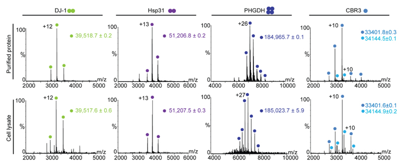
Mass spectra were recorded on an Orbitrap platform for dimeric DJ-1 and Hsp31, tetrameric phosphoglycerate dehydrogenase, and monomeric CBR3 in its apo- and NADPH bound forms. Spectra recorded from purified samples are shown on the top and those generated directly from lysates are at the bottom. The oligomeric states of each of the proteins are indicated by a cartoon of circles beside the protein’s name.

(a) Time-course analysis monitoring the overexpression rate of DJ-1 (top) and CBR3 (bottom) post-induction. The recombinant proteins’ concentrations were calculated based on relative band intensity, by densitometry, using known amounts of BSA as a standard. Arrows point to the band of the overexpressed protein. (b) Mass spectra of DJ-1 and CBR3 collected 0.5, 1 and 1.5 h post-induction.

Different volumes of CBR3 and Hsp31 cell cultures, ranging from 25 to 1 ml, were lysed in 2 ml buffer and analyzed directly by native MS. Clear charge state series of apo CBR3 and its NADPH-bound form were detected in all cell culture volumes. The homodimeric Hsp31 protein was detected in all tested culture volumes, however, dissociation into monomers occurred in correlation with the reduction in sample volume, probably due to denaturing during the sonication lysis procedure.
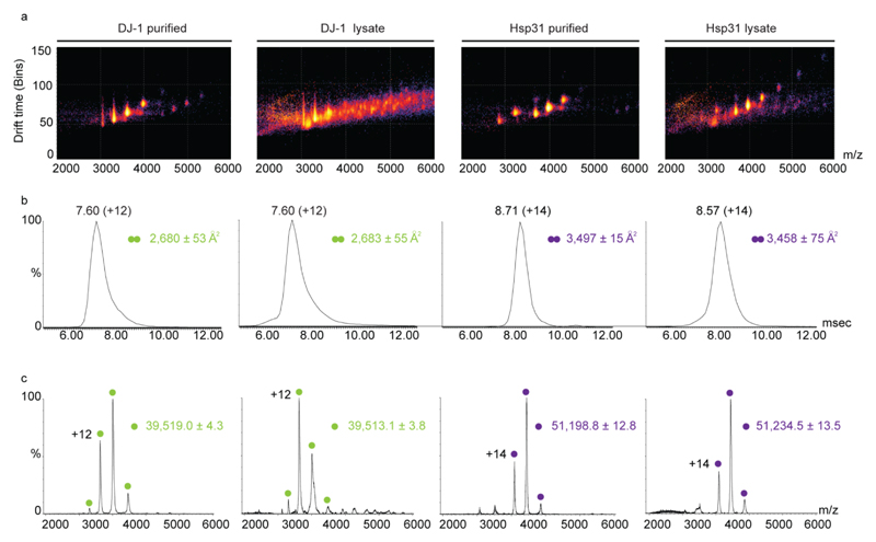
(a) Three dimensional IM-MS spectra that separate the ions based on their size and shape were measured for DJ-1 and CBR3 in crude cell lysates and in purified samples. Data was collected on a Synapt G2 mass spectrometer. (b) Representative ion mobility arrival time distributions for the 12+ and 14+ charge states of the homodimeric proteins DJ-1 and Hsp31, respectively. Calculated collision cross sections values, from at least three spectra, are indicated. (c) Representative two-dimensional plots of m/z versus intensity of DJ-1 and CBR3 from cell lysate and purified samples. Overall, comparable data were obtained for both samples, regardless if cell lysates or purified proteins were analyzed.

(a) Direct-MS spectra recorded for DJ-1WT and its mutants, DJ-1A104T, DJ-1C106A and DJ-1D149A, indicating that all mutants are expressed and soluble and maintain the homodimeric structure. (b) Simultaneous MS measurement of crude cell lysate mix of the three DJ-1 variants: DJ-1WT DJ-1A104T and DJ-1D149A. (c) Characterization of computationally designed RuBisCO variants, RBC17 and RBC31, and WT RuBisCO. The measured mass and charge state distribution reveal that RBC17 and RBC31 are partially unfolded monomers, unlike the WT protein that is a folded homodimer. (d) Activity assays monitoring the 3-phosphoglycerate product of RuBisCO using a coupled-assay with phosphoglycerate kinase and glyceraldehyde 3-phosphate dehydrogenase. The latter’s conversion of NADH to NAD+ was spectroscopically monitored. The assay indicated that RBC17 and RBC31 are completely inactive proteins in comparison to the WT protein, as expected from the results obtained in (c). Error bars represent standard error of three experimental replicates.
References
-
- Structural Genomics C. China Structural Genomics C. Northeast Structural Genomics C. Graslund S, Nordlund P, Weigelt J, Hallberg BM, Bray J, Gileadi O, Knapp S, Oppermann U, et al. Nat Methods. 2008;5:135–146.
-
- Sharon M. Science. 2013;340:1059–1060. - PubMed
-
- Liko I, Allison TM, Hopper JT, Robinson CV. Curr Opin Struct Biol. 2016;40:136–144. - PubMed
-
- Hernandez H, Robinson CV. Nat Protoc. 2007;2:715–726. - PubMed
Publication types
MeSH terms
Substances
Grants and funding
LinkOut - more resources
Full Text Sources
Other Literature Sources
