. 2017 Sep;284(3):725-736.
doi: 10.1148/radiol.2017162020. Epub 2017 Mar 27.

Detection of Clinically Significant Prostate Cancer: Short Dual-Pulse Sequence versus Standard Multiparametric MR Imaging-A Multireader Study

Affiliations

Detection of Clinically Significant Prostate Cancer: Short Dual-Pulse Sequence versus Standard Multiparametric MR Imaging-A Multireader Study

Borna K Barth et al. Radiology. 2017 Sep.

Abstract

Purpose To compare the diagnostic performance of a short dual-pulse sequence magnetic resonance (MR) imaging protocol versus a standard six-pulse sequence multiparametric MR imaging protocol for detection of clinically significant prostate cancer. Materials and Methods This HIPAA-compliant study was approved by the regional ethics committee. Between July 2013 and March 2015, 63 patients from a prospectively accrued study population who underwent MR imaging of the prostate including transverse T1-weighted; transverse, coronal, and sagittal T2-weighted; diffusion-weighted; and dynamic contrast material-enhanced MR imaging with a 3-T imager at a single institution were included in this retrospective study. The short MR imaging protocol image set consisted of transverse T2-weighted and diffusion-weighted images only. The standard MR imaging protocol image set contained images from all six pulse sequences. Three expert readers from different institutions assessed the likelihood of prostate cancer on a five-point scale. Diagnostic performance on a quadrant basis was assessed by using areas under the receiver operating characteristic curves, and differences were evaluated by using 83.8% confidence intervals. Intra- and interreader agreement was assessed by using the intraclass correlation coefficient. Transperineal template saturation biopsy served as the standard of reference. Results At histopathologic evaluation, 84 of 252 (33%) quadrants were positive for cancer in 38 of 63 (60%) men. There was no significant difference in detection of tumors larger than or equal to 0.5 mL for any of the readers of the short MR imaging protocol, with areas under the curve in the range of 0.74-0.81 (83.8% confidence interval [CI]: 0.64, 0.89), and for readers of the standard MR imaging protocol, areas under the curve were 0.71-0.77 (83.8% CI: 0.62, 0.86). Ranges for sensitivity were 0.76-0.95 (95% CI: 0.53, 0.99) and 0.76-0.86 (95% CI: 0.53, 0.97) and those for specificity were 0.84-0.90 (95% CI: 0.79, 0.94) and 0.82-0.90 (95% CI: 0.77, 0.94) for the short and standard MR protocols, respectively. Ranges for interreader agreement were 0.48-0.60 (83.8% CI: 0.41, 0.66) and 0.49-0.63 (83.8% CI: 0.42, 0.68) for the short and standard MR imaging protocols. Conclusion For the detection of clinically significant prostate cancer, no difference was found in the diagnostic performance of the short MR imaging protocol consisting of only transverse T2-weighted and diffusion-weighted imaging pulse sequences compared with that of a standard multiparametric MR imaging protocol. © RSNA, 2017 Online supplemental material is available for this article.


Figures

Figure 1:
Figure 1:
Flowchart shows patient selection process according to Standards for the Reporting of Diagnostic Accuracy Guidelines, or STARD. mpMRIshort = short multiparametric MR imaging protocol. mpMRIstandard = standard MR imaging sequence.
Figure 2a:
Figure 2a:
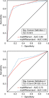
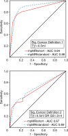
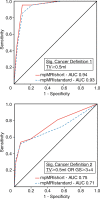
ROC curves for readers (a) 1, (b) 2, and (c) 3 for the short and standard MR imaging protocols. AUCs for clinically significant prostate cancer, according to definition 1 (top row) and definition 2 (bottom row), are shown. GS = Gleason score, mpMRIshort = short multiparametric MR imaging protocol, mpMRIstandard = standard multiparametric MR imaging protocol, Sig. = significant, TV = tumor volume.
Figure 2b:
Figure 2b:
ROC curves for readers (a) 1, (b) 2, and (c) 3 for the short and standard MR imaging protocols. AUCs for clinically significant prostate cancer, according to definition 1 (top row) and definition 2 (bottom row), are shown. GS = Gleason score, mpMRIshort = short multiparametric MR imaging protocol, mpMRIstandard = standard multiparametric MR imaging protocol, Sig. = significant, TV = tumor volume.
Figure 2c:
Figure 2c:
ROC curves for readers (a) 1, (b) 2, and (c) 3 for the short and standard MR imaging protocols. AUCs for clinically significant prostate cancer, according to definition 1 (top row) and definition 2 (bottom row), are shown. GS = Gleason score, mpMRIshort = short multiparametric MR imaging protocol, mpMRIstandard = standard multiparametric MR imaging protocol, Sig. = significant, TV = tumor volume.
Figure 3a:
Figure 3a:
Multiparametric MR images of the prostate in a 61-year-old man show sharply demarcated T2-weighted hypointense lesion (arrow) on (a) transverse, (b) coronal, and (c) sagittal imaging planes with correspondingly restricted diffusion on (d) apparent diffusion coefficient map and (e) high-b-value (b = 1400 sec/mm2) image, and (f) DCE image shows increased early contrast media uptake in left anterior apex of gland (arrow). All readers assigned a score of 5 of 5 to lesion, with both short and standard MR imaging protocols. This lesion was assigned Gleason score 4+5 (cancer core length, 27.5 mm) at histologic evaluation.
Figure 3b:
Figure 3b:
Multiparametric MR images of the prostate in a 61-year-old man show sharply demarcated T2-weighted hypointense lesion (arrow) on (a) transverse, (b) coronal, and (c) sagittal imaging planes with correspondingly restricted diffusion on (d) apparent diffusion coefficient map and (e) high-b-value (b = 1400 sec/mm2) image, and (f) DCE image shows increased early contrast media uptake in left anterior apex of gland (arrow). All readers assigned a score of 5 of 5 to lesion, with both short and standard MR imaging protocols. This lesion was assigned Gleason score 4+5 (cancer core length, 27.5 mm) at histologic evaluation.
Figure 3c:
Figure 3c:
Multiparametric MR images of the prostate in a 61-year-old man show sharply demarcated T2-weighted hypointense lesion (arrow) on (a) transverse, (b) coronal, and (c) sagittal imaging planes with correspondingly restricted diffusion on (d) apparent diffusion coefficient map and (e) high-b-value (b = 1400 sec/mm2) image, and (f) DCE image shows increased early contrast media uptake in left anterior apex of gland (arrow). All readers assigned a score of 5 of 5 to lesion, with both short and standard MR imaging protocols. This lesion was assigned Gleason score 4+5 (cancer core length, 27.5 mm) at histologic evaluation.
Figure 3d:
Figure 3d:
Multiparametric MR images of the prostate in a 61-year-old man show sharply demarcated T2-weighted hypointense lesion (arrow) on (a) transverse, (b) coronal, and (c) sagittal imaging planes with correspondingly restricted diffusion on (d) apparent diffusion coefficient map and (e) high-b-value (b = 1400 sec/mm2) image, and (f) DCE image shows increased early contrast media uptake in left anterior apex of gland (arrow). All readers assigned a score of 5 of 5 to lesion, with both short and standard MR imaging protocols. This lesion was assigned Gleason score 4+5 (cancer core length, 27.5 mm) at histologic evaluation.
Figure 3e:
Figure 3e:
Multiparametric MR images of the prostate in a 61-year-old man show sharply demarcated T2-weighted hypointense lesion (arrow) on (a) transverse, (b) coronal, and (c) sagittal imaging planes with correspondingly restricted diffusion on (d) apparent diffusion coefficient map and (e) high-b-value (b = 1400 sec/mm2) image, and (f) DCE image shows increased early contrast media uptake in left anterior apex of gland (arrow). All readers assigned a score of 5 of 5 to lesion, with both short and standard MR imaging protocols. This lesion was assigned Gleason score 4+5 (cancer core length, 27.5 mm) at histologic evaluation.
Figure 3f:
Figure 3f:
Multiparametric MR images of the prostate in a 61-year-old man show sharply demarcated T2-weighted hypointense lesion (arrow) on (a) transverse, (b) coronal, and (c) sagittal imaging planes with correspondingly restricted diffusion on (d) apparent diffusion coefficient map and (e) high-b-value (b = 1400 sec/mm2) image, and (f) DCE image shows increased early contrast media uptake in left anterior apex of gland (arrow). All readers assigned a score of 5 of 5 to lesion, with both short and standard MR imaging protocols. This lesion was assigned Gleason score 4+5 (cancer core length, 27.5 mm) at histologic evaluation.
Figure 4a:
Figure 4a:
Multiparametric MR images of the prostate in a 56-year-old man show hypointense lesion (arrow) in peripheral zone of right midgland on T2-weighted images in (a) transverse, (b) coronal, and (c) sagittal imaging planes, best depicted in the transverse plane (a). (d) Mild diffusion restriction (apparent diffusion coefficient, 0.98 × 10⁻³ sec/mm2) may be seen on apparent diffusion coefficient map and (e) high-b-value (b = 1400 sec/mm2) image. (f) DCE image shows early uptake in corresponding location, accurately matching observation on transverse T2-weighted images. Two of three readers scored lesion as greater than or equal to 4 with short MR imaging protocol image set, whereas all readers scored lesion as greater than or equal to 4 with standard protocol set. Lesion was assigned Gleason score of 4+3 (cancer core length, 24.2 mm) at histologic evaluation. Most likely, DCE images provided incremental information for detection of tumor for one of the readers.
Figure 4b:
Figure 4b:
Multiparametric MR images of the prostate in a 56-year-old man show hypointense lesion (arrow) in peripheral zone of right midgland on T2-weighted images in (a) transverse, (b) coronal, and (c) sagittal imaging planes, best depicted in the transverse plane (a). (d) Mild diffusion restriction (apparent diffusion coefficient, 0.98 × 10⁻³ sec/mm2) may be seen on apparent diffusion coefficient map and (e) high-b-value (b = 1400 sec/mm2) image. (f) DCE image shows early uptake in corresponding location, accurately matching observation on transverse T2-weighted images. Two of three readers scored lesion as greater than or equal to 4 with short MR imaging protocol image set, whereas all readers scored lesion as greater than or equal to 4 with standard protocol set. Lesion was assigned Gleason score of 4+3 (cancer core length, 24.2 mm) at histologic evaluation. Most likely, DCE images provided incremental information for detection of tumor for one of the readers.
Figure 4c:
Figure 4c:
Multiparametric MR images of the prostate in a 56-year-old man show hypointense lesion (arrow) in peripheral zone of right midgland on T2-weighted images in (a) transverse, (b) coronal, and (c) sagittal imaging planes, best depicted in the transverse plane (a). (d) Mild diffusion restriction (apparent diffusion coefficient, 0.98 × 10⁻³ sec/mm2) may be seen on apparent diffusion coefficient map and (e) high-b-value (b = 1400 sec/mm2) image. (f) DCE image shows early uptake in corresponding location, accurately matching observation on transverse T2-weighted images. Two of three readers scored lesion as greater than or equal to 4 with short MR imaging protocol image set, whereas all readers scored lesion as greater than or equal to 4 with standard protocol set. Lesion was assigned Gleason score of 4+3 (cancer core length, 24.2 mm) at histologic evaluation. Most likely, DCE images provided incremental information for detection of tumor for one of the readers.
Figure 4d:
Figure 4d:
Multiparametric MR images of the prostate in a 56-year-old man show hypointense lesion (arrow) in peripheral zone of right midgland on T2-weighted images in (a) transverse, (b) coronal, and (c) sagittal imaging planes, best depicted in the transverse plane (a). (d) Mild diffusion restriction (apparent diffusion coefficient, 0.98 × 10⁻³ sec/mm2) may be seen on apparent diffusion coefficient map and (e) high-b-value (b = 1400 sec/mm2) image. (f) DCE image shows early uptake in corresponding location, accurately matching observation on transverse T2-weighted images. Two of three readers scored lesion as greater than or equal to 4 with short MR imaging protocol image set, whereas all readers scored lesion as greater than or equal to 4 with standard protocol set. Lesion was assigned Gleason score of 4+3 (cancer core length, 24.2 mm) at histologic evaluation. Most likely, DCE images provided incremental information for detection of tumor for one of the readers.
Figure 4e:
Figure 4e:
Multiparametric MR images of the prostate in a 56-year-old man show hypointense lesion (arrow) in peripheral zone of right midgland on T2-weighted images in (a) transverse, (b) coronal, and (c) sagittal imaging planes, best depicted in the transverse plane (a). (d) Mild diffusion restriction (apparent diffusion coefficient, 0.98 × 10⁻³ sec/mm2) may be seen on apparent diffusion coefficient map and (e) high-b-value (b = 1400 sec/mm2) image. (f) DCE image shows early uptake in corresponding location, accurately matching observation on transverse T2-weighted images. Two of three readers scored lesion as greater than or equal to 4 with short MR imaging protocol image set, whereas all readers scored lesion as greater than or equal to 4 with standard protocol set. Lesion was assigned Gleason score of 4+3 (cancer core length, 24.2 mm) at histologic evaluation. Most likely, DCE images provided incremental information for detection of tumor for one of the readers.
Figure 4f:
Figure 4f:
Multiparametric MR images of the prostate in a 56-year-old man show hypointense lesion (arrow) in peripheral zone of right midgland on T2-weighted images in (a) transverse, (b) coronal, and (c) sagittal imaging planes, best depicted in the transverse plane (a). (d) Mild diffusion restriction (apparent diffusion coefficient, 0.98 × 10⁻³ sec/mm2) may be seen on apparent diffusion coefficient map and (e) high-b-value (b = 1400 sec/mm2) image. (f) DCE image shows early uptake in corresponding location, accurately matching observation on transverse T2-weighted images. Two of three readers scored lesion as greater than or equal to 4 with short MR imaging protocol image set, whereas all readers scored lesion as greater than or equal to 4 with standard protocol set. Lesion was assigned Gleason score of 4+3 (cancer core length, 24.2 mm) at histologic evaluation. Most likely, DCE images provided incremental information for detection of tumor for one of the readers.

Similar articles

Cited by

References

    1. Blomqvist L, Carlsson S, Gjertsson P, et al. . Limited evidence for the use of imaging to detect prostate cancer: a systematic review. Eur J Radiol 2014;83(9):1601–1606. - PubMed
    1. Johnson LM, Turkbey B, Figg WD, Choyke PL. Multiparametric MRI in prostate cancer management. Nat Rev Clin Oncol 2014;11(6):346–353. - PMC - PubMed
    1. Scheenen TW, Rosenkrantz AB, Haider MA, Fütterer JJ. Multiparametric magnetic resonance imaging in prostate cancer management: current status and future perspectives. Invest Radiol 2015;50(9):594–600. - PubMed
    1. Rosenkrantz AB, Shanbhogue AK, Wang A, Kong MX, Babb JS, Taneja SS. Length of capsular contact for diagnosing extraprostatic extension on prostate MRI: Assessment at an optimal threshold. J Magn Reson Imaging 2016;43(4):990–997. - PubMed
    1. Rosenkrantz AB, Triolo MJ, Melamed J, Rusinek H, Taneja SS, Deng FM. Whole-lesion apparent diffusion coefficient metrics as a marker of percentage Gleason 4 component within Gleason 7 prostate cancer at radical prostatectomy. J Magn Reson Imaging 2015;41(3):708–714. - PMC - PubMed

Publication types

LinkOut - more resources