Review
. 2017 Mar 27;7(2):34.
doi: 10.3390/biom7020034.

The Process and Regulatory Components of Inflammation in Brain Oncogenesis

Affiliations
Review

The Process and Regulatory Components of Inflammation in Brain Oncogenesis

A G M Mostofa et al. Biomolecules. .

Abstract

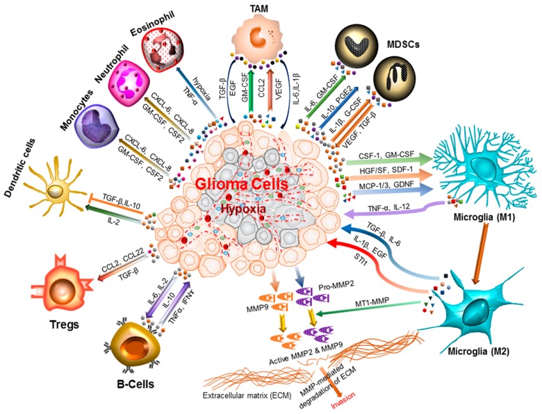
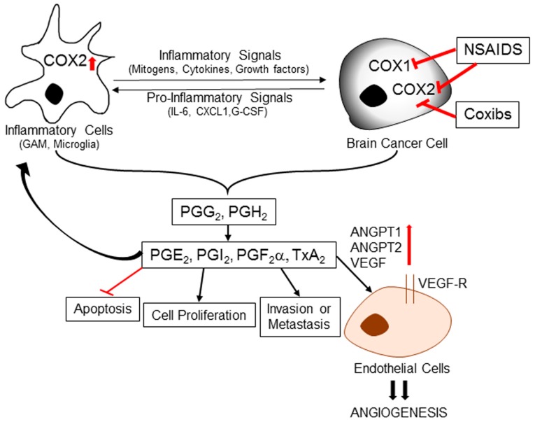
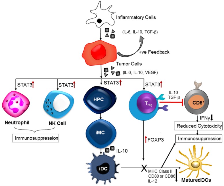
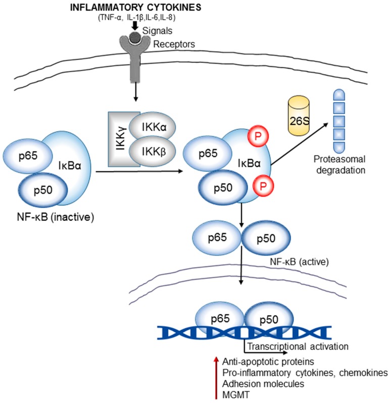
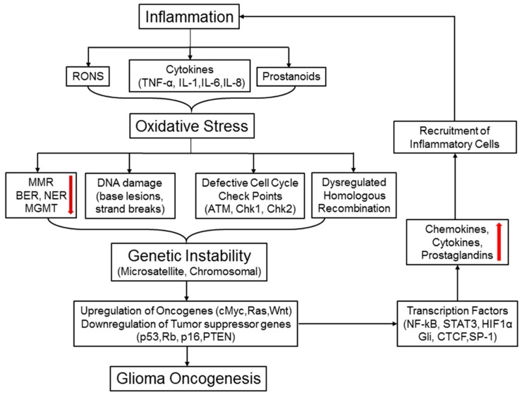
Central nervous system tumors comprising the primary cancers and brain metastases remain the most lethal neoplasms and challenging to treat. Substantial evidence points to a paramount role for inflammation in the pathology leading to gliomagenesis, malignant progression and tumor aggressiveness in the central nervous system (CNS) microenvironment. This review summarizes the salient contributions of oxidative stress, interleukins, tumor necrosis factor-α (TNF-α), cyclooxygenases, and transcription factors such as signal transducer and activator of transcription 3 (STAT3) and nuclear factor kappa-light-chain-enhancer of activated B-cells (NF-κB) and the associated cross-talks to the inflammatory signaling in CNS cancers. The roles of reactive astrocytes, tumor associated microglia and macrophages, metabolic alterations, microsatellite instability, O⁶-methylguanine DNA methyltransferase (MGMT) DNA repair and epigenetic alterations mediated by the isocitrate dehydrogenase 1 (IDH1) mutations have been discussed. The inflammatory pathways with relevance to the brain cancer treatments have been highlighted.

Keywords: MGMT DNA repair; STAT3; epigenetic deregulation; gliomas; inflammation; interleukins.


Conflict of interest statement

The authors declare no conflict of interest.

Figures

Figure 1
Figure 1
Inflammatory microenvironment prevalent in brain cancers. The glioma microenvironment is heavily infiltrated with different inflammatory cells, including microglia, macrophage, neutrophil, eosinophil, monocytes, dendritic cells, T-cells, B-cells, and myeloid derived suppressor cells (MDSCs) [18]. Upon activation, these cells release an array of mediators that promote cancer cell proliferation, survival, migration and invasion. These include the pro-inflammatory and cytotoxic cytokines, growth factors, bioactive lipids, hydrolytic enzymes, matrix metalloproteinases, reactive oxygen intermediates, and nitric oxide [19,20]. The cell types involved and the cytokines produced are shown. CCL: C–C Motif Chemokine Ligand 2/monocyte chemoattractant protein 1 (MCP1); TNF-α: Tumor necrosis factor-alpha; CXCL: Chemokine (C–X–C motif) ligand; INF-ɣ: Interferon gamma; TAM: Tumor-associated macrophages; Tregs: Regulatory T-cells; IL-1β: Interleukin 1 beta; IL-2/6/10/12: Interleukin 2/6/10/12; CSF-1: Colony stimulating factor 1; GM-CSF: Granulocyte-macrophage colony-stimulating factor; HGF/SF: Hepatocyte growth factor or scatter factor; SDF-1: Stromal cell-derived factor 1; MCP-1/3 : Monocyte chemoattractant protein 1 or 3; GDNF: Glial cell-derived neurotrophic factor; TGF-β: Transforming growth factor beta; EGF: Epidermal growth factor; STI1: Stress inducible protein 1; MT1-MMP: Membrane type 1-matrix metalloproteinase; MMP2/9: Matrix metalloproteinase 2 or 9.
Figure 2
Figure 2
Role of inflammation-induced cyclooxygenase in brain cancer development. Inflammatory signals like mitogens, cytokines and growth factors can promote overexpression of COX-2 in tumor-associated cells immune cells and endothelial cells. COX enzymes catalyze the conversion of arachidonic acid into prostaglandin H2 (PGH2) and prostaglandin G2 (PGG2), precursors of all prostaglandins (PGs) and thromboxanes (TXs). PGH2 is further converted into various effector molecules by specific synthases. These effector molecules can mediate a diverse array of functions, including inflammation, inhibition of apoptosis, cell proliferation, invasion, and metastasis. Some downstream mediators also induce expression of angiopoetins (ANGPT1 and ANGPT2) and vascular endothelial growth factor (VEGF) to promote angiogenic potential of endothelial cells. NSAIDs and COXIBs can inhibit prostaglandin synthesis and thereby prevent pro-carcinogenic downstream effects. NSAIDs: Non-steroidal anti-inflammatory drugs; COXIBs: selective COX-2 inhibitors; CXCL1: CXCL1 chemokine; G-CSF: Granulocyte colony stimulating factor.
Figure 3
Figure 3
Inflammation-induced immune dysregulation contributes to brain cancer pathogenesis. Cytokines released by inflammatory cells cause upregulation of signal transducer and activation of transcription 3 (STAT3). Increased STAT3 activity negatively affects the immune responses in the tumor stroma. Further, STAT3 signaling in haematopoietic progenitor cells (HPCs) cause generation of immature myeloid cells (iMC) followed by formation of immature dendritic cells (iDC). Dendritic cell (DC) maturation is prevented due to STAT3-mediated reduction in the expression of MHC class II molecules, CD80, CD86, and IL-12. Persistent activation of STAT3 in natural killer (NK) cells and neutrophils inhibits the tumor killing activity of those effector cells. Tumor-associated regulatory T (Treg) cells exhibit increased level of forkhead box P3 (FOXP3), transforming growth factor-β (TGFβ) and interleukin-10 (IL-10), these factors restrain CD8+ effector T-cells and DC maturation.
Figure 4
Figure 4
Inflammation and NF-κB signaling cross-talk is an important link in brain cancer pathogenesis. The inflammatory cytokines (TNF-α, IL-1β, IL-6, and IL-8) released in the tumor microenvironment generate signals that lead to activation of IκB kinases (IKKs). IKKs induce phosphorylation and subsequent proteosomal degradation of NF-κB inhibitor, IκBα. The resulting free NF-κB (e.g., heterodimer of p65 and p50 subunits) then translocates to the nucleus and activate the transcription of various target genes that encode anti-apoptotic proteins (e.g., B-cell lymphoma 2/ Bcl-2), pro-inflammatory cytokines and chemokines, adhesion molecules (integrins and cadherins), proteases (MMPs), DNA repair proteins such as the MGMT (O6-methylguanine DNA methyltransferase). Many of these regulators contribute significantly to the oncogenesis of gliomas.
Figure 5
Figure 5
Inflammation, oxidative stress, and chromosomal instability: a vicious cycle that promotes brain tumorigenesis. Inflammatory cells produce various reactive oxygen and nitrogen species (RONS) and other factors, such as cytokines, chemokines, and prostanoids. All these mediators promote oxidative stress in cells leading to deregulated DNA repair systems MMR (DNA mismatch repair), NER (nucleotide excision repair), BER (base excision repair), and MGMT), increased DNA damage, defective cell cycle checkpoints, and dysregulated homologous recombination (HR) pathway. Oxidative stress-induced MMR silencing often causes formation of error-prone microsatellites in the coding regions of various genes and repeated DNA sequences, termed as microsatellite instability (MSI). RONS-mediated DNA damage and strand breaks along with abnormal mitotic checkpoints and homologous recombination (HR) collectively engender genetic instability (GI). Both MSI and GI can spawn random genetic diversification in precancerous cells. Clones harboring the optimal combination of activated oncogenes and inactivated tumor suppressor genes will eventually proceed to initiate malignancy. Upon activation, some oncogenes may turn on the transcription factors like NF-κB, STAT3, and HIF-1α, followed by production of pro-inflammatory cytokines and chemokines.

Similar articles

Cited by

References

    1. PDQ Adult Treatment Editorial Board . PDQ Cancer Information Summaries. National Cancer Institute; Bethesda, MD, USA: 2002. Adult Treatment Editorial Board. Adult central nervous system tumors treatment (PDQ®): Health professional version. - PubMed
    1. Huttner A. Overview of primary brain tumors: Pathologic classification, epidemiology, molecular biology, and prognostic markers. Hematol. Oncol. Clin. N. Am. 2012;26:715–732. doi: 10.1016/j.hoc.2012.05.004. - DOI - PubMed
    1. Berghoff A.S., Preusser M. The inflammatory microenvironment in brain metastases: Potential treatment target? Chin. Clin. Oncol. 2015;4:21. - PubMed
    1. Chamberlain M.C., Baik C.S., Gadi V.K., Bhatia S., Chow L.Q. Systemic therapy of brain metastases: Non-small cell lung cancer, breast cancer, and melanoma. Neuro Oncol. 2017;19:i1–i24. doi: 10.1093/neuonc/now197. - DOI - PMC - PubMed
    1. Huse J.T., Holland E.C. Targeting brain cancer: Advances in the molecular pathology of malignant glioma and medulloblastoma. Nat. Rev. Cancer. 2010;10:319–331. doi: 10.1038/nrc2818. - DOI - PubMed

Publication types