Review
. 2017 Jun;97(6):669-697.
doi: 10.1038/labinvest.2017.25. Epub 2017 Mar 27.

T-lymphocyte homing: an underappreciated yet critical hurdle for successful cancer immunotherapy

Affiliations
Review

T-lymphocyte homing: an underappreciated yet critical hurdle for successful cancer immunotherapy

Robert Sackstein et al. Lab Invest. 2017 Jun.

Abstract

Advances in cancer immunotherapy have offered new hope for patients with metastatic disease. This unfolding success story has been exemplified by a growing arsenal of novel immunotherapeutics, including blocking antibodies targeting immune checkpoint pathways, cancer vaccines, and adoptive cell therapy (ACT). Nonetheless, clinical benefit remains highly variable and patient-specific, in part, because all immunotherapeutic regimens vitally hinge on the capacity of endogenous and/or adoptively transferred T-effector (Teff) cells, including chimeric antigen receptor (CAR) T cells, to home efficiently into tumor target tissue. Thus, defects intrinsic to the multi-step T-cell homing cascade have become an obvious, though significantly underappreciated contributor to immunotherapy resistance. Conspicuous have been low intralesional frequencies of tumor-infiltrating T-lymphocytes (TILs) below clinically beneficial threshold levels, and peripheral rather than deep lesional TIL infiltration. Therefore, a Teff cell 'homing deficit' may arguably represent a dominant factor responsible for ineffective immunotherapeutic outcomes, as tumors resistant to immune-targeted killing thrive in such permissive, immune-vacuous microenvironments. Fortunately, emerging data is shedding light into the diverse mechanisms of immune escape by which tumors restrict Teff cell trafficking and lesional penetrance. In this review, we scrutinize evolving knowledge on the molecular determinants of Teff cell navigation into tumors. By integrating recently described, though sporadic information of pivotal adhesive and chemokine homing signatures within the tumor microenvironment with better established paradigms of T-cell trafficking under homeostatic or infectious disease scenarios, we seek to refine currently incomplete models of Teff cell entry into tumor tissue. We further summarize how cancers thwart homing to escape immune-mediated destruction and raise awareness of the potential impact of immune checkpoint blockers on Teff cell homing. Finally, we speculate on innovative therapeutic opportunities for augmenting Teff cell homing capabilities to improve immunotherapy-based tumor eradication in cancer patients, with special focus on malignant melanoma.


Figures

Figure 1
Figure 1. Multi-step homing mechanism for Teff cell recruitment to the skin
Teff cells primed by Ag in regional LN draining skin (not shown) become imprinted with skin homing molecules, among which include adhesive glycoproteins CLA and CD43E, chemokine receptors CCR4 (Th2) and CCR10 (Th22), and integrins LFA-1 and VLA-4. (Step 1) Circulating Teff cells in postcapillary venules of the dermis tether and roll in blood flow via engagement of CLA with E/P-selectins and CD43E with E-selectin. These interactions, which slow Teff velocity thereby prepping cells for step 2, are facilitated by the tetrasaccharide moiety, sLeX, which is synthesized by α1,3FT during skin imprinting. Of note, non-inflamed dermal endothelium constitutively expresses low levels of E-selectin, VCAM-1 and ICAM-1, all of which can be elevated in response to cytokine insult, thereby permitting Teff skin homing under both resting and inflammatory conditions. (Steps 2–3) Chemokine CCL17, which is secreted by Langerhans cells and keratinocytes in the epidermis and by fibroblasts and endothelial cells in the dermis, as well as CCL27, which is secreted by keratinocytes, bind Teff-CCR4 and CCR10 receptors, respectively. CCL17 and CCL27 may be concentrated on glycosaminoglycans (GAG) expressed on endothelial apical, basal or basement membrane surfaces to allow for enhanced chemokine receptor binding. Chemokine ligation of CCR4 and CCR10 elicits Gαi-signaling, switches VLA-4 from an intermediate to highly active structure and LFA-1 from inactive to highly active, and eventuates in VLA-4/VCAM-1 and LFA-1/ICAM-1 mediated firm adhesion (arrows; solid = known signaling, dotted = speculated signaling). Integrins may also undergo activation independently of chemokine receptor signaling via a ‘Step 2 bypass’ circuit involving bimolecular association of E-selectin ligands (i.e. CLA) directly with VLA-4 (arrow). (Step 4) Firmly adherent Teff cells undergo VLA-4/LFA-1 and CCR4/CCR10 mediated transendothelial migration into the dermis and then potential further recruitment into the epidermis.
Figure 2
Figure 2. Multi-step homing mechanism for Teff cell recruitment to the gut (small intestine)
Teff cells primed by Ag in Peyer’s patches and mesenteric LN draining gut (not shown) become imprinted with gut homing molecules, among which include chemokine receptors CCR9 and CXCR4 and integrin α4β7. (Step 1) Circulating Teff cells in postcapillary venules of the gut (small intestine) engage CCR9-CCL25 and CXCR4-CXCL12 thereby eliciting Gαi-dependent signaling, activation of α4β7, and subsequent tethering and rolling on intestinal endothelial MAdCAM-1 (arrows; solid = known signaling, dotted = speculated signaling). –,,– CCL25 and CXCL12 are produced constitutively by epithelial cells (enterocytes) of the small intestine while MAdCAM-1 is expressed constitutively in HEV of gut Peyer’s patches and mesenteric lymph nodes (not shown) and by intestinal endothelium of the lamina propria.–,, (Steps 2–3) Rolling of α4β7 on MAdCAM-1 eventuates in Teff cell firm adhesion. (Step 4) Teff cells undergo transendothelial migration into the lamina propria, facilitated by concentration gradients of immobilized CCL25 and CXCL12 on apical and basal endothelial GAGs, on epithelial GAGs, and within the lamina propria.–, Accessory support of steps 2–4 may involve CXCR3-CXCL10 signaling (boxed). A subset of Teff cells traverse the lamina propria and then embed themselves as intraepithelial lymphocytes (IEL) into the epithelial cell layer of the intestinal lumen., This latter process is accompanied by concurrently decreased α4β7 and increased αEβ7 expression, CCL25-CCR9 signaling and activation of αEβ7 (arrow), and αEβ7 binding to E-cadherin.,, During inflammatory reactions, the gut homing repertoire is expanded to cause increased Teff cell recruitment. This involves elevation in the expression of E/P-selectins, MAdCAM-1, VCAM-1, and ICAM on intestinal endothelium, along with CCR6-CCL20 signaling to increase Teff cell adhesive interactions through selectin ligands, VLA-4, and LFA-1 (not shown).,–
Figure 3
Figure 3. Native homing circuitry for Teff (CD8+) cell entry into melanoma and other lesional tissues
Melanoma-infiltrating Teff cells natively express homing molecules at variable and suboptimal levels, including E-selectin ligands, VLA-4 and LFA-1 integrins, CXCR3 and CCR5 chemokine receptors, along with a TCR specific for a melanoma antigen. (Step 1) Circulating Teff cells tether and roll in blood flow via engagement of undefined E-selectin ligands (synthesized by α1,3FT) with tumor endothelial-E-selectin. This interaction slows Teff velocity, thereby prepping Teff cells for step 2. (Steps 2–3) Chemokines CXCL9, CXCL10, CCL3, CCL4, and CCL5 are secreted directly by melanoma and/or stromal cells of the tumor microenvironment and then bound by Teff-CXCR3 and CXCR5 receptors. Chemokines may be concentrated on GAGs expressed on tumor microvessel apical, basal or basement membrane surfaces for enhanced chemokine receptor binding. CXCR3/CCR5-chemokine ligation elicits Gαi-signaling, switches VLA-4 from an intermediate to highly active structure and LFA-1 from inactive to highly active, and eventuates in VLA-4/VCAM-1 and LFA-1/ICAM-1 mediated firm adhesion (arrows; solid = known signaling, dotted = speculated signaling). Integrins may also undergo activation independently of chemokine receptor signaling via a ‘Step 2 bypass’ circuit involving bimolecular association of E-selectin ligands directly with VLA-4 (arrow). (Step 4) Firmly adherent Teff cells undergo VLA-4/LFA-1 and CXCR3/CCR5 mediated transendothelial migration and directly contact heterogeneous tumor cell subsets, including malignant melanoma-initiating stem (MMIC) and non-stem subsets, via TCR-based recognition of melanoma antigens displayed on HLA. Accessory homing mediators supporting Teff cell infiltration into melanoma (boxed, no question marks) or other cancer types (boxed, question marks) are listed. Question marks indicate determinants which might be employed in Teff trafficking into melanoma though for which direct data is lacking. Also listed are various blood and lymphatic channels that traverse the tumor parenchyma to provide access routes for circulating Teff cells, including peritumoral and angiogenic vessels, vasculogenic mimicry channels, ectopic lymphoid venules, and lymphatic venules.
Figure 4
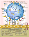
Figure 4. Optimization of ACTeff cells for broad delivery into widespread metastases (melanoma and others)
Bioengineering of CD8+ or CD4+ ACTeff cells with vastly improved capacity for homing into widespread, metastatic tissues is now possible by combinatorially leveraging and integrating new glycoengineering and genetic engineering technologies with the latest knowledge on immune cell homing and cancer metastatic circuitries. As shown, suboptimal and/or minimal native glycosylation of CD44 and PSGL-1 on ACTeff cells could be compensated for using a (1) cell-extrinsic GPS approach with requisite α1,3FT (e.g. FTVI or others) in generation of (2) E-selectin-binding CD44-sLeX (HCELL) and E/P-selectin binding PSGL-1-sLeX (CLA) homing determinants. GPS may advantageously generate additional, unidentified selectin-binding glycoprotein and glycolipid homing determinants (not shown). Consequent bimolecular association of HCELL with VLA-4 via a Rap/Rac signaling mechanism, or of PSGL-1 with VLA-4 (not shown), would activate VLA-4 adhesion to VCAM-1 via a ‘step-2 bypass’. (3–4) Cell-intrinsic creation of HCELL and CLA is shown, whereby viral transduction or transfection of mod-RNA, cDNA, or CRISPR-based platforms encoding α1,3FT (e.g. FTVI or others) would result in its cytoplasmic translation, insertion into the golgi compartment, and heightened synthesis of sLeX-selectin binding moieties on CD44 and PSGL-1 (and possibly other glycoproteins and glycolipids, not shown) transiting the secretory pathway. Genetically introduced (5) CXCR1 or CXCR2, normally low or absent on ACTeff cells, would prime Gαi signaling and homing responses when bound by cognate chemokines, CXCL1 or CXCL8, expressed by melanoma cells (or by other cancer types). (6) Genetic overexpression of αVβ3 or Mac-1 (αMβ2), also normally absent or low on ACTeff cells would, when rendered fully active potentially by (7) HCELL/PSGL-1 ‘step-2 bypass’ biomolecular association or by (8) CXCR1/CXCR2 chemokine receptor signaling, bind a plethora of diverse tumor endothelial adhesive proteins as shown. (9) Lesional targeting and homing specificity could be improved through positive selection and/or genetic overexpression of multiple different TCR, TCRgm or CAR receptors (and co-stimulators) recognizing diverse TA’s and with capacities to activate integrins as shown. (10) Preconditioning regimens applied either prior to and/or following ACTeff cell infusion could synergistically enhance trafficking capabilities through augmentation of tumor endothelial or ACTeff pro-homing determinants, including adhesion molecules, chemokines and chemokine receptors, and TA. Incorporation of inducible suicide genes (to limit ACTeff-associated cytokine storms and inflammation), immune checkpoint blockers, and inhibitors of immune-evasive mechanisms, these innovative homing and effector enhancement strategies could vastly improve immunotherapeutic outcomes in advanced cancer patients with widespread metastases.

References

    1. Papaioannou NE, Beniata OV, Vitsos P, et al. Harnessing the immune system to improve cancer therapy. Ann Transl Med. 2016;4:261. - PMC - PubMed
    1. Khalil DN, Smith EL, Brentjens RJ, et al. The future of cancer treatment: immunomodulation, CARs and combination immunotherapy. Nat Rev Clin Oncol. 2016;13:273–290. - PMC - PubMed
    1. Houot R, Schultz LM, Marabelle A, et al. T-cell-based Immunotherapy: Adoptive Cell Transfer and Checkpoint Inhibition. Cancer Immunol Res. 2015;3:1115–1122. - PubMed
    1. Lee N, Zakka LR, Mihm MC, Jr, et al. Tumour-infiltrating lymphocytes in melanoma prognosis and cancer immunotherapy. Pathology. 2016;48:177–187. - PubMed
    1. Fu H, Ward EJ, Marelli-Berg FM. Mechanisms of T cell organotropism. Cell Mol Life Sci. 2016 - PMC - PubMed

Publication types

Substances