. 2017 Mar 27:2:17037.
doi: 10.1038/nmicrobiol.2017.37.

Dengue virus NS2B protein targets cGAS for degradation and prevents mitochondrial DNA sensing during infection

Affiliations

Dengue virus NS2B protein targets cGAS for degradation and prevents mitochondrial DNA sensing during infection

Sebastian Aguirre et al. Nat Microbiol. .

Abstract

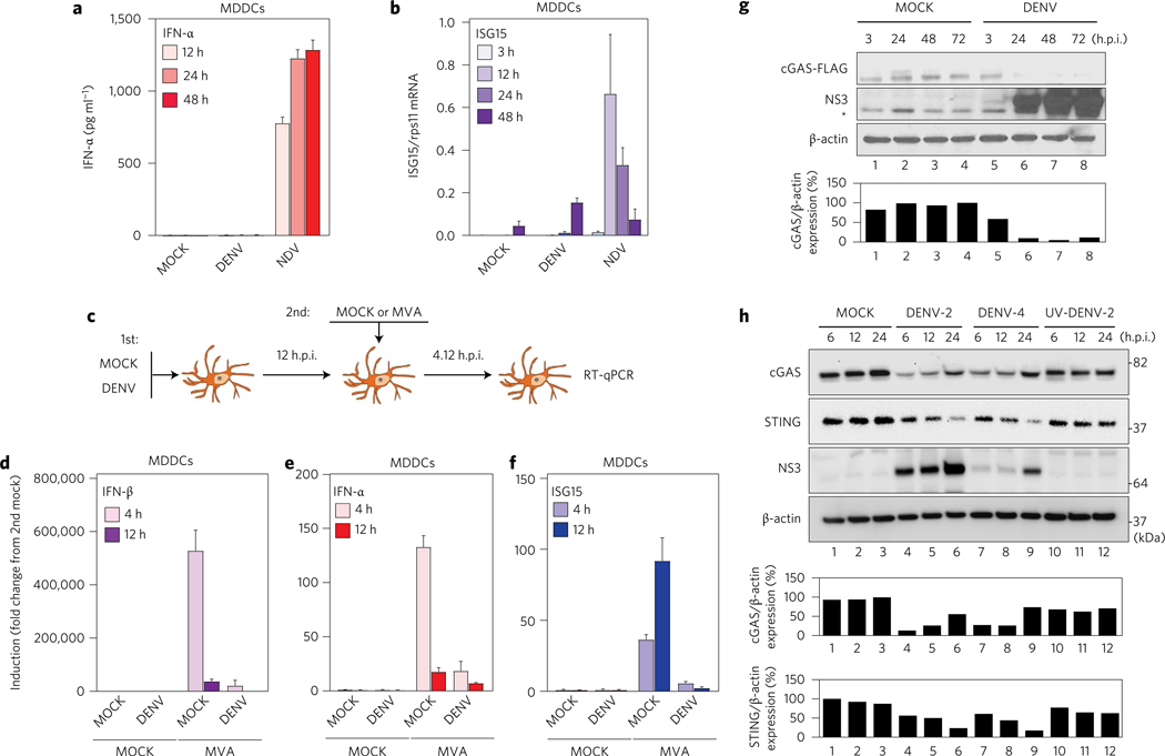
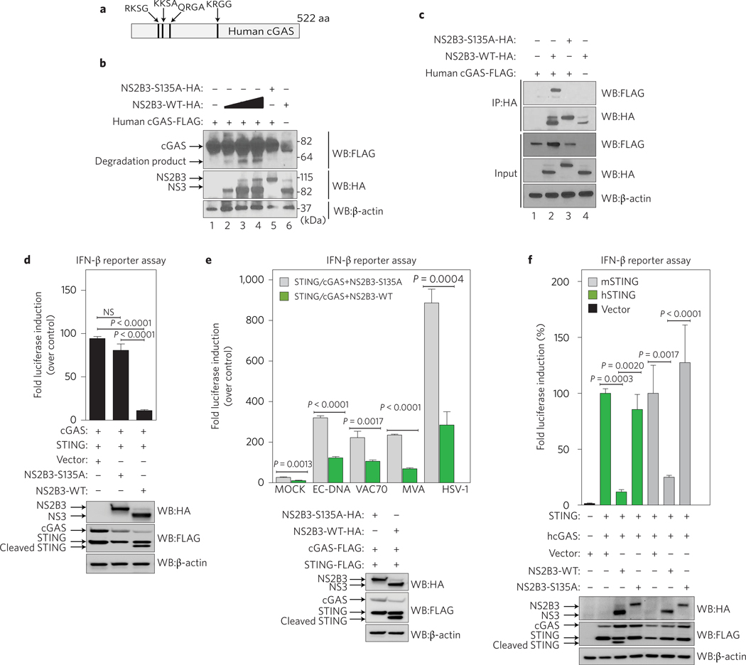
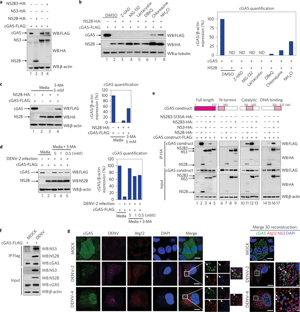
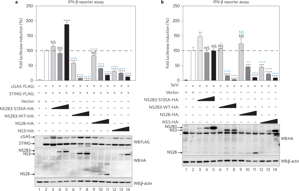
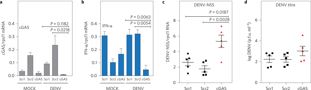
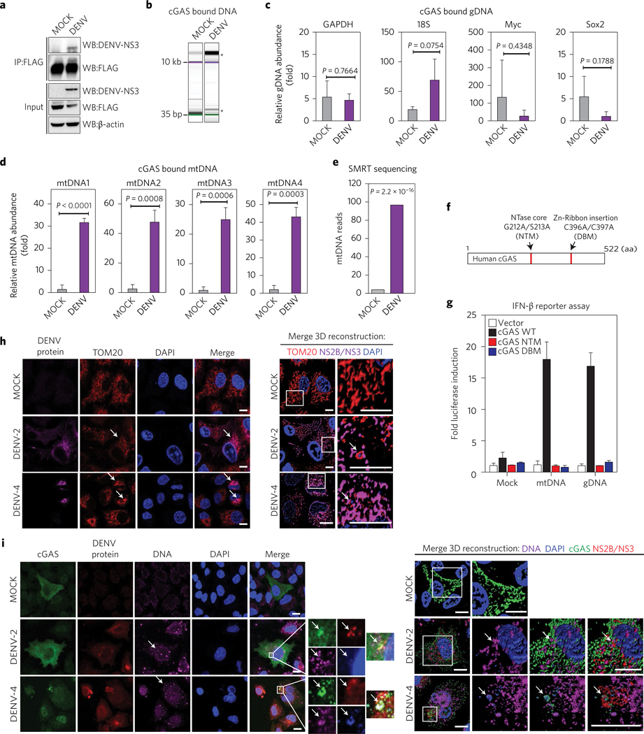
During the last few decades, the global incidence of dengue virus (DENV) has increased dramatically, and it is now endemic in more than 100 countries. To establish a productive infection in humans, DENV uses different strategies to inhibit or avoid the host innate immune system. Several DENV proteins have been shown to strategically target crucial components of the type I interferon system. Here, we report that the DENV NS2B protease cofactor targets the DNA sensor cyclic GMP-AMP synthase (cGAS) for lysosomal degradation to avoid the detection of mitochondrial DNA during infection. Such degradation subsequently results in the inhibition of type I interferon production in the infected cell. Our data demonstrate a mechanism by which cGAS senses cellular damage upon DENV infection.


Conflict of interest statement

Competing interests

The authors declare no competing financial interests.

Figures

Figure 1 |
Figure 1 |. cGAS is degraded during DENV infection.
Human MDDCs were infected with MOCK, DENV or NDV at MOI = 0.5, and samples of cells and supernatants were collected at 3, 12, 24 and 48 h.p.i. a, Quantification of IFN-α protein in cell supernatants by ELISA. b, Quantification of ISG15 mRNA by RT-qPCR (relative to rps11 mRNA). Results are expressed as mean ± s.e.m. from three biological replicates. c, Scheme for co-infection experiment. d, IFN-β mRNA quantification by RT–qPCR (relative to rps11). e, As in d, for IFN-α mRNA. f, As in d, for ISG15 mRNA. Results are representative of two independent experiments and are expressed as mean ± s.e.m. from three biological replicates. g, Western blot analysis of human cGAS during MOCK or DENV-2 infection (MOI = 5) conditions. cGAS-FLAG tagged was detected using anti-FLAG antibody. DENV infection was detected using a polyclonal rabbit serum raised against the helicase domain of DENV-2 NS3 protein. *Unspecific band. Antibody anti-β-actin was used to measure total loaded protein in western blot. h, Protein analysis by western blot of primary MDDCs treated with MOCK, DENV-2 (MOI = 10), DENV-4 (MOI = 10) or same number of UV-DENV-2 particles. Detection of endogenous human cGAS, human STING and DENV NS3 protein (anti-NS3 serum was raised against DENV-2 NS3 but can also detect DENV-4 NS3, although less efficiently). Antibody anti-β-actin was used to measure total loaded protein in western blot. Densitometry analysis of cGAS and/or STING relative to β-actin is expressed as a percentage using ImageJ software for g and h, and plotted as bar graphs.
Figure 2 |
Figure 2 |. DENV protease complex counteracts the cGAS/STING pathway.
a, Graphic representation of human cGAS protein with the predicted DENV NS2B3 potential cleavage sites indicated. b, Co-expression of human cGAS and different amounts of DENV NS2B3 WT (0.2, 1 and 2 μg) or NS2B3–S135A (1 μg) analysed by western blot using anti-HA, anti-FLAG and anti-β-actin antibodies. c, Co-immunoprecipitation of cGAS by DENV NS2B3 WT or NS2B3–S135A and analysis by western blot using the same antibodies as in b. d, Evaluation of DENV NS2B3 protease complex as an antagonist of the cGAS/STING pathway by IFN-β reporter assay. 293T–IFN-β–FFLuc cells were transfected with plasmids encoding human STING, cGAS and either empty vector, DENV NS2B3 WT or proteolytically inactive mutant NS2B3–S135A. Results are representative of two independent experiments and are expressed as mean ± s.e. m. from three biological replicates (one-way ANOVA). e, IFN-β reporter assay. Cells expressing the same plasmids as in d were treated with MOCK, or purified E. coli DNA (EC DNA), synthetic 70mer of Vaccinia virus DNA (VAC70), infection by modified vaccinia Ankara virus (MVA), or infection by HSV-1 to trigger the cGAS/STING pathway. Results are representative of two independent experiments and are expressed as mean ± s.e.m. from three biological replicates. (unpaired Student’s t-test). f, IFN-β reporter assay using cells expressing (human cGAS and human STING) or (human cGAS and murine STING) to induce the promoter in the presence of empty vector, DENV NS2B3 WT or NS2B3–S135A for each case. Results are representative of two independent experiments and are expressed as mean ± s.e.m. from three biological replicates (one-way ANOVA). WB, western blot; IP, immunoprecipitation; NS, not significant.
Figure 3 |
Figure 3 |. NS2B protease cofactor degrades cGAS in an autophagy–lysosome-dependent mechanism.
a, Evaluation of cGAS degradation by NS2B3 protease or its components. Expression analysis of human cGAS and empty vector, DENV NS2B, NS3 or NS2B3, performed in 293T cells (48 h) by SDS–PAGE/western blot. b, Evaluation of the mechanism of cGAS degradation by DENV NS2B. Co-expression of cGAS-FLAG and DENV NS2B-HA in 293T cells in the presence of DMSO, Z-VAD-FMK (5uM), MG-132 (5 μM), clasto-lactacystin β-lactone (lactacystin) (1 μM), DBeQ (5 μM), chloroquine (5 μM) or NH4Cl (2 mM). cGAS, DENV NS2B and α-tubulin expression levels were analysed by western blot. Bar graph: densitometry analysis of cGAS protein by ImageJ software. c,d, Analysis of cGAS degradation by NS2B (c) or DENV-2 infection (MOI = 10) (d) in the presence of 3-MA (autophagosome formation inhibitor). Densitometry analysis of cGAS protein by ImageJ software. e, Analysis of the interaction between DENV HA-tagged NS2B, NS3, NS2B3 protease complex WT or S135A mutant with human full-length cGAS-FLAG or cGAS domains by co-immunoprecipitation in 293T cells. f, Analysis of cGAS and viral proteins interaction during infection. Immunoprecipitation of cGAS-FLAG in 293T cells MOCK or DENV-2 infected for 12 h. Immunodetection of viral proteins NS3 and NS2B by western blot, using specific antibodies. g, Analysis of cGAS in DENV-infected cells by immunofluorescence: A549 cells expressing cGAS-V5 were infected MOCK or with DENV-2 or DENV-4. At 24 or 48 h.p.i., cells were fixed and processed for indirect immunofluorescence (see Methods). Primary antibodies against V5 (cGAS), autophagy marker (Atg12) or DENV NS3 protein were used. Alexa Fluor-conjugated secondary antibodies −488 (green), −568 (red) and −647 (magenta) were used to detect the primary antibodies, respectively. Nuclei were stained with DAPI (blue). Images on the right correspond to the squared zoomed images in each panel. Scale bars, 10 μm. A detailed three-dimensional reconstruction of the Z-stacks is shown top right. Scale bars, 0.5 μm. Arrows mark co-localization of cGAS with autophagosomes and viral protein. ND, not detected.
Figure 4 |
Figure 4 |. DENV protease complex and its components counteract the cGAS/STING pathway.
Evaluation of the DENV protease components as antagonists of the cGAS/STING pathway by IFN-β reporter assay. 293T reporter cells were co-transfected with plasmids encoding human cGAS, STING, plus empty vector, proteolytically inactive NS2B3 mutant (NS2B3–S135A), DENV WT protease (NS2B3 WT), NS2B or NS3 expressed individually. In every case, three different plasmids concentrations were used to evaluate a dose-dependent effect. a, The cGAS/STING pathway was activated by co-expression of the two proteins. Results are representative of two independent experiments and are expressed as mean ± s.e.m. from three biological replicates. P values: lanes 2–3, P = 0.2783; lanes 2–4, P = 0.8536; lanes 2–5, 2–6, 2–7 and 2–8, P < 0.0001; lanes 2–9, P = 0.0822; lanes 2–10, 2–11, 2–12, 2–13 and 2–14, P < 0.0001; lanes 3–6, 3–9 and 3–12, P < 0.0001; lanes 4–7, 4–10 and 4–13, P < 0.0001; lanes 5–8, 5–11 and 5–14, P < 0.0001 (one-way ANOVA). b, Infection with Sendai virus (SeV) as an inducer of the RIG-I-like receptors pathway. Results are representative of two independent experiments and are expressed as mean ± s.e.m. from three biological replicates. P values: lanes 2–3, P = 0.0022; lanes 2–4, P > 0.999; lanes 2–5, P > 0.999; lanes 2–6, P > 0.999; lanes 2–7 and 2–8, P < 0.0001; lanes 2–9, P = 0.4737; lanes 2–10, P = 0.0003; lanes 2–11, P < 0.0001; lanes 2–12, P = 0.0002; lanes 2–13 and 2–14, P < 0.0001; lanes 3–6, P = 0.0152; lanes 3–9, P = 0.4481; lanes 3–12, P < 0.0001; lanes 4–7, P < 0.0001; lanes 4–10, P = 0.0018; lanes 4–13, P < 0.0001; lanes 5–8, 5–11 and 5–14, P < 0.0001 (one-way ANOVA). NS, not significant. Protein expression control for a and b is shown. *Unspecific band. Asterisk in black represents specific P values obtained against empty vector control (lane 2 condition). Asterisk in blue represents P values obtained against the corresponding concentration of NS2B3–S135A plasmid (lanes 3 to 5 conditions).
Figure 5 |
Figure 5 |. cGAS restricts DENV replication.
Silencing of endogenous cGAS in primary human MDDCs. MDDCs from five independent donors were transfected with a specific cGAS siRNA or two different scramble siRNA as described in the Methods. At 48 h.p.i., cells were MOCK or DENV-2-infected (MOI = 0.5). a, Relative levels of cGAS mRNA were quantified by RT–qPCR at 4 h.p.i. b, Relative levels of IFN-α mRNA were quantified by RT–qPCR at 24 h.p.i. (data shows one representative donor out of five). Results are one representative of five independent experiments and are expressed as mean ± s.e.m. from three biological replicates (one-way ANOVA). c, Accumulation of DENV RNA was measured by RT–qPCR at 48 h.p.i. for the five donors tested. Results are expressed as mean from each of the five independent donors. Error bars represent mean ± s.e.m. d, Infectious DENV particles were quantified in the supernatant at 24 h.p.i. by plaque assay. Data show the mean ± s.e.m for five independent donors tested and showed a normal distribution.
Figure 6 |
Figure 6 |. cGAS binds mitochondrial DNA (mtDNA) during DENV infection.
a, cGAS immunoprecipitation (IP). Total lysates and IP fractions were used to detect cGAS-FLAG, DENV NS3 and β-actin by SDS–PAGE/western blot. b, Analysis of DNA bound to IP cGAS from MOCK, DENV conditions by DNA electrophoresis (Bioanalyzer). *Enriched DNA bands. Relative enrichment of genomic DNA in cGAS pulldown from MOCK and DENV conditions. c, RT–qPCR for GAPDH, 18S, Myc and Sox2 DNAs, relative to EGFP DNA. d, Relative enrichment of mtDNA in cGAS pulldown from both conditions. RT–qPCR for four sets of primers that amplify fragments from different regions of the human mtDNA. Data represent mean ± s.e.m. from three IP experiments, (unpaired Student’s t-test). e, SMRT sequencing analysis of purified DNA from cGAS pulldowns in MOCK or DENV conditions (Fisher’s exact test). f, Schematic representation of human cGAS aminoacidic sequence, NTase mutant (G212A/S213A) or DNA binding core domain mutant (C396A/C397A). NTM, nucleotidyltransferase mutant; DBM, DNA binding mutant. g, IFN-reporter assay. 293T–STING–IFN-β–Luc expressing cGAS WT, or cGAS mutants were stimulated with MOCK, mtDNA or gDNA and luciferase induction was measured. Data are expressed as mean ± s.e.m. from three biological replicates h, Analysis of mitochondria distribution and morphology during DENV infection by immunofluorescence. A549 cells were infected MOCK or with DENV-2 or DENV-4. At 24 h.p.i. cells were fixed and stained with TOM20, DENV-2 NS2B or DENV-4 NS3. Alexa Fluor-conjugated secondary antibodies −568 (red) and −647 (magenta) were used to detect the primary antibodies, respectively. Nuclei were stained with DAPI (blue). Arrows mark co-localization of aggregated mitochondria and viral proteins. A detailed three-dimensional reconstruction of the Z-stacks is shown on the right. i, Detection of cytoplasmic DNA by immunofluorescence. A549 cells expressing cGAS-V5 were infected MOCK or with DENV-2 or DENV-4. Primary antibodies against V5 (cGAS), single-stranded DNA, DENV-2 NS2B or DENV-4 NS3 protein were used. Alexa Fluor-conjugated secondary antibodies −488 (green), −647 (magenta) and −568 (red) were used to detect the primary antibodies, respectively. Nuclei were stained with DAPI (blue). Images on the right indicate the squared zoom in each panel. A detailed three-dimensional reconstruction of the Z-stacks is shown on the right. Scale bars, 10 μm. Arrows mark co-localization of cGAS with viral protein and DNA.

Comment in

References

    1. Uchida L. et al. The dengue virus conceals double-stranded RNA in the intracellular membrane to escape from an interferon response. Sci. Rep 4, 7395 (2014). - PMC - PubMed
    1. Espada-Murao LA & Morita K. Delayed cytosolic exposure of Japanese encephalitis virus double-stranded RNA impedes interferon activation and enhances viral dissemination in porcine cells. J. Virol 85, 6736–6749 (2011). - PMC - PubMed
    1. Goubau D, Deddouche S & Reis e Sousa C. Cytosolic sensing of viruses. Immunity 38, 855–869 (2013). - PMC - PubMed
    1. Bhatt S. et al. The global distribution and burden of dengue. Nature 496, 504–507 (2013). - PMC - PubMed
    1. Welsch S. et al. Composition and three-dimensional architecture of the dengue virus replication and assembly sites. Cell Host Microbe 5, 365–375 (2009). - PMC - PubMed

MeSH terms