. 2017 Mar 27;13(3):e1006687.
doi: 10.1371/journal.pgen.1006687. eCollection 2017 Mar.

Reduced dosage of β-catenin provides significant rescue of cardiac outflow tract anomalies in a Tbx1 conditional null mouse model of 22q11.2 deletion syndrome

Affiliations

Reduced dosage of β-catenin provides significant rescue of cardiac outflow tract anomalies in a Tbx1 conditional null mouse model of 22q11.2 deletion syndrome

Silvia E Racedo et al. PLoS Genet. .

Abstract

The 22q11.2 deletion syndrome (22q11.2DS; velo-cardio-facial syndrome; DiGeorge syndrome) is a congenital anomaly disorder in which haploinsufficiency of TBX1, encoding a T-box transcription factor, is the major candidate for cardiac outflow tract (OFT) malformations. Inactivation of Tbx1 in the anterior heart field (AHF) mesoderm in the mouse results in premature expression of pro-differentiation genes and a persistent truncus arteriosus (PTA) in which septation does not form between the aorta and pulmonary trunk. Canonical Wnt/β-catenin has major roles in cardiac OFT development that may act upstream of Tbx1. Consistent with an antagonistic relationship, we found the opposite gene expression changes occurred in the AHF in β-catenin loss of function embryos compared to Tbx1 loss of function embryos, providing an opportunity to test for genetic rescue. When both alleles of Tbx1 and one allele of β-catenin were inactivated in the Mef2c-AHF-Cre domain, 61% of them (n = 34) showed partial or complete rescue of the PTA defect. Upregulated genes that were oppositely changed in expression in individual mutant embryos were normalized in significantly rescued embryos. Further, β-catenin was increased in expression when Tbx1 was inactivated, suggesting that there may be a negative feedback loop between canonical Wnt and Tbx1 in the AHF to allow the formation of the OFT. We suggest that alteration of this balance may contribute to variable expressivity in 22q11.2DS.


Conflict of interest statement

The authors have declared that no competing interests exist.

Figures

Fig 1
Fig 1. Constitutive β-catenin expression in the AHF promotes differentiation.
(A) Whole mount in situ hybridization (WMISH) of Wnt2 in a wild type (Tbx1+/+) embryo at embryonic day 9.5 (E9.5) and a sagittal section from the heart region of the embryo is shown in B. (C) Mef2c-AHF-Cre lineage in a wild type embryo in a sagittal section. (D) Visualization of β-catenin signaling using a TCF/Lef:H2B-GFP reporter allele in a sagittal section of a wild type embryo. (E) WMISH of Tbx1 in a wild type embryo. (F) Mef2c-AHF-Cre lineage shown by green fluorescence in a wild type embryo (Mef2c-AHF-Cre/+;ROSA26-GFP f/+) at E9.5. Numbers denote the pharyngeal arches 1, 2 and 3. (G) Whole mount embryo showing the region that has been dissected for the experiments (rectangle in dorsal to the heart). (H) The micro-dissected region containing the Mef2c-AHF-Cre lineage is shown from a frontal view. (I) Comparison of global gene expression changes in micro-dissected tissues of β-catenin GOF and β-catenin LOF embryos at E9.5. Differentially expressed genes (p < 0.05 and FC > 1.5) in at least one of the two comparisons, β-catenin LOF vs control (x-axis) or β-catenin GOF vs control (y-axis), were plotted. Red dots denote cardiac differentiation genes. Abbreviations: heart (H), pharyngeal arch (PA), outflow tract (OFT), ventricle (V), atrium (A), FC = fold change.
Fig 2
Fig 2. Congenital heart defects in Tbx1 LOF embryos.
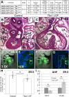
(A) Heart phenotype analysis of Tbx1 LOF embryos at E14.5 generated from two different crosses. PTA-VSD refers to a PTA associated with a ventricular septal defect (VSD) while PTA refers to hearts that did not show a VSD. Partial septation in Tbx1 LOF embryos means a PTA with presence of a short septum at some level of the OFT and complete septation between the ventricles. N = total number of hearts observed per group. Significance between Tbx1 LOF and controls calculated by Fisher’s exact test (p < 0.001). Note that Mef2c-AHF-Cre;Tbx1flox/flox embryos had additional phenotypes (three with double outlet right ventricle, DORV and one with tetralogy of Fallot, TOF, as indicated); (B) H&E histological sections of the heart of a control embryo at E14.5, with a typical ventricular septum is shown in the inset on the lower right part of the image. (C) Tbx1 LOF embryo with a PTA-VSD. (D) Mef2c-AHF-Cre lineage tracing by using a GFP reporter allele in a control embryo at E9.5 and a representative sagittal section is shown in E. (F) Mef2c-AHF-Cre lineage tracing in a Tbx1 LOF embryo and a representative sagittal section of the embryo is shown in G. DAPI fluorescent stain to visualize nuclei and identify the tissue is shown in blue. (H) Mef2c-AHF-Cre lineage quantification from the area shown in the inset in G for control and Tbx1 LOF embryos. (I) Detection of Tbx1 and β-catenin by qRT-PCR in control and Tbx1 LOF embryos. Statistical significance of the difference in gene expression was estimated using two-tailed t-test, FC = fold change, p values < 0.05. Error bars = standard deviation (SD). Abbreviations: aorta (Ao), pulmonary trunk (PT), left atrium (LA), right atrium (RA), left ventricle (LV), right ventricle (RV), pharyngeal arch (PA), outflow tract (OFT), 1, 2 and 3 indicate the first, second and third pharyngeal arches (PA), respectively.
Fig 3
Fig 3. Opposing β-catenin and Tbx1 conditional mutants have same effect on expression of pro-differentiation genes in the AHF.
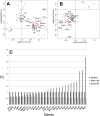
(A) Comparison of global gene expression changes in the micro-dissected AHF of β-catenin GOF and Tbx1 LOF embryos at E9.5. Plotted are differentially expressed genes (p < 0.05 and FC > 1.5) in at least one of the two comparisons, Tbx1 LOF vs controls (x-axis) or β-catenin GOF vs controls (y-axis). (B) Comparison of global gene expression changes in the micro-dissected AHF from Tbx1 GOF and β-catenin LOF embryos at E9.5. Plotted are differentially expressed genes (p < 0.05 and FC > 1.5) in at least one of the two comparisons, Tbx1 GOF vs controls (x-axis) or β-catenin LOF vs controls (y-axis). Red dots denote cardiac differentiation genes. (C) qRT-PCR analysis of Tbx1 LOF versus β-catenin LOF (FC = Fold Change) of selected genes demonstrating the opposite gene expression changes in these mutant embryos.
Fig 4
Fig 4. Phenotype analysis of significantly rescued Tbx1 LOF embryos.
Phenotypes in embryos in which one β-catenin loss of function allele to either Mef2c-AHF-Cre/+;Tbx1f/- embryos (A) or to Mef2c-AHF-Cre/+;Tbx1f/f embryos (B) was done to lower the dosage of β-catenin within the AHF. Significant rescue (p < 0.001, Fisher’s exact test) was obtained in both sets of double mutant embryos. Middle panels show the percent within the groups with PTA (C and C’), DORVor TOF associated with a VSD or PTA without VSD (dark grey) and those ones with partial septation or a normal heart (light grey). Partial septation in Tbx1 LOF embryos means a PTA with presence of a short septum at some level of the OFT (D and D’) and complete septation between the ventricles (8/50) while partial septation in the rescue genotype embryos (34/56) means a range of noticeably less severe phenotypes including: a longer partial OFT septation (E and E’) and either complete septation between ventricles (15/56) or VSD (10/56) or normal OFT (F and F’) with a VSD (4/56) or normal heart [5/56] (F and F’). Abbreviations: aorta (Ao), pulmonary trunk (PT), right ventricle (RV), outflow tract (OFT), PTA (persistent truncus arteriosus).
Fig 5
Fig 5. Histology analysis of representative embryos with the “rescue” genotype.
Transverse H&E histological sections of hearts from E14.5 significantly rescued embryos (Tbx1 LOF with loss of one allele of β-catenin in the Mef2c-AHF-Cre domain). (A) An embryonic heart with a thin septum formed that separates the Ao and the PT (top black arrow), and a small VSD compared to the usual PTA-VSD in Tbx1 LOF embryos (Fig 2C). (B) Example showing a DORV (middle black arrow), with a separate Ao and PT; normal ventricular septation is present. (C) Example of a heart showing a partially rescued septation between the Ao and PT (middle black arrow) and normal septation between the two ventricles (right black arrow). (D) Rescued septation between the Ao and PT. (E) Mef2c-AHF-Cre lineage quantification from the same area shown in Fig 2G for control, Tbx1 LOF and rescued embryos. (F) Detection of Tbx1 and β-catenin by qRT-PCR in control, Tbx1 LOF and rescued embryos. Statistical significance of the difference in gene expression was estimated using two-tailed t-test, FC = fold change, p values < 0.05. Error bars = standard deviation (SD). Abbreviations: RA = right atrium, RV = right ventricle, LA = left atrium, LV = left ventricle, Ao = Aorta, PT = pulmonary trunk, VS = ventricular septum, PTA = persistent truncus arteriosus, VSD = ventricular septal defect, DORV = double outlet right ventricle, OFT = outflow tract.
Fig 6
Fig 6. Gene expression of cardiac muscle differentiation genes are normalized in rescued embryos.
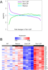
(A) Comparison of global expression changes. Differentially expressed genes (p < 0.05; n = 3,636) in either Tbx1 LOF or β-catenin LOF were sorted by their FC between Tbx1 LOF and controls (x-axis). The sorted genes were grouped (50 genes per group) and then the average expression changes for each group in the Tbx1 LOF, β-catenin LOF or rescue embryos (vs controls) were plotted in the y-axis. (B) Heatmap showing the expression of selected key genes increased in expression in Tbx1 LOF embryos but decreased in expression in β-catenin LOF embryos.
Fig 7
Fig 7. Model for Tbx1 and β-catenin function in the SHF.
In the model, the triangle represents the Mef2c-AHF-Cre lineage and the Tbx1 expression pattern, before migrating into the heart tube at E9.5. Left panel: Tbx1 expression is strongest in the AHF and weakest in the posterior SHF (pSHF) while Wnt/β-catenin expression is opposite. Left panel depicts a possible double negative feedback loop between the two genes in the SHF, required for normal cardiac outflow tract (OFT) formation. Middle panel shows increased differentiation in the AHF when Tbx1 is inactivated or β-catenin is constitutively active in the AHF. This results in premature differentiation within this tissue. Right panel depicts the rescue genotype in which both alleles of Tbx1 and one allele of β-catenin was inactivated in the AHF. Significant rescue of heart defects was obtained. Abbreviations: A = anterior, P = posterior, OFT = outflow tract.

Similar articles

Cited by

References

    1. Gross SJ, Stosic M, McDonald-McGinn DM, Bassett AS, Norvez A, Dhamankar R, et al. Clinical Experience with Single-Nucleotide Polymorphism-Based Noninvasive Prenatal Screening for 22q11.2 Deletion Syndrome. Ultrasound Obstet Gynecol. 2015. - PMC - PubMed
    1. Burn J, Goodship J. Developmental genetics of the heart. Curr Opin Genet Dev. 1996;6(3):322–5. - PubMed
    1. Goldmuntz E, Driscoll D, Budarf ML, Zackai EH, McDonald-McGinn DM, Biegel JA, et al. Microdeletions of chromosomal region 22q11 in patients with congenital conotruncal cardiac defects. J Med Genet. 1993;30(10):807–12. PubMed Central PMCID: PMCPMC1016559. - PMC - PubMed
    1. Chapman DL, Garvey N, Hancock S, Alexiou M, Agulnik SI, Gibson-Brown JJ, et al. Expression of the T-box family genes, Tbx1-Tbx5, during early mouse development. Dev Dyn. 1996;206(4):379–90. 10.1002/(SICI)1097-0177(199608)206:4<379::AID-AJA4>3.0.CO;2-F - DOI - PubMed
    1. Jerome LA, Papaioannou VE. DiGeorge syndrome phenotype in mice mutant for the T-box gene, Tbx1. Nat Genet. 2001;27(3):286–91. 10.1038/85845 - DOI - PubMed

Publication types

MeSH terms