. 2017 Mar 27;12(3):e0174136.
doi: 10.1371/journal.pone.0174136. eCollection 2017.

EBV induces persistent NF-κB activation and contributes to survival of EBV-positive neoplastic T- or NK-cells

Affiliations

EBV induces persistent NF-κB activation and contributes to survival of EBV-positive neoplastic T- or NK-cells

Honami Takada et al. PLoS One. .

Erratum in

Abstract

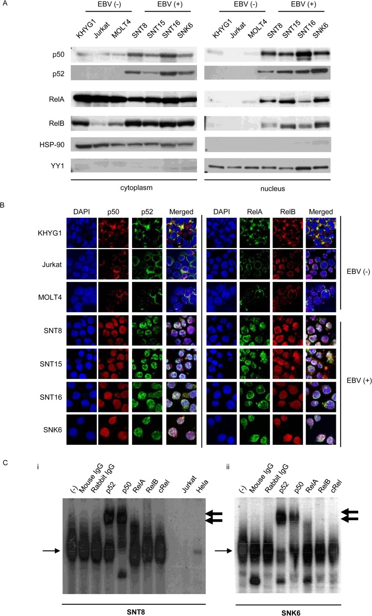
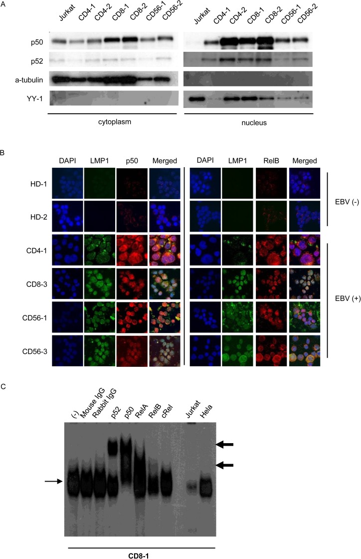
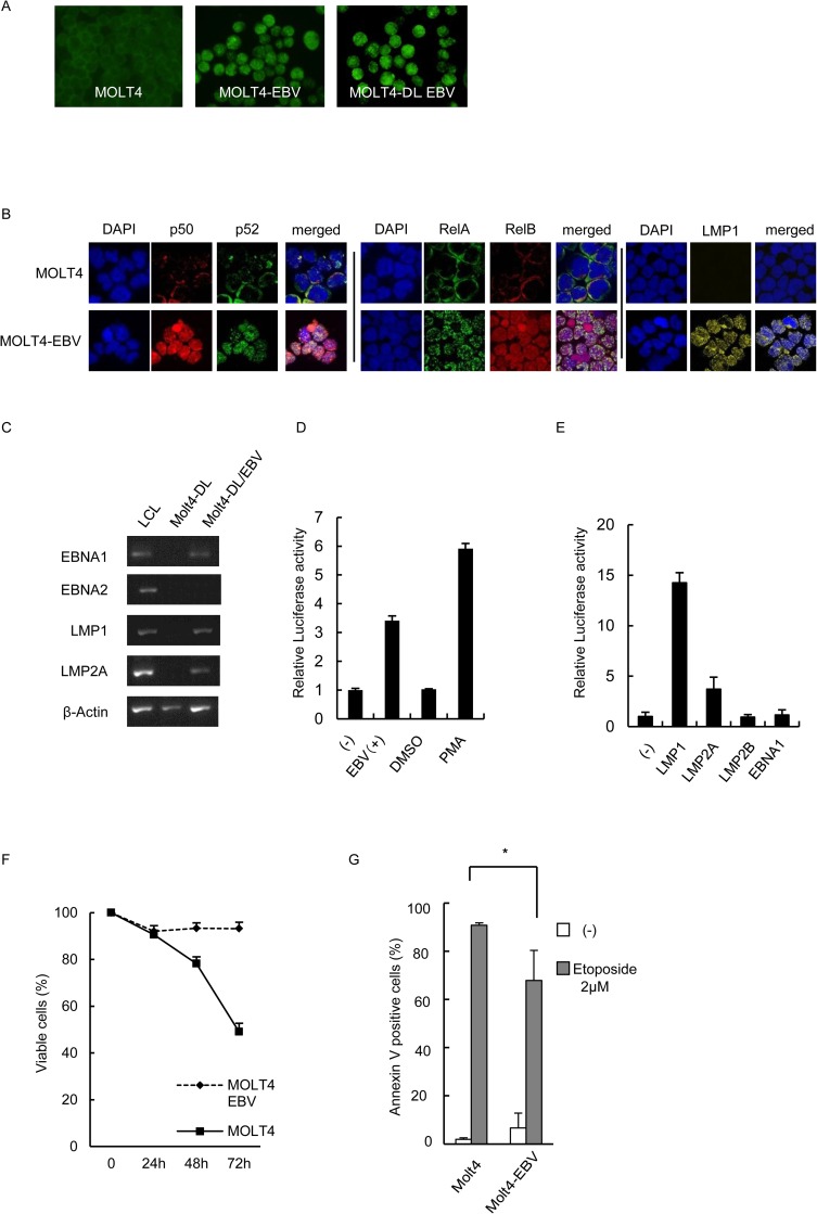
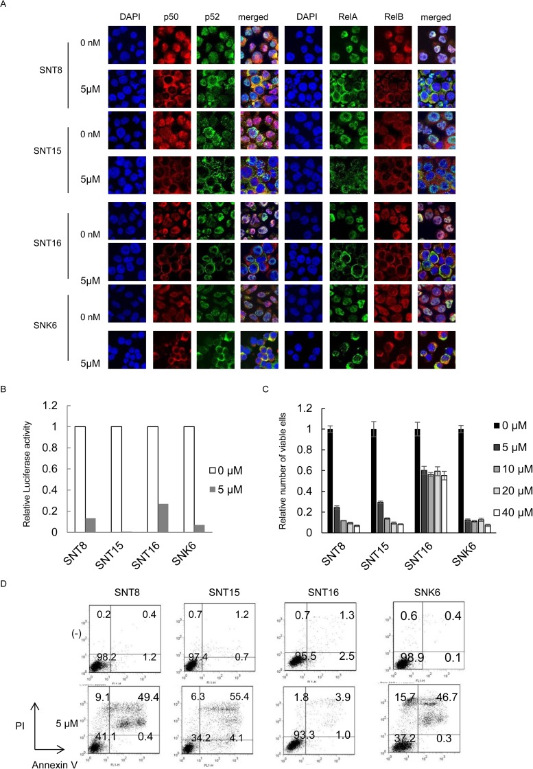
Epstein-Barr virus (EBV) has been detected in several T- and NK-cell neoplasms such as extranodal NK/T-cell lymphoma nasal type, aggressive NK-cell leukemia, EBV-positive peripheral T-cell lymphoma, systemic EBV-positive T-cell lymphoma of childhood, and chronic active EBV infection (CAEBV). However, how this virus contributes to lymphomagenesis in T or NK cells remains largely unknown. Here, we examined NF-κB activation in EBV-positive T or NK cell lines, SNT8, SNT15, SNT16, SNK6, and primary EBV-positive and clonally proliferating T/NK cells obtained from the peripheral blood of patients with CAEBV. Western blotting, electrophoretic mobility shift assays, and immunofluorescent staining revealed persistent NF-κB activation in EBV-infected cell lines and primary cells from patients. Furthermore, we investigated the role of EBV in infected T cells. We performed an in vitro infection assay using MOLT4 cells infected with EBV. The infection directly induced NF-κB activation, promoted survival, and inhibited etoposide-induced apoptosis in MOLT4 cells. The luciferase assay suggested that LMP1 mediated NF-κB activation in MOLT4 cells. IMD-0354, a specific inhibitor of NF-κB that suppresses NF-κB activation in cell lines, inhibited cell survival and induced apoptosis. These results indicate that EBV induces NF-κB-mediated survival signals in T and NK cells, and therefore, may contribute to the lymphomagenesis of these cells.


Conflict of interest statement

Competing Interests: The authors have declared that no competing interests exist.

Figures

Fig 1
Fig 1. NF-κB activation in Epstein-Barr virus positive T or NK cells.
(A) Western blotting for NF-κB protein localization in Epstein-Barr virus (EBV)-positive T or NK cell (EBV-T/NK cell) lines. The same samples were electrophoresed twice and transferred onto two membranes: one membrane for anti-p50 staining, and the other for p52, RelA, and RelB antibody staining. HSP90 and YY1 were proteins that localized to the cytoplasm and nucleus, respectively. (B) Immunofluorescent staining for NF-κB protein localization in EBV-T/NK cell lines. Immunofluorescent staining with anti-p50, p52, RelA, and RelB antibodies was performed as indicated. DAPI was used for nuclear staining. The EBV-negative cell lines, KHYG1, Jurkat, and MOLT4, were used as negative controls. Cells were analyzed via confocal microscopy. (C) Electrophoretic mobility shift assay of SNT8 (i) and SNK6 (ii) cells. Nuclear extracts were obtained, incubated with a KBF-1 probe containing the NF-κB binding sequence with or without indicated antibodies, and subjected to the assay. The thin, black arrows indicate KBF-1-binding NF-κB proteins. The thick, black arrows indicate supershifted bands. Nuclear extracts of Jurkat cells and HeLa cells treated with 10 ng/ml of TNF-α for 30 min were used as controls.
Fig 2
Fig 2. NF-κB activation in Epstein-Barr virus positive T or NK cells from patients with EBV-positive T or NK cell lymphoproliferative disorders.
(A) Western blotting for p50 and p52 protein localization in EBV-positive T or NK cells purified from patient peripheral blood mononuclear cells (PBMCs) using antibody-conjugated magnetic beads and immediately used in the assay. (B) Immunofluorescent staining for NF-κB protein localization in patient PBMCs. EBV-positive T or NK cells purified from patient PBMCs using antibody-conjugated magnetic beads were immediately subjected to immunofluorescent staining with anti-LMP1 and anti-p50 or anti-LMP1 and RelB antibodies. DAPI was used for nuclear staining. PBMCs from healthy donors were used as negative controls. Cells were analyzed via confocal microscopy. (C) Electrophoretic mobility shift assay of PBMCs from CD8-1. Nuclear extracts were obtained, incubated with KBF-1 containing the NF-κB binding sequence with or without indicated antibodies, and subjected to the assay. Thin, black arrows indicate KBF-1-binding NF-κB proteins. Thick, black arrows indicate supershifted bands. Nuclear extracts of Jurkat cells and HeLa cells treated with 10 ng/ml of TNF-α for 30 min were used as controls.
Fig 3
Fig 3. The effects of Epstein-Barr virus infection on MOLT4 cells
(A) The original (left), Epstein-Barr virus (EBV)-infected MOLT4 (middle), and EBV-infected MOLT4-DL cells (right) were obtained for EBNA1 staining. Green fluorescent cells are EBNA1-positive. (B) Immunofluorescent staining for NF-κB protein localization in EBV-infected MOLT4 cells (MOLT4-EBV). The original and MOLT4-EBV cells were subjected to immunofluorescent staining with anti-p50, p52, RelA, and RelB antibodies as indicated. DAPI was used for nuclear staining. Cells were analyzed via confocal microscopy. (C) Reverse-transcriptase PCR analysis of EBV protein-encoding gene expression in EBV-infected MOLT4-DL cells. Infection was confirmed via detection of mRNAs encoding the viral proteins EBNA1, LMP1, LMP2A, and LMP2B. EBNA2 expression was not detected, and the infection type was considered latency type 2. (D) Dual luciferase assay for NF-κB in EBV-infected MOLT4-DL cells. As a positive control, MOLT4-DL cells were treated with 20 nM phorbol 12-myristate 13-acetate (PMA) for 18 h. (E) Luciferase reporter gene assay using expression vectors for EBV proteins in MOLT4 cells. MOLT4 cells were transfected with 10 μg of EBNA1, LMP1, LMP2A, LMP2B, or empty vector as indicated, along with 10 μg of pNF-κB-luc and 1 μg of pRLSV40. Twelve hours after transfection, cells were harvested for the dual luciferase assay. Luciferase activity was normalized to Renilla luciferase activity and expressed as an increase relative to the control. Data are shown as the means ± standard deviations of 3 independent experiments. (F) The original and EBV-infected MOLT4 cells were cultured in 10% FCS–RPMI or FCS-free RPMI. The time-dependent viable cell numbers were examined by trypan-blue staining. Data are shown as the means ± standard deviations of 3 independent experiments. (G) The original and EBV-infected MOLT4 cells were cultured for 24 h in 10% FCS–RPMI with or without 2 μM VP16. The viable and apoptotic cell numbers were then determined by Annexin V staining, and the percentage of Annexin V-positive cells was determined. * p = 0.03245. Each experiment was independently performed 3 times, and the average data are presented.
Fig 4
Fig 4. The effects of IMD-0354 on NF-κB activity and cell survival in Epstein-Barr virus positive T- or NK-cells.
(A) Dual luciferase assay for NF-κB in SNT8, SNT15, SNT16, and SNK6 cells treated with IMD-0354. The cells were transfected with 10 μg of pNF-κB-luc and 1 μg of pRLSV40. Twelve hours after transfection, cells were treated with IMD-0354 for 24 hours and harvested for the dual luciferase assay. Luciferase activity was normalized to Renilla luciferase activity and expressed as an increase relative to the control. (B) SNT8, SNT15, SNT16, and SNK6 cells were treated with IMD-0354 for 24 hours and the viable cell numbers were estimated using the XTT assay and expressed in arbitrary units. Data are shown as the means ± standard deviations of 3 independent experiments. (C) SNT8, SNT15, SNT16, and SNK6 cells were treated with IMD-0354 as indicated for 24 h and subjected to immunofluorescent staining. NF-κB protein expression and localization were examined by immunofluorescent staining using anti-p50, p52, RelA, and RelB antibodies as indicated. DAPI was used for nuclear staining. Cells were analyzed via confocal microscopy. (D) SNT8, SNT15, SNT16, and SNK6 cells were treated with IMD-0354 as indicated for 24h and used in the assay. Cells were stained with Annexin V and PI and subsequently analyzed by flow cytometry.

Similar articles

Cited by

References

    1. Chan JKC, Quintanilla-Martinez L, Ferry JA, Peh S-C. Extranodal NK/T-cell lymphoma, nasal type In: Jaffe E, Harris N, Stein H, editors. WHO Classification of Tumors of Haematopoietic and Lymphoid Tissues. Lyon IARC Press; 2008. 285–288.
    1. Chan JKC, Jaffe ES, Ralfkiaer E, Ko Y-H. Aggressive NK-cell leukemia In: In: Jaffe E, Harris N, Stein H, editors. WHO Classification of Tumors of Haematopoietic and Lymphoid Tissues. Lyon. France: IARC Pre; 2008. 276–277.
    1. Swerdlow SH, Campo E, Pileri SA, Harris NL, Stein H, Siebert R, et al. The 2016 revision of the World Health Organization classification of lymphoid neoplasms. Blood. 2016;127(20):2375–90. PubMed Central PMCID: PMCPMC4874220. 10.1182/blood-2016-01-643569 - DOI - PMC - PubMed
    1. Quintanilla-Martinez L, Kimura H, Jaffe ES. EBV-positive T-cell lymphoproliferative disorders of childhood In: Jaffe E, Harris N, Stein H, editors. WHO Classification of Tumors of Haematopoietic and Lymphoid Tissues. Lyon IARC Press; 2008. 278–280.
    1. Kimura H, Ito Y, Kawabe S, Gotoh K, Takahashi Y, Kojima S, et al. EBV-associated T/NK-cell lymphoproliferative diseases in nonimmunocompromised hosts: prospective analysis of 108 cases. Blood. 2012;119(3):673–86. 10.1182/blood-2011-10-381921 - DOI - PubMed

MeSH terms

Substances

LinkOut - more resources