Review
. 2017 Mar 27;36(1):34.
doi: 10.1186/s40880-017-0201-z.

Progressive multifocal exophytic pontine glioblastoma: a case report with literature review

Affiliations
Review

Progressive multifocal exophytic pontine glioblastoma: a case report with literature review

Fanfan Chen et al. Chin J Cancer. .

Abstract

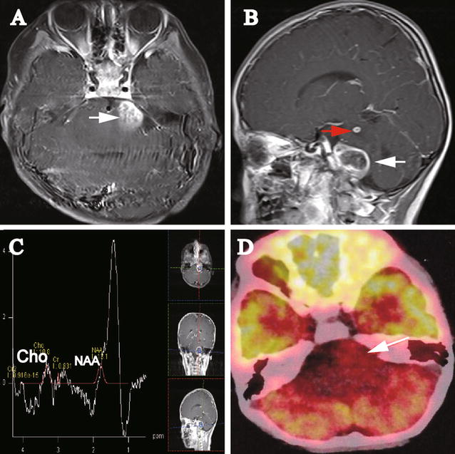
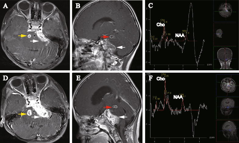
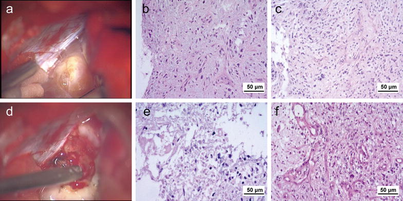
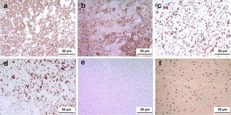
Multifocal pontine glioblastoma exhibiting an exophytic growth pattern in the cerebello-pontine angle (CPA) is rare. We present a case of a 5-year-old girl with consecutive neurological imaging and other clinical findings indicating progressive multifocal exophytic pontine glioblastoma. Three lesions were reported, of which two were initially presented, and one was developed 2 months later. One lesion demonstrated a progressing exophytic extension in the cistern of the left side of the CPA. The other two lesions were located and confined within the pons. Initial magnetic resonance imaging and positron emission tomography-computed tomography indicated low-grade glioma or inflammatory disease. However, 2 and 3 months later, subsequent magnetic resonance spectroscopy (MRS) displayed elevated choline and depressed N-acetyl aspartate peaks compared with the peaks on the initial MRS, indicating a high-grade glioma. Subtotal resection was performed for the CPA lesion. Histopathologic examination showed discrepant features of different parts of the CPA lesion. The patient received no further chemotherapy or radiotherapy and died 2 months after surgery. The multifocal and exophytic features of this case and the heterogeneous manifestations on neurological images were rare and confusing for both diagnosis and surgical decision-making. Our case report may contribute knowledge and helpful guidance for other medical doctors.

Keywords: Brainstem; Cerebello-pontine angle; Glioma; Multiple lesion; Pontine.


Figures

Fig. 1
Fig. 1
Images of the initial magnetic resonance imaging (MRI) and positron emission tomography–computed tomography (PET–CT) on Oct 8, 2015 for the 5-year-old girl with multiple posterior fossa lesions. A Axial gadolinium-enhanced MRI displays a lesion (2.0 cm × 2.6 cm; white arrow) in the cerebello-pontine angle (CPA). No relatively clear margin was observed between the lesion and the pons. B Sagittal gadolinium-enhanced MRI shows two lesions. The upper left pontine lesion (0.5 cm × 0.6 cm) was significantly gadolinium-enhanced with relatively clear margin (red arrow). From the sagittal view, the CPA lesion (white arrow) seemingly demonstrates clear boundary to the pons. C Magnetic resonance spectroscopy (MRS) of the lesion shows a decrease in choline (Cho) and N-acetyl aspartate (NAA). D PET–CT image demonstrates a hypometabolic lesion (white arrow) in the left side of the CPA
Fig. 2
Fig. 2
Images of repeated MRI on Dec 11, 2015 and Jan 12, 2016 for the 5-year-old girl with multiple posterior fossa lesions. A Axial gadolinium-enhanced MRI (Dec 11, 2015) displays the occurrence of a new lesion (0.8 cm × 0.9 cm, yellow arrow). The left CPA lesion is enlarged and extended along the cistern of the CPA (3.1 cm × 1.2 cm, white arrow). B Sagittal gadolinium-enhanced MRI (Dec 11, 2015) shows that the CPA lesion was enlarged and extended along the cistern of CPA (white arrow) and the upper left pontine lesion was enlarged (red arrow). C MRS (Dec 11, 2015) shows increased Cho and decreased NAA expression. The Cho:NAA ratio was also significantly increased. D Axial gadolinium-enhanced MRI (Jan 12, 2016) displays that both the CPA lesion (white arrow) and the new lesion in the right side of the pons are enlarged (1.1 cm × 1.3 cm, yellow arrow). The CPA lesion is extended along the cistern of the CPA. E Sagittal gadolinium-enhanced MRI (Jan 12, 2016) shows that both the CPA lesion (white arrow) and upper left pontine lesion (red arrow) were enlarged. F MRS (Jan 12, 2016) shows further increased Cho and decreased NAA expression. The Cho:NAA ratio was 2.33
Fig. 3
Fig. 3
Histopathologic examination of the CPA lesion with hematoxylin–eosin staining. a Macroscopically, the outer layer of CPA lesion is rubbery and nodule-like. b Under a microscope, the outer layer of the CPA lesion displays moderate mitoses and necrosis. c Under a microscope, the outer layer of the CPA lesion displays moderate vascularization. d Macroscopically, the internal portion of the lesion is softer and has a higher blood supply than the outer layer. e Under a microscope, the internal portion of the CPA lesion displays obvious mitoses and necrosis. f Under a microscope, the internal portion of the CPA lesion displays hyper-vascularization
Fig. 4
Fig. 4
Immunohistochemical examination of the tumor with diaminobenzidine staining. Glial fibrillary acidic protein (a), S-100 (b), P53 (c), and Ki-67 (d) are diffusely expressed. The expression of methylated O6-methylguanine-DNA methyltransferase (e) and isocitrate dehydrogenase 1 (f) is weak

Similar articles

Cited by

References

    1. Rubin G, Michowitz S, Horev G, Herscovici Z, Cohen IJ, Shuper A, et al. Pediatric brain stem gliomas: an update. Childs Nerv Syst. 1998;14(4–5):167–173. doi: 10.1007/s003810050205. - DOI - PubMed
    1. Freeman CR, Farmer JP. Pediatric brain stem gliomas: a review. Int J Radiat Oncol Biol Phys. 1998;40(2):265–271. doi: 10.1016/S0360-3016(97)00572-5. - DOI - PubMed
    1. Tomita T, Grahovac G. Cerebellopontine angle tumors in infants and children . Childs Nerv Syst. 2015;31(10):1739–1750. doi: 10.1007/s00381-015-2747-x. - DOI - PMC - PubMed
    1. Holman MA, Schmitt WR, Carlson ML, Driscoll CL, Beatty CW, Link MJ. Pediatric cerebellopontine angle and internal auditory canal tumors: clinical article. J Neurosurg Pediatr. 2013;12(4):317–324. doi: 10.3171/2013.6.PEDS1383. - DOI - PubMed
    1. Ragab A, Emara A, Shouker M, Ebied O. Prospective evaluation of the clinical profile and referral pattern differences of vestibular schwannomas and other cerebellopontine angle tumors. Otol Neurotol. 2012;33(5):863–870. doi: 10.1097/MAO.0b013e318255dd59. - DOI - PubMed

MeSH terms