. 2017 Oct;1863(10 Pt B):2601-2613.
doi: 10.1016/j.bbadis.2017.03.015. Epub 2017 Mar 25.

Global assessment of oxidized free fatty acids in brain reveals an enzymatic predominance to oxidative signaling after trauma

Affiliations

Global assessment of oxidized free fatty acids in brain reveals an enzymatic predominance to oxidative signaling after trauma

Tamil S Anthonymuthu et al. Biochim Biophys Acta Mol Basis Dis. 2017 Oct.

Abstract

Traumatic brain injury (TBI) is a major health problem associated with significant morbidity and mortality. The pathophysiology of TBI is complex involving signaling through multiple cascades, including lipid peroxidation. Oxidized free fatty acids, a prominent product of lipid peroxidation, are potent cellular mediators involved in induction and resolution of inflammation and modulation of vasomotor tone. While previous studies have assessed lipid peroxidation after TBI, to our knowledge no studies have used a systematic approach to quantify the global oxidative changes in free fatty acids. In this study, we identified and quantified 244 free fatty acid oxidation products using a newly developed global liquid chromatography tandem-mass spectrometry (LC-MS/MS) method. This methodology was used to follow the time course of these lipid species in the contusional cortex of our pediatric rat model of TBI. We show that oxidation peaked at 1h after controlled cortical impact and was progressively attenuated at 4 and 24h time points. While enzymatic and non-enzymatic pathways were activated at 1h post-TBI, enzymatic lipid peroxidation was the predominant mechanism with 15-lipoxygenase (LOX) contributing to the majority of total oxidized fatty acid content. Pro-inflammatory lipid mediators were significantly increased at 1 and 4h after TBI with return to basal levels by 24h. Anti-inflammatory lipid mediators remained significantly increased across all three time points, indicating an elevated and sustained anti-inflammatory response following TBI.

Keywords: Cardiolipin; Docosanoids; Eicosanoids; Lipid peroxidation; Octadecanoids; Pro-resolving mediators.


Figures

Figure 1
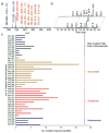
Figure 1. A new global LC-MS/MS method for oxidized FFA analysis
(a) Representative spectrum showing oxidized FFA identified using the newly developed LC-MS/MS method. (b) Extracted ion chromatogram of m/z 359.22 (DHA + 2O) – top layer – and m/z 335.222 (arachidonic acid + 2O) – bottom layer – showing multiple lipid species. (c) Number of species identified for each m/z based on the exact mass and retention time. With this methodology, 244 distinct oxidized FFA were identified. The identity of 40 species was confirmed by MS2 analysis.
Figure 2
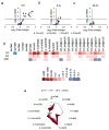
Figure 2. Octadecanoid production and temporal profile after TBI
Volcano plots showing changes in octadecanoids at 1 hr (a), 4 hr (b), and 24 hr (c) after TBI with respect to naïve. The species above the dotted line are significantly increased (p<0.05). (d) Heat map showing fold increase of octadecanoids at 1, 4, and 24 hr after TBI compared to naïve. Species confirmed with MS2 analysis are listed by name, whereas species that did not undergo fragmentation are marked with m/z at the aligned retention time (*p<0.05). (e) Radar plot showing the quantitation of confirmed octadecanoids after TBI. The radial axis is shown in log10 scale of pmol per nmol of phospholipid.
Figure 3
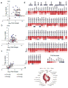
Figure 3. Eicosanoid production and temporal profile after TBI
Volcano plots showing changes in eicosanoids at 1 hr (a), 4 hr (b), and 24 hr (c) after TBI with respect to naïve. The species above the dotted line are significantly increased (p<0.05). (d) Heat map showing fold increase of eicosanoids at 1, 4, and 24 hr after TBI compared to naïve. Species confirmed with MS2 analysis are listed by name, whereas species that did not undergo fragmentation are marked with m/z at the aligned retention time (*p<0.05). (e) Radar plot showing the quantitation of confirmed eicosanoids after TBI. The radial axis is shown in log10 scale of pmol per nmol of phospholipid.
Figure 4
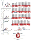
Figure 4. Docosanoid production and temporal profile after TBI
Volcano plots showing changes in docosanoids at 1 hr (a), 4 hr (b), and 24 hr (c) after TBI with respect to naïve. The species above the dotted line are significantly increased (p<0.05). (d) Heat map showing fold increase of docosanoids at 1, 4, and 24 hr after TBI compared to naïve. Species confirmed with MS2 analysis are listed by name, whereas species that did not undergo fragmentation are marked with m/z at the aligned retention time (*p<0.05). (e) Radar plot showing the quantitation of confirmed docosanoids after TBI. The radial axis is shown in log10 scale of pmol per nmol of phospholipid.
Figure 5
Figure 5. Production and temporal course of other oxidized fatty acids after TBI
Volcano plots showing changes in other oxidized fatty acids at 1 hr (a), 4 hr (b), and 24 hr (c) after TBI with respect to naïve. The species above the dotted line are the significantly increased (p<0.05). (c) Heat map showing fold increase of other oxidized fatty acids at 1, 4, and 24 hr after TBI compared to naïve. Individual species are marked with m/z at the aligned retention time (*p<0.05).
Figure 6
Figure 6. Source and inflammatory signaling of oxidized fatty acids after TBI
(a) Bar graph showing the amount of oxidized fatty acids (pmol per nmol of phospholipid) generated from enzymatic (COX, LOX, Cyt P450) or non-enzymatic mechanisms. (b) Time course quantifying pro- and anti-inflammatory oxidized free fatty acids after TBI. Inset showing increased anti-inflammatory lipid mediators at 24 hr compared to naïve with a return of pro-inflammatory lipid mediators to basal levels at this time point.
Figure 7
Figure 7
Pathways involved in oxidized FFA production after TBI.

References

    1. Faul XLM, Wald MM, Coronado V. N.C.o.I.P.a.C. Centers for Disease Control and Prevention, editor. Traumatic Brain Injury in the United States: Emergency Department Visits, Hospitalizations and Deaths, 2002–2006. Atlanta, GA: 2010.
    1. C.f.D.C.a. Prevention; N.C.f.I.P.a.C.D.o.U.I. Prevention, editor. Report to Congress on Traumatic Brain Injury in the United States: Epidemiology and Rehabilitation. Atlanta, GA: 2015. - PubMed
    1. Thurman DJ, Alverson C, Dunn KA, Guerrero J, Sniezek JE. Traumatic brain injury in the United States: A public health perspective. The Journal of head trauma rehabilitation. 1999;14:602–615. - PubMed
    1. Zaloshnja E, Miller T, Langlois JA, Selassie AW. Prevalence of long-term disability from traumatic brain injury in the civilian population of the United States, 2005. The Journal of head trauma rehabilitation. 2008;23:394–400. - PubMed
    1. Masel BE, DeWitt DS. Traumatic brain injury: a disease process, not an event. Journal of neurotrauma. 2010;27:1529–1540. - PubMed

Publication types

MeSH terms

Substances