. 2017 Jun:506:64-72.
doi: 10.1016/j.virol.2017.03.009. Epub 2017 Mar 24.

Porcine reproductive and respiratory syndrome virus (PRRSV) up-regulates IL-8 expression through TAK-1/JNK/AP-1 pathways

Affiliations

Porcine reproductive and respiratory syndrome virus (PRRSV) up-regulates IL-8 expression through TAK-1/JNK/AP-1 pathways

Yihao Liu et al. Virology. 2017 Jun.

Abstract

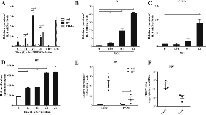
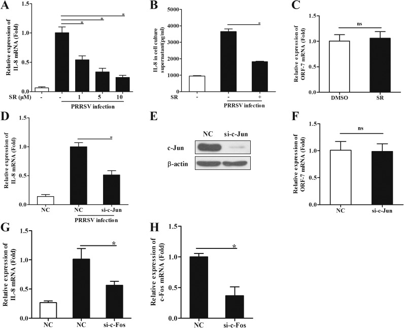
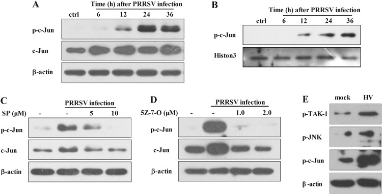
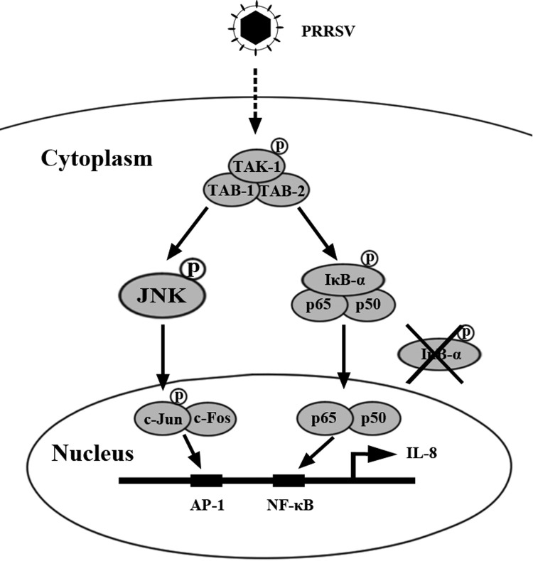
The acute phase of respiratory distress caused by porcine reproductive and respiratory syndrome virus (PRRSV) is likely a consequence of the release of inflammatory cytokines in the lung. IL-8, the main chemokine and activator of neutrophils, might be related to the lung injury upon PRRSV infection. In this study, we showed that PRRSV induced IL-8 expression in vivo and in vitro. Subsequently, we demonstrated that JNK and NF-κB pathways were activated upon PRRSV infection and required for the enhancement of IL-8 expression. We further verified that PRRSV-activated TAK-1 was essential for the activation of JNK and NF-κB pathways and IL-8 expression. Moreover, we revealed an AP-1 binding motif in the cloned porcine IL-8 (pIL-8) promoter, and deletion of this motif abolished the pIL-8 promoter activity. Finally, we found that the JNK-activated AP-1 subunit c-Jun was critical for the up-regulation of IL-8 expression by PRRSV. These data suggest that PRRSV-induced IL-8 production is likely through the TAK-1/JNK/AP-1 pathways.

Keywords: AP-1; IL-8; JNK; PRRSV; TAK-1.


Figures

Fig. 1.
Fig. 1
PRRSV induces IL-8 expression in vitro and in vivo. (A) PAMs were inoculated with HP-PRRSV isolate HV, CH-1a strain, heat inactivated HV (h-HV) at a multiplicity of infection (MOI) of 1, LPS, or medium alone. Total RNA was extracted from cell lysates at 6, 12, 24, and 36 h post inoculation. (B, C) PAMs were infected with HV or CH-1a at an MOI of 0.02, 0.1 and 1, and then the Total RNA was extracted from cell lysates at 24 and 36 hpi. IL-8 mRNA was quantified by real-time PCR, and results were normalized to GAPDH and expressed as fold induction over medium alone. (D) Supernatants were harvested at 6, 12, 24, and 36 hpi post PRRSV infection (MOI=1), and levels of IL-8 (pg/ml) released were determined by ELISA. (E) Pigs were infected intranasally with 2 ml (105 TCID50 virus/ml) HV. Samples were collected at 5 days post infection. IL-8 mRNA was quantified by real-time PCR. And results were normalized to GAPDH and expressed as fold induction over samples from uninfected pigs. (F) PRRSV ORF-7 mRNA was quantified by real-time PCR. Differences were evaluated by Student's t-test (*, P<0.05).
Fig. 2.
Fig. 2
PRRSV up-regulates IL-8 expression through JNK and NF-κB pathways. (A) PAMs were pretreated with inhibitors of JNK (SP600125 (SP), 10 μM), NF-κB (BAY11-7082 (BAY), 1 μM), P38 (SB202190 (SB), 5 μM), MEK (PD98059 (PD), 10 μM), PKC (GF-109203X (GF), 1 μM), PI3K (LY294002 (LY), 5 μM), or DMSO control for 1 h, and then cells were inoculated with or without PRRSV (MOI=0.1). Twenty-four hours later, total RNAs were extracted for IL-8 mRNA analysis by real-time PCR. (B) PAMs were pretreated with JNK or NF-κB inhibitor at different doses (1, 5, 10 μM for SP and 0.1, 0.5, 1 μM for BAY) or DMSO control for 1 h, and then cells were inoculated with or without PRRSV (MOI=0.1). Twenty-four hours later, total RNAs were extracted for IL-8 mRNA analysis by real-time PCR. (C) PAMs were pretreated with JNK (5 μM) or NF-κB (0.5 μM) inhibitor or DMSO control for 1 h, and then cells were inoculated with or without PRRSV (MOI=0.1). IL-8 production in cell supernatants was detected at 24 hpi by ELISA. (D) PRRSV ORF-7 mRNA was analyzed by real-time PCR. (E, F) PAMs were inoculated with PRRSV (MOI=0.1), and cells were harvested at 6, 12, 24, and 36 hpi for p-JNK, JNK (D), p-IκB and IκB (E) analysis by Western blotting. Differences were evaluated by Student's t-test (*, P<0.05).
Fig. 3.
Fig. 3
TAK-1 is required in PRRSV- induced IL-8 expression. (A) PAMs were pretreated with the inhibitor of TAK-1 ((5Z)-7-Oxozeaenol) at different concentrations (0.5 and 1 μM), or DMSO control for 1 h, and then cells were inoculated with or without PRRSV (MOI=0.1). Twenty-four hours later, total RNAs were extracted for IL-8 mRNA analysis by real-time PCR. (B) PAMs were pretreated with TAK-1 inhibitor at 1 μM. IL-8 production in cell supernatants was detected at 24 hpi using ELISA. (C) PAMs were pretreated with 1 μM (5Z)-7-Oxozeaenol or DMSO for 1 h, and then cells were inoculated with or without PRRSV (MOI=0.1). Twenty-four hours later, PRRSV ORF-7 mRNA was analyzed by real-time PCR. (D) PAMs were inoculated with PRRSV (MOI=0.1). Cells were then harvested at 6, 12, 24, and 36 h for p-TAK-1 and TAK-1 analysis by Western blotting. (E) PAMs were pretreated with the TAK-1 inhibitor at doses of 1 or 2 μM for 1 h, and then infected with HP-PRRSV (MOI=0.1). At 24 hpi, cells were harvested for p-JNK and JNK analysis by Western blotting. (F) PAMs were pretreated with the TAK-1 inhibitor at doses of 0.1, 0.5, 1 or 2 μM for 1 h, and then infected with HV (MOI=0.1). At 24 hpi, cells were harvested for p-IκB and IκB analysis by Western blotting. Differences were evaluated by Student's t-test (*, P<0.05).
Fig. 4.
Fig. 4
Cloning, sequence analysis, and characterization of porcine IL-8 promoter. (A) Cloning and sequence analysis of the 2751-bp porcine IL-8 promoter. The positions of the putative regulatory motifs are relative to the transcription initiation site. Schematic representation of the porcine IL-8 promoter and promoter deletion mutants inserted into pGL3 basic luciferase vectors: −2628/+123-luc, −1982/+123-luc, −1278/+123-luc, −939/+123-luc, −515/+123-luc, −187/+123-luc, −108/+123-luc, and −76/+123-luc porcine IL-8 promoter vectors. The relative lengths and positions of the 5′-ends of these fragments are indicated. (B) The porcine IL-8 promoter vectors or pGL3 empty vector were transfected into Marc-145 cells. Twenty-four hours later, cells were inoculated with PRRSV (MOI=1) or medium. Cells were harvested to determine luciferase activity at 36 hpi. (C) Schematic diagram represents porcine IL-8 deletion mutant constructs including −187/123(ΔAP-1)-luc, −187/123(ΔNF-κB)-luc, −187/123(ΔC/EBP β)-luc, −187/123(ΔAP-1, NF-κB)-luc, −187/123(ΔAP-1, C/EBP β)-luc, −187/123(ΔC/EBP β, NF-κB)-luc and −187/123(ΔAP-1, NF-κB, C/EBP β)-luc. (D) Marc-145 cells were transfected with IL-8 deletion mutant promoter or pGL3 empty vector. Twenty-four hours later, cells were inoculated with PRRSV (MOI=1) or medium control and then harvested for luciferase activity analysis at 24 hpi. Differences were evaluated by Student's t-test (*, P<0.05).
Fig. 5.
Fig. 5
c-Jun is required for PRRSV- induced IL-8 expression. (A) PAMs were pretreated with the inhibitor of c-Jun (SR11302) at different doses (1, 5, and 10 μM), or DMSO for 1 h, and then cells were inoculated with or without PRRSV (MOI=0.1). Twenty-four hours later, total RNAs were extracted for IL-8 mRNA analysis by real-time PCR. (B) PAMs were pretreated with the inhibitor SR11302 at 10 μM. IL-8 production in cell supernatants was analyzed at 24 hpi by ELISA. (C) PAMs were pretreated with the inhibitor SR11302 at 10 μM or DMSO for 1 h, and then cells were inoculated with or without PRRSV (MOI=0.1). Twenty-four hours later, PRRSV ORF-7 mRNA was analyzed by real-time PCR. (D) PAMs were transfected with siRNA targeting c-Jun for 48 h and then infected with HV (MOI=0.1). Total RNAs were extracted for IL-8 mRNA analysis by real-time PCR at 24 hpi. (E) C-Jun knockdown efficiency was determined by Western blotting. (F) ORF-7 mRNA analysis by real-time PCR. (G, H) PAMs were transfected with siRNA targeting c-Fos for 48 h and then infected with HV (MOI=0.1). Total RNAs were extracted for IL-8 and c-Fos mRNA analysis by real-time PCR at 24 hpi. Differences were evaluated by Student's t-test (*, P<0.05).
Fig. 6.
Fig. 6
PRRSV activates c-Jun through JNK. (A) PAMs were inoculated with PRRSV (MOI=0.1), and cells were harvested at 6, 12, 24, and 36 hpi for p-c-Jun and c-Jun analysis by Western blotting. (B) PAMs were inoculated with HV (MOI=0.1), and nuclear protein fractions were separated for p-c-Jun analysis at 6, 12, 24, and 36 hpi. (C) PAMs were pretreated with the JNK inhibitor at a dose of 5 or 10 μM for 1 h, and then infected with HV (MOI=0.1). At 24 hpi, cells were harvested for p-c-Jun and c-Jun analysis by Western blotting. (D) PAMs were pretreated with the TAK-1 inhibitor at a dose of 1 or 2 μM for 1 h, and then infected with HV (MOI=0.1). At 24 hpi, cells were harvested for p-c-Jun analysis by Western blotting. (E) Pigs were infected intranasally with 2 ml (105 TCID50 virus/ml) HV. PAMs were collected at 5 days post infection for p-TAK-1, P-JNK, and p-c-Jun analysis by Western blotting.
Fig. 7.
Fig. 7
A model showing that PRRSV induces IL-8 production mainly through TAK-1/JNK/AP-1 and NF-κB pathways.

Similar articles

Cited by

References

    1. Anderson P., Kedersha N. RNA granules: post-transcriptional and epigenetic modulators of gene expression. Nat. Rev. Mol. Cell Biol. 2009;10(6):430–436. - PubMed
    1. Avasarala S., Zhang F., Liu G., Wang R., London S.D., London L. Curcumin modulates the inflammatory response and inhibits subsequent fibrosis in a mouse model of viral-induced acute respiratory distress syndrome. PLoS One. 2013;8(2):e57285. - PMC - PubMed
    1. Bohnet S., Kotschau U., Braun J., Dalhoff K. Role of interleukin-8 in community-acquired pneumonia: relation to microbial load and pulmonary function. Infection. 1997;25(2):95–100. - PMC - PubMed
    1. Bottero V., Kerur N., Sadagopan S., Patel K., Sharma-Walia N., Chandran B. Phosphorylation and polyubiquitination of transforming growth factor beta-activated kinase 1 are necessary for activation of NF-kappaB by the Kaposi's sarcoma-associated herpesvirus G protein-coupled receptor. J. Virol. 2011;85(5):1980–1993. - PMC - PubMed
    1. Cargnello M., Roux P.P. Activation and function of the MAPKs and their substrates, the MAPK-activated protein kinases. Microbiol. Mol. Biol. Rev. 2011;75(1):50–83. - PMC - PubMed

Publication types

MeSH terms

Substances