Review
. 2017 Aug:45:58-68.
doi: 10.1016/j.gde.2017.03.002. Epub 2017 Mar 24.

The role of metabolic states in development and disease

Affiliations
Review

The role of metabolic states in development and disease

Matthew H Sieber et al. Curr Opin Genet Dev. 2017 Aug.

Abstract

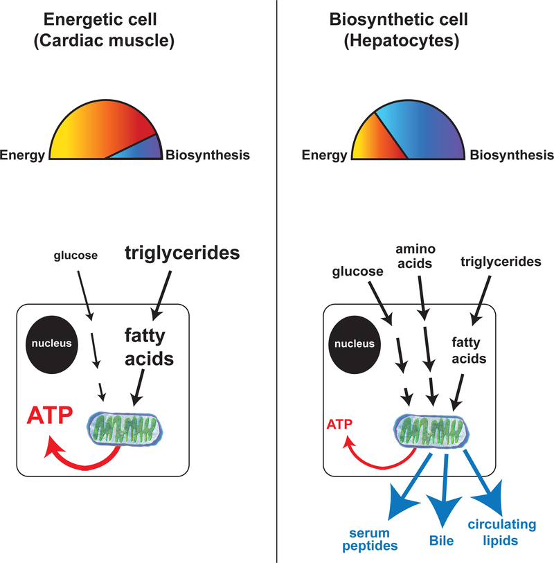
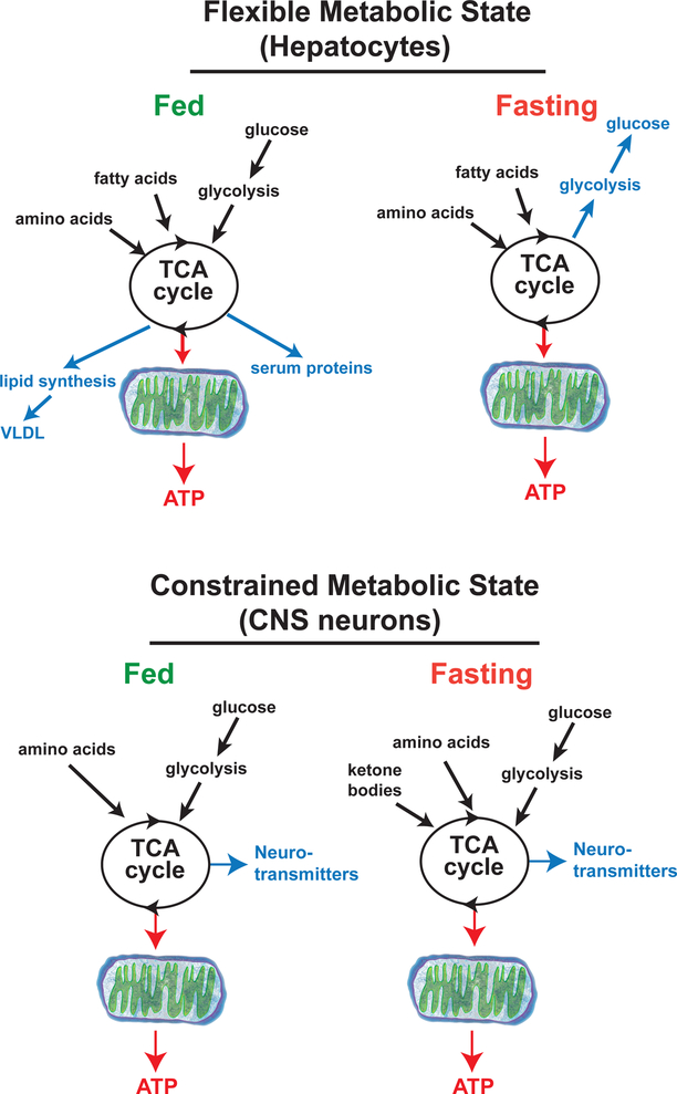
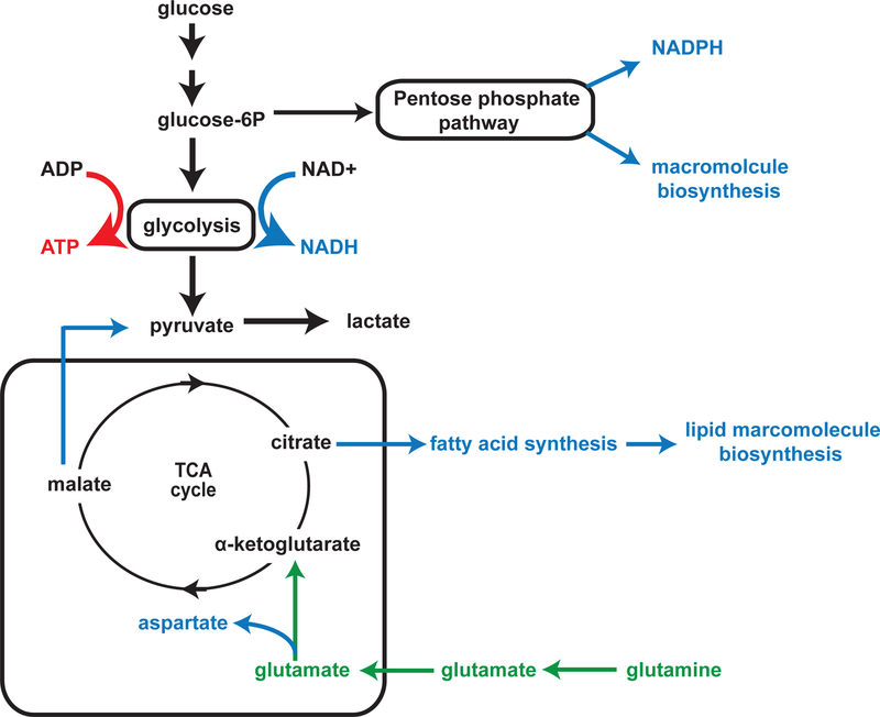
During development, cells adopt distinct metabolic strategies to support growth, produce energy, and meet the demands of a mature tissue. Some of these metabolic states maintain a constrained program of nutrient utilization, while others providing metabolic flexibility as a means to couple developmental progression with nutrient availability. Here we discuss our understanding of metabolic programs, and how they support specific aspects of animal development. During phases of rapid proliferation a subset of metabolic programs provide the building blocks to support growth. During differentiation, metabolic programs shift to support the unique demands of each tissue. Finally, we discuss how a model system, such as Drosophila egg development, can provide a versatile platform to discover novel mechanisms controlling programmed shift in metabolism.


Figures

Figure 1.
Figure 1.. Metabolic states: the examples of heart and liver
Diagrams show the major metabolic inputs and outputs of cardiac myocytes (A) and liver parenchymal cells (B). Cardiac cells primarily take up fatty acids and use them to generate ATP for muscle contraction. In contrast, liver parenchymal cells take up carbohydrates, lipids and amino acids from the circulation and convert them into a variety of outputs, including glycogen, fat, blood proteins and diverse biosynthetic products for storage or export to other organs throughout the body.
Figure 2.
Figure 2.. Flexible vs constrained metabolic states
This diagram depicts how nutrients are consumed by the hepatocytes(A) and neurons of the CNS (B). Upon fasting the highly flexible metabolic state of hepatocytes shifts dramatically and suppresses both glycolysis and the production of several biosynthetic products while at the same time activating gluconeogenesis. In contrast, upon fasting the constrained metabolic state of neurons changes very little by increasing the consumption of ketone bodies to compensate for reduced uptake of glucose.
Figure 3.
Figure 3.. Aerobic Glycolysis and Glutaminolysis work together to support rapid growth.
This diagram depicts a generalized metabolic program that supports growth. Under these conditions, aerobic glycolysis consumes glucose to produce cytosolic ATP for energy and NADH to feed main biosynthetic process in the cytosol. Upon activation of aerobic glycolysis carbon from glucose is shuttled into lactate production. To make up for the decreased pyruvate import into the TCA cycle glutaminolysis is activated to supplement the TCA cycle with carbon from glutamate. By supplementing the TCA cycle glutaminolysis helps maintain mitochondrial transport and support the biosynthesis of many compounds such as fatty acids and maintain transport in and out of the mitochondria.
Figure 4
Figure 4. Metabolic transitions during Drosophila germline development.
(A) The diagram shows the stepwise accumulation of yolk proteins, lipids, and glycogen during Drosophila oocyte development. (B) This diagram depicts how steroid signaling functions during mid-oogenesis to activate SREBP mediated lipid storage during stage 10 of oogenesis. After lipid loading during stage-10 AKT signaling is inactivated leading to a GSK3 mediated suppression of mitochondrial function and subsequent glycogen storage. (C) Depicts the how once activated GSK3 induces changes in the mitochondrial proteome that stimulate a remodeling of the ETC. The GSK3 mediate ETC remodeling cause mitochondrial to enter a state of mitochondrial respiratory quiescence that suppresses the consumption of nutrients and promotes glycogen and lipid storage.

Similar articles

Cited by

References

    1. Tennessen JM, et al., The Drosophila estrogen-related receptor directs a metabolic switch that supports developmental growth. Cell Metab, 2011. 13(2): p. 139–48. - PMC - PubMed
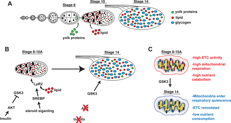
    2. Using a combination of metabolomics and gene expression analysis the authors of this study describe a pronounced shift in metabolism during mid-embryogenesis that favors high rates of glycolysis. This shift to aerobic glycolysis functions to support rapid growth during early Drosophila development.

    1. Tennessen JM, et al., Coordinated metabolic transitions during Drosophila embryogenesis and the onset of aerobic glycolysis. G3 (Bethesda), 2014. 4(5): p. 839–50. - PMC - PubMed
    1. Sieber MH, Thomsen MB, and Spradling AC, Electron Transport Chain Remodeling by GSK3 during Oogenesis Connects Nutrient State to Reproduction. Cell, 2016. 164(3): p. 420–32. - PMC - PubMed
    2. In the final stages of oogenesis oocytes display a very low level of mitochondrial activity. In this study the authors show that establish a form of mitochondrial respiratory quiescence through a pronounce shift in mitochondrial proteins that leads to remodeling of the electron transport chain (ETC). Interestingly, this remodeling of the ETC it regulated in a nutrient-dependent manner by the insulin/AKT signaling pathway which couple systemic metabolism in the germline.

    1. Villanueva CJ, et al., TLE3 is a dual-function transcriptional coregulator of adipogenesis. Cell Metab, 2011. 13(4): p. 413–27. - PMC - PubMed
    1. Seale P, et al., PRDM16 controls a brown fat/skeletal muscle switch. Nature, 2008. 454(7207): p. 961–7. - PMC - PubMed

LinkOut - more resources