. 2017 May 15;6(5):698-705.
doi: 10.1242/bio.021717.

WRN conditioned media is sufficient for in vitro propagation of intestinal organoids from large farm and small companion animals

Affiliations

WRN conditioned media is sufficient for in vitro propagation of intestinal organoids from large farm and small companion animals

Robin H Powell et al. Biol Open. .

Abstract

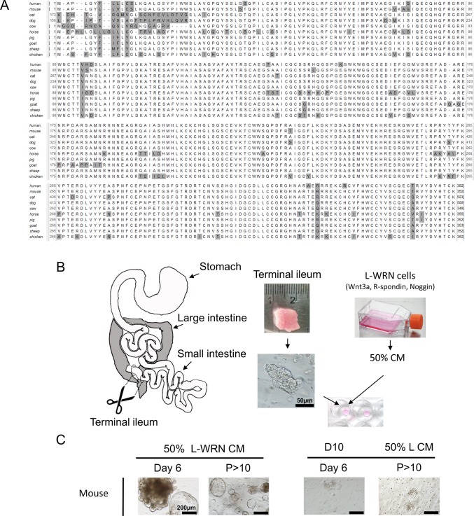
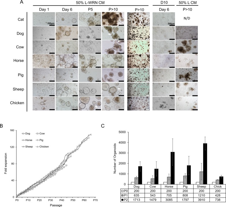
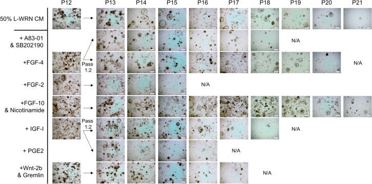
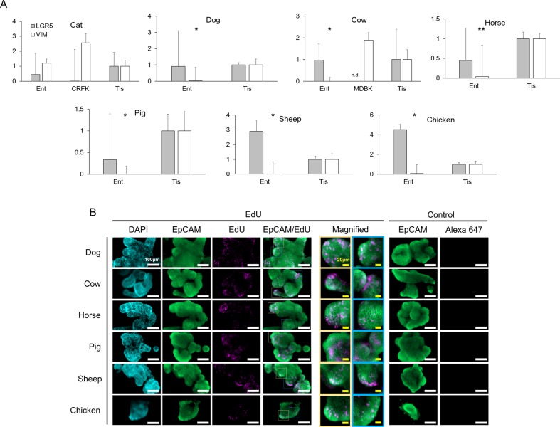
Recent years have seen significant developments in the ability to continuously propagate organoids derived from intestinal crypts. These advancements have been applied to mouse and human samples providing models for gastrointestinal tissue development and disease. We adapt these methods for the propagation of intestinal organoids (enteroids) from various large farm and small companion (LF/SC) animals, including cat, dog, cow, horse, pig, sheep and chicken. We show that LF/SC enteroids propagate and expand in L-WRN conditioned media containing signaling factors Wnt3a, R-spondin-3, and Noggin (WRN). Multiple successful isolations were achieved for each species, and the growth of LF/SC enteroids was maintained to high passage number. LF/SC enteroids expressed crypt stem cell marker LGR5 and low levels of mesenchymal marker VIM. Labeling with EdU also showed distinct regions of cell proliferation within the enteroids marking crypt-like regions. The ability to grow and maintain LF/SC enteroid cell lines provides additional models for the study of gastrointestinal developmental biology as well as platforms for the study of host-pathogen interactions between intestinal cells and zoonotic enteric pathogens of medical importance.

Keywords: Companion animal; Conditioned media; Crypt; Enteroid; Farm animal; Intestine; Organoid.


Conflict of interest statement

Competing interestsThe authors declare no competing or financial interests.

Figures

Fig. 1.
Fig. 1.
Conservation of crypt signaling molecules suggests mouse intestine enteroid culture may apply to LF/SC animals. (A) Alignment of Wnt3a protein orthologs from human, mouse, and LF/SC animals shows broad sequence conservation, non-consensus amino acid (gray). The non-consensus N-terminal region of cat and dog Wnt3a are not shown. (B) Schematic of isolation and growth of enteroids. Tissue samples from the terminal ileum were harvested, trimmed to 1 cm2, and crypts were isolated with collagenase digestion. Primary media was incubated with the L-WRN cell line which secrete Wnt3a, R-spondin-3 and Noggin to create 50% L-WRN CM. Isolated crypts were suspended in Matrigel matrix and grown in 50% L-WRN CM. (C) Isolation and growth of mouse crypt enteroids demonstrates successful preparation of 50% L-WRN CM from L-WRN cells. Mouse enteroids only grow in 50% L-WRN CM after isolation (Day 6) to high passage (P>10) number. Mouse enteroids fail to grow when cultured in D10 after isolation (Day 6) or when passed to 50% L CM after high passage (P>10) growth in 50% L-WRN CM.
Fig. 2.
Fig. 2.
50% L-WRN CM is required for LF/SC enteroid growth and expansion. (A) Enteroids from LF/SC animals cultured with 50% L-WRN CM expand soon after crypt isolation (Day 1), growing to large size after passage (Day 6), and continue to expand and obtain crypt-like morphology at higher passage (white arrows) (P5 and P>10). Lower magnification shows enteroids were maintained at high density throughout passage history (P>10, 500 µm). Enteroids fail to grow when cultured in D10 after isolation (Day 6) or when passed to 50% L CM after high passage (P>10) growth in 50% L-WRN CM, no data (N/D). (B) Fold expansion of wells containing LF/SC enteroids. LF/SC enteroids grown in 24-well plates were maintained at high density. The ratio of per well expansion was recorded at each passage throughout the passage history. (C) Number of enteroids expands with each passage. A total of 200 high passage (P>10) enteroids were seeded to two wells and the number of enteroids was counted after 3-4 days of growth, and again after subsequent passage splitting 1:2, n=3. Cat samples are not included in B or C due to the lower passage and density of enteroids. Error bars indicate ±s.d.
Fig. 3.
Fig. 3.
Supplementation of 50% L-WRN CM with crypt niche factors. Cat enteroids were passed to multiple wells at P12 and cultured with 50% L-WRN CM or with additional crypt niche factor(s) shown to modulate organoid growth in various platforms. The individual cultures were passed every 3-4 days and monitored for growth. Cultures were terminated when the number and growth of the enteroids dissipated (N/A). None of the crypt niche factors rescued the cat enteroids much beyond those grown with 50% L-WRN CM.
Fig. 4.
Fig. 4.
LGR5 expression in LF/SC enteroids. (A) LGR5 RNA expression in LF/SC enteroids: cat (P11-12), dog (P27-34), cow (P18-27), horse (P29-35), pig (P48-49), sheep (P28-35), chicken (P18-28). qPCR of LGR5 and VIM expression in enteroids (Ent) and control tissue (Tis), student's t-test: *P≤0.05, **P≤0.005. Most LF/SC enteroids express LGR5 above the control tissue. LF/SC enteroids also have very low expression of mesenchymal marker VIM as compared to control tissue and kidney epithelial cells [cat (CRFK) and cow (MDBK)]. Enteroid values are fold change of control tissue; mean±se.m.; n.d., not detected; n=3. (B) EdU incorporation using the Click-iT EdU Alexa Fluor 647 assay in LF/SC enteroids confirms active proliferation with crypt-like regions. IFA of LF/SC enteroids probed with multi-species reactive α-EpCam (green, Alexa Fluor 488), Click-iT EdU (pink, Alexa Fluor 647), and nuclei stained with DAPI (blue). Merged EpCam and EdU images shows distinct regions of proliferation along crypt-like structures. Enteroids probed with α-EpCam (green) fail to stain pink (Alexa 647) in negative control IFAs lacking the Click-iT EdU.

Similar articles

Cited by

References

    1. Aoki R., Shoshkes-Carmel M., Gao N., Shin S., May C. L., Golson M. L., Zahm A. M., Ray M., Wiser C. L., Wright C. V. E. et al. (2016). Foxl1-expressing mesenchymal cells constitute the intestinal stem cell niche. Cell Mol. Gastroenterol. Hepatol. 2, 175-188. 10.1016/j.jcmgh.2015.12.004 - DOI - PMC - PubMed
    1. Barker N., Huch M., Kujala P., van de Wetering M., Snippert H. J., van Es J. H., Sato T., Stange D. E., Begthel H., van den Born M. et al. (2010). Lgr5(+ve) stem cells drive self-renewal in the stomach and build long-lived gastric units in vitro. Cell Stem Cell 6, 25-36. 10.1016/j.stem.2009.11.013 - DOI - PubMed
    1. Biswas S., Davis H., Irshad S., Sandberg T., Worthley D. and Leedham S. (2015). Microenvironmental control of stem cell fate in intestinal homeostasis and disease. J. Pathol. 237, 135-145. 10.1002/path.4563 - DOI - PMC - PubMed
    1. Boj S. F., Hwang C.-I., Baker L. A., Chio I. I. C., Engle D. D., Corbo V., Jager M., Ponz-Sarvise M., Tiriac H., Spector M. S. et al. (2015). Organoid models of human and mouse ductal pancreatic cancer. Cell 160, 324-338. 10.1016/j.cell.2014.12.021 - DOI - PMC - PubMed
    1. Clevers H. (2016). Modeling development and disease with organoids. Cell 165, 1586-1597. 10.1016/j.cell.2016.05.082 - DOI - PubMed