Review
. 2018 Feb 1;10(2):a022210.
doi: 10.1101/cshperspect.a022210.

TGF-β Signaling in Control of Cardiovascular Function

Affiliations
Review

TGF-β Signaling in Control of Cardiovascular Function

Marie-José Goumans et al. Cold Spring Harb Perspect Biol. .

Abstract

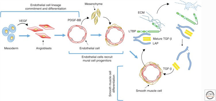
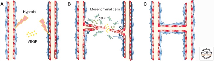
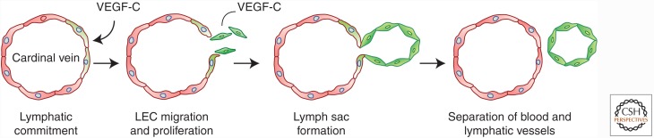
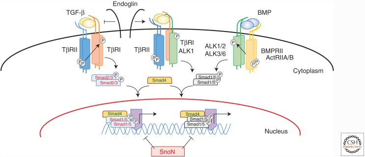
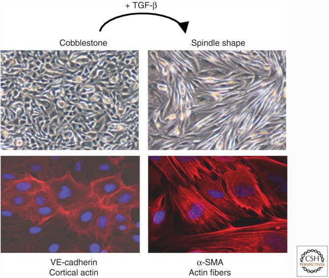
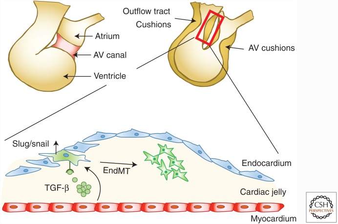
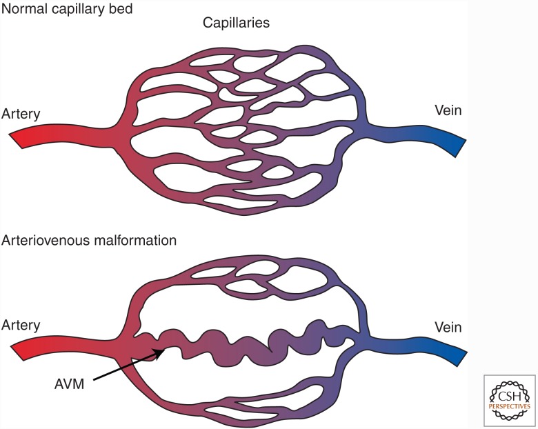
Genetic studies in animals and humans indicate that gene mutations that functionally perturb transforming growth factor β (TGF-β) signaling are linked to specific hereditary vascular syndromes, including Osler-Rendu-Weber disease or hereditary hemorrhagic telangiectasia and Marfan syndrome. Disturbed TGF-β signaling can also cause nonhereditary disorders like atherosclerosis and cardiac fibrosis. Accordingly, cell culture studies using endothelial cells or smooth muscle cells (SMCs), cultured alone or together in two- or three-dimensional cell culture assays, on plastic or embedded in matrix, have shown that TGF-β has a pivotal effect on endothelial and SMC proliferation, differentiation, migration, tube formation, and sprouting. Moreover, TGF-β can stimulate endothelial-to-mesenchymal transition, a process shown to be of key importance in heart valve cushion formation and in various pathological vascular processes. Here, we discuss the roles of TGF-β in vasculogenesis, angiogenesis, and lymphangiogenesis and the deregulation of TGF-β signaling in cardiovascular diseases.


Figures

Figure 1.
Figure 1.
Process of vasculogenesis. Vasculogenesis starts with the differentiation and proliferation of mesodermal cells into angioblasts followed by their differentiation into endothelial cells (ECs) in response to vascular endothelial growth factor (VEGF). These ECs fuse and form a lumen in a honeycomb structure. Platelet-derived growth factor (PDGF) secreted by ECs induces recruitment of mesenchymal cells that will differentiate into smooth muscle cells (SMCs) or pericytes. When SMCs or pericytes adhere to ECs, the ECs start to produce TGF-β. TGF-β is produced as a prepropolypeptide, which is proteolytically processed, and forms a small latent TGF-β complex consisting of the mature TGF-β dimer associated with two latency-associated peptides (LAPs). The small latent TGF-β complex binds to latent transforming growth factor β binding protein (LTBP), and this complex is secreted as the large latent TGF-β complex. LTBP binds to extracellular matrix (ECM) proteins such as fibronectin. Activation of TGF-β by, for example, proteolytic release from LAP will induce growth arrest and terminal differentiation of SMCs.
Figure 2.
Figure 2.
Process of angiogenesis. (A) Angiogenesis begins when endothelial cells are activated, for example, by hypoxia. These cells proliferate and the tip cells migrate into the perivascular space toward a vascular endothelial growth factor (VEGF) gradient (B). During the resolution phase, Endothelial cells stop dividing and vascular sprouts fuse, rebuild their extracellular matrix, and attract pericytes and smooth muscle cells in a platelet-derived growth factor (PDGF)-dependent manner. (C) These latter cells will stabilize the newly formed sprout.
Figure 3.
Figure 3.
Process of lymphangiogenesis. Lymph vessels originate from blood vessels when and where some of the cardinal vein endothelial cells start to express lymphatic vessel endothelial hyaluronan receptor 1 (LYVE-1) and prospero homeobox transcription factor (Prox)-1 (green cells). The Prox-1-expressing lymphatic endothelial cells (LECs) migrate out of the vessel, proliferate, and form the lymphatic sac, the first lymphatic structure.
Figure 4.
Figure 4.
TGF-β signaling pathways. TGF-βs signal by binding to a specific, heteromeric complex that includes types I and II kinase receptors (TβRI/ALK-5 and TβRII, respectively). In most cells, TGF-β binds to a complex of TβRII and TβRI/ALK-5 complex, but in endothelial cells, they can also bind to a complex of TβRII and ALK-1. The coreceptor endoglin inhibits TGF-β binding to the TβRII-ALK-5 complex and promotes TGF-β binding to the TβRII-ALK-1 complex, thus activating ALK-1 signaling. In a similar manner, bone morphogenetic proteins (BMPs) also bind to complexes of two types of receptors—that is, the type I receptors ALK-1, -2, -3, or -6, and the type II receptors BMPRII, ActRIIA, or ActRIIB. Intracellular signaling through Smad activation can be divided into two pathways. In one pathway, ALK-5 phosphorylates, and thus activates, Smad2 and Smad3, and in the other one, ALK-1, -2, -3, and -6 activate Smad1, Smad5, and Smad8. These receptor-activated Smads form heteromeric complexes with the common mediator Smad, Smad4. These complexes translocate into the nucleus, where they act as transcription factor complexes and regulate the expression of specific target genes.
Figure 5.
Figure 5.
Endothelial-to-mesenchymal transition. When stimulated with TGF-β, endothelial cells undergo EndMT. They lose their cobble-stone morphology (upper panels, bright field), reduce the expression of endothelial cell markers such as platelet/endothelial cell adhesion molecule-1 (PECAM1), and start to express mesenchymal markers such as α-smooth muscle actin (α-SMA). Cortical actin (in red) is reorganized into stress fibers. The nucleus is stained with DAPI in blue.
Figure 6.
Figure 6.
Cardiac cushion formation. The developmental formation of cardiac cushions involves endothelial-to-mesenchymal transition (EndMT). The endothelial cells lose their cobblestone morphology, adopt a mesenchymal phenotype, and invade the cardiac jelly. AV, Arteriovenous.
Figure 7.
Figure 7.
Arteriovenous malformations (AVMs) in hereditary hemorrhagic telangiectasia (HHT). One hallmark of HHT is the development of an AVM. In the AVM, the capillary bed is lost, and the artery drains directly into the vein via a tortuous, weak vessel, which is prone to rupture.

References

    1. Abdalla SA, Letarte M. 2006. Hereditary haemorrhagic telangiectasia: Current views on genetics and mechanisms of disease. J Med Genet 43: 97–110. - PMC - PubMed
    1. Adams RH, Alitalo K. 2007. Molecular regulation of angiogenesis and lymphangiogenesis. Nat Res Mol Cell Biol 8: 464–478. - PubMed
    1. Akhurst RJ, Hata A. 2012. Targeting the TGF-β signalling pathway in disease. Nat Rev Drug Discov 11: 790–811. - PMC - PubMed
    1. Akhurst RJ, Lehnert SA, Faissner A, Duffie E. 1990. TGF-β in murine morphogenetic processes: The early embryo and cardiogenesis. Development 108: 645–656. - PubMed
    1. Alam MA, Sami S, Babu S. 2011. Successful treatment of bleeding gastro-intestinal angiodysplasia in hereditary haemorrhagic telangiectasia with thalidomide. BMJ Case Rep 2011: pii: bcr0820114585. - PMC - PubMed

Publication types

MeSH terms

Substances