. 2017 May 23;85(6):e00091-17.
doi: 10.1128/IAI.00091-17. Print 2017 Jun.

Iron Efflux by PmtA Is Critical for Oxidative Stress Resistance and Contributes Significantly to Group A Streptococcus Virulence

Affiliations

Iron Efflux by PmtA Is Critical for Oxidative Stress Resistance and Contributes Significantly to Group A Streptococcus Virulence

Arica R VanderWal et al. Infect Immun. .

Abstract

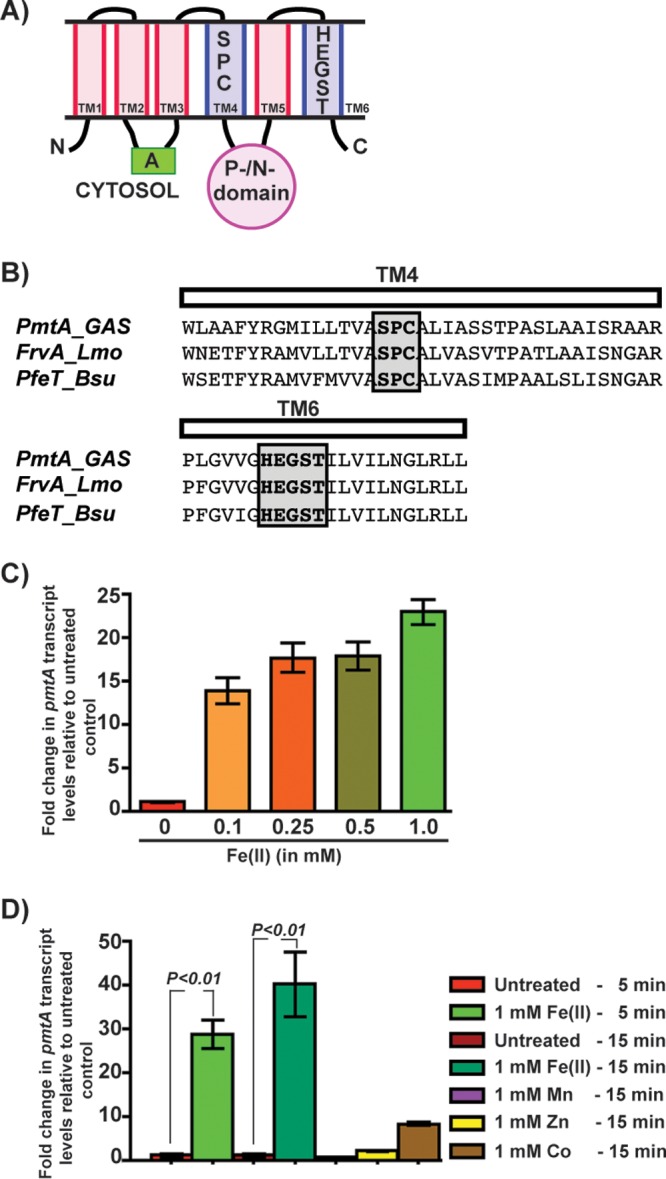
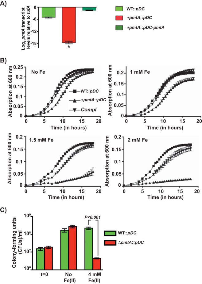
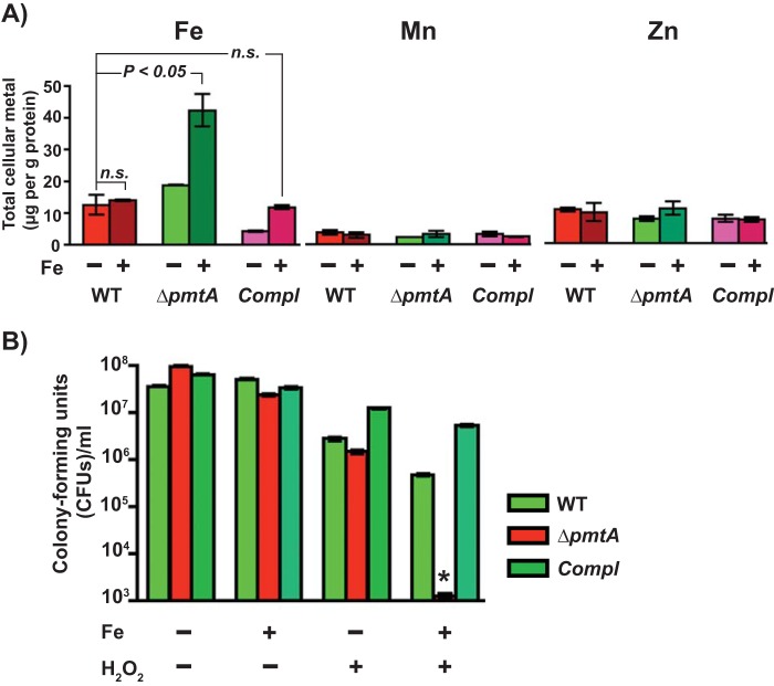
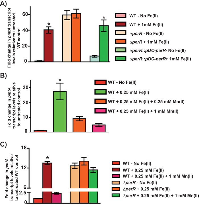
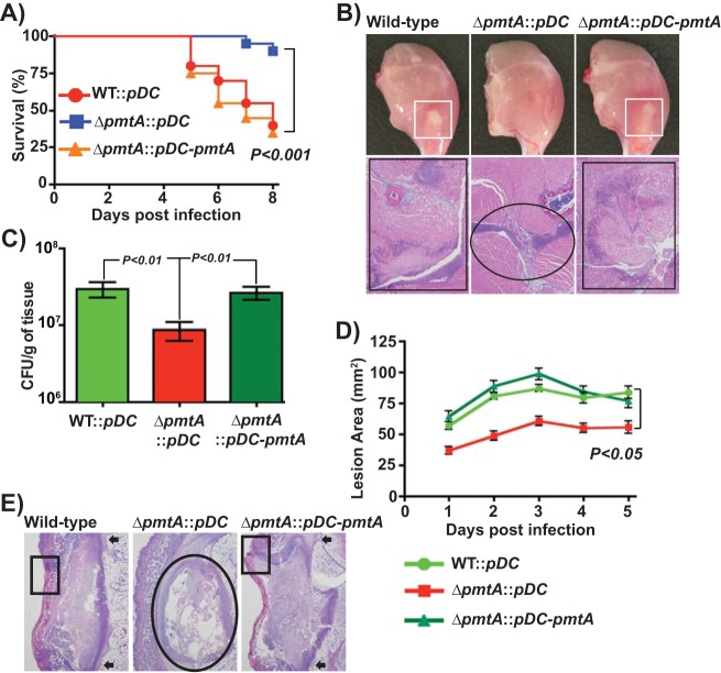
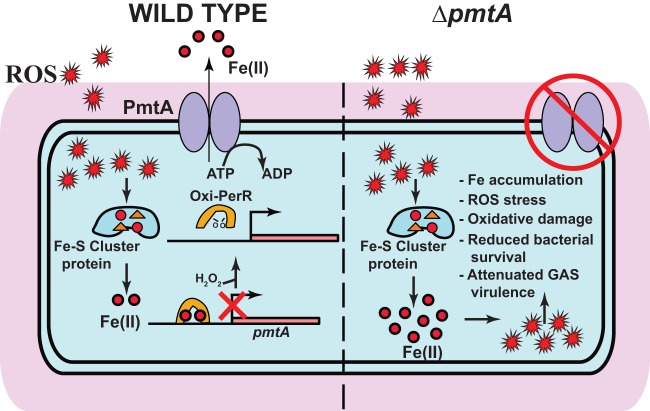
Group A Streptococcus (GAS) is a human-only pathogen that causes a spectrum of disease conditions. Given its survival in inflamed lesions, the ability to sense and overcome oxidative stress is critical for GAS pathogenesis. PerR senses oxidative stress and coordinates the regulation of genes involved in GAS antioxidant defenses. In this study, we investigated the role of PerR-controlled metal transporter A (PmtA) in GAS pathogenesis. Previously, PmtA was implicated in GAS antioxidant defenses and suggested to protect against zinc toxicity. Here, we report that PmtA is a P1B4-type ATPase that functions as an Fe(II) exporter and aids GAS defenses against iron intoxication and oxidative stress. The expression of pmtA is specifically induced by excess iron, and this induction requires PerR. Furthermore, a pmtA mutant exhibited increased sensitivity to iron toxicity and oxidative stress due to an elevated intracellular accumulation of iron. RNA-sequencing analysis revealed that GAS undergoes significant alterations in gene expression to adapt to iron toxicity. Finally, using two mouse models of invasive infection, we demonstrated that iron efflux by PmtA is critical for bacterial survival during infection and GAS virulence. Together, these data demonstrate that PmtA is a key component of GAS antioxidant defenses and contributes significantly to GAS virulence.

Keywords: bacterial pathogenesis; gene regulation; iron efflux; metal homeostasis; oxidative stress.


Figures

FIG 1
FIG 1
Expression of the pmtA gene, encoding a P1B-4-type ATPase, is upregulated during GAS growth in the presence of excess iron. (A) Schematic representation of the topology and domain architecture of the P1B-4-type ATPase. The predicted six transmembrane helices are labeled. The amino and carboxy termini are marked with N and C, respectively. The cytosolic actuator (A) and the phosphorylation/nucleotide-binding (P-/N-) domains are indicated. The transmembrane metal-binding residues within the transmembrane 4 (TM4) and TM6 helices that are conserved among members of the P1B-4 subgroup of the P1 family of ATPases are shown. (B) Amino acid sequence alignment of the predicted TM4 and TM6 helices from PmtA of GAS and its paralogs FrvA of L. monocytogenes (Lmo) and PfeT of B. subtilis (Bsu). The conserved metal-binding residues are shaded and boxed. (C) Transcript levels of pmtA in GAS grown in THY-C medium supplemented with increasing concentrations of Fe(II) compared to GAS grown in unsupplemented medium measured by qRT-PCR. (D) Transcript levels of pmtA in GAS grown in THY-C medium supplemented with different metals compared to the untreated sample measured by qRT-PCR. Three biological replicates were performed and analyzed in triplicate. Data graphed are means ± standard deviations. Average values for unsupplemented samples were used as a reference, and fold changes in transcript levels of the indicated strains relative to the reference sample are shown.
FIG 2
FIG 2
PmtA is critical for GAS growth under iron toxicity. (A) Transcript levels of pmtA in the indicated strains as measured by qRT-PCR. (B) Growth kinetics of the indicated strains in THY-C medium supplemented with increasing concentrations of FeSO4. Three biological replicates were performed, and the graph represents means ± standard deviations. Compl, complemented strain. (C) GAS strains were grown to mid-exponential phase in THY-C broth, incubated with 4 mM FeSO4 for 15 min, and grown for an additional 3 h in THY broth. The mean CFU recovered after a 3-h recovery period are shown, with P values as determined by a t test. Triplicate cultures were grown under the indicated conditions, and each biological replicate was assessed in duplicate. *, P < 0.0001.
FIG 3
FIG 3
The ΔpmtA mutant is defective in iron efflux and sensitive to oxidative stress. (A) The WT, ΔpmtA, and trans-complemented strains were grown in THY-C broth with (+) or without (−) 1 mM FeSO4 to the mid-exponential phase of growth, and the intracellular metal content was measured by ICP-MS. n.s., not significant. (B) GAS strains were grown to mid-exponential phase in THY-C broth, incubated with 4 mM FeSO4 for 15 min, and grown in THY broth in the presence or absence of 1 mM H2O2. Mean CFU recovered after the 6-h recovery period are shown, with P values (*, P < 0.0001) as determined by a t test. Triplicate cultures were grown under the indicated conditions, and each biological replicate was assessed in duplicate.
FIG 4
FIG 4
Upregulation of pmtA is dependent on the metallated state of PerR. (A) Transcript levels of pmtA in the indicated GAS strains grown in THY-C medium supplemented with 1 mM Fe(II) compared to those in untreated WT GAS as measured by qRT-PCR. (B) Transcript level analysis of pmtA in WT GAS grown in medium supplemented with 0.25 mM FeSO4 and challenged with increasing concentrations of Mn(II). (C) Transcript level analysis of pmtA in WT GAS and the ΔperR mutant supplemented with 0.25 mM FeSO4 and challenged with increasing concentrations of Mn(II). Three biological replicates were performed and analyzed in triplicate. Data graphed are means ± standard deviations. Average values for the growth of the WT in unsupplemented medium were used as a reference, and fold changes in the transcript levels in the indicated strains relative to those in the reference sample are shown. *, P < 0.0001.
FIG 5
FIG 5
PmtA is critical for survival during infection and GAS virulence. (A) Twenty outbred CD-1 mice per strain were injected intramuscularly with 1 × 10⁷ CFU of each strain. Shown is a Kaplan-Meier survival curve with P values derived by a log rank test. (B) Macroscopic (top) and histopathological (bottom) analyses of hind-limb lesions from mice infected with the indicated strains at 48 h postinfection. Areas of host tissue damage are boxed (white boxes). Areas of disseminated lesions in the WT and trans-complemented strains are boxed (black box), whereas confined, less destructive lesions are circled. (C) Twenty mice were infected intramuscularly, and mean CFU recovered from infected muscle tissue at 96 h postinfection are shown, with P values as determined by a t test. (D) Twenty immunocompetent hairless mice were infected with the indicated strains, and the lesion area produced by each strain was determined. The lesion area was measured daily and graphed (means ± standard error of the means). The P value was derived by a log rank test. (E) Histopathological analysis of subcutaneous lesions of mice infected with the indicated strains at 48 h postinfection. Areas of disseminated lesions and ulcerations on the skin surfaces of mice infected with the WT and trans-complemented strains are boxed, whereas confined, less destructive lesions in tissues infected with the ΔpmtA mutant are circled.
FIG 6
FIG 6
Proposed model for regulation and contribution of PmtA to GAS pathogenesis. In wild-type GAS (left), iron released from Fe-S cluster-containing proteins by ROS stress causes an increase in the GAS intracellular iron concentration. As a result, the iron-metallated form of PerR senses ROS, and oxidized PerR (Oxi-PerR) causes the derepression of pmtA by its dissociation from the pmtA promoter. PmtA aids GAS antioxidant defense by exporting iron out of the cytosol using the energy derived from ATP hydrolysis. In the ΔpmtA mutant, a failure to prevent the cytosolic accumulation of iron results in increased sensitivity to oxidative stress, increased oxidative damage, reduced bacterial survival, and attenuated GAS virulence.

References

    1. Nairz M, Schroll A, Sonnweber T, Weiss G. 2010. The struggle for iron—a metal at the host-pathogen interface. Cell Microbiol 12:1691–1702. doi:10.1111/j.1462-5822.2010.01529.x. - DOI - PubMed
    1. Weinberg ED. 2000. Modulation of intramacrophage iron metabolism during microbial cell invasion. Microbes Infect 2:85–89. doi:10.1016/S1286-4579(00)00281-1. - DOI - PubMed
    1. Nakashige TG, Zhang B, Krebs C, Nolan EM. 2015. Human calprotectin is an iron-sequestering host-defense protein. Nat Chem Biol 11:765–771. doi:10.1038/nchembio.1891. - DOI - PMC - PubMed
    1. Flo TH, Smith KD, Sato S, Rodriguez DJ, Holmes MA, Strong RK, Akira S, Aderem A. 2004. Lipocalin 2 mediates an innate immune response to bacterial infection by sequestrating iron. Nature 432:917–921. doi:10.1038/nature03104. - DOI - PubMed
    1. Cassat JE, Skaar EP. 2013. Iron in infection and immunity. Cell Host Microbe 13:509–519. doi:10.1016/j.chom.2013.04.010. - DOI - PMC - PubMed

Publication types

MeSH terms

LinkOut - more resources