. 2017 May 23;85(6):e01067-16.
doi: 10.1128/IAI.01067-16. Print 2017 Jun.

Analysis of Shigella flexneri Resistance, Biofilm Formation, and Transcriptional Profile in Response to Bile Salts

Affiliations

Analysis of Shigella flexneri Resistance, Biofilm Formation, and Transcriptional Profile in Response to Bile Salts

Kourtney P Nickerson et al. Infect Immun. .

Abstract

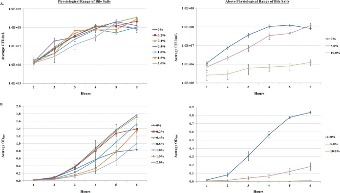
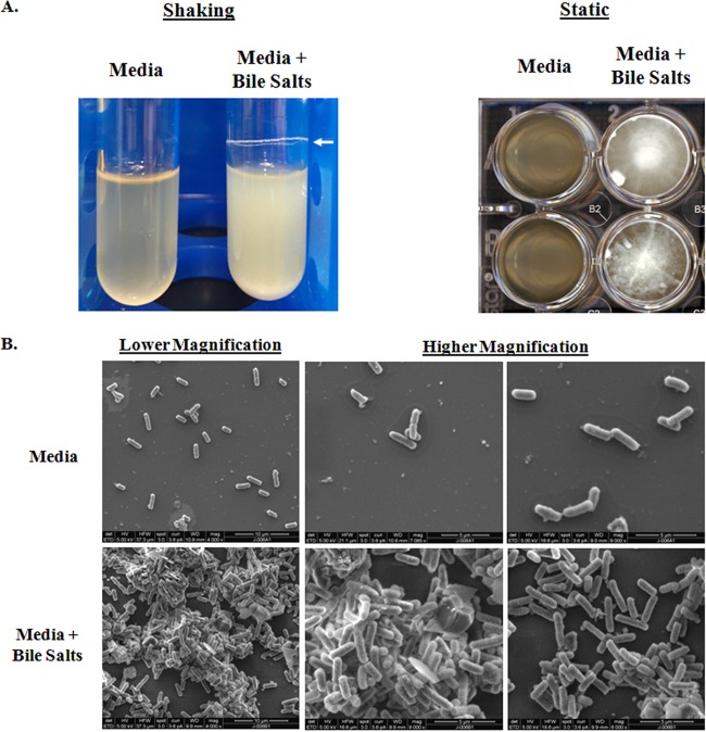
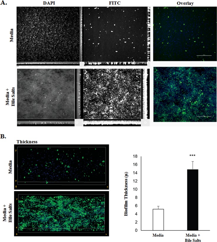
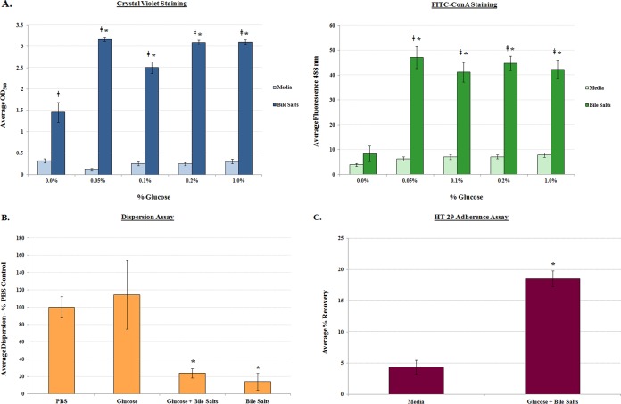
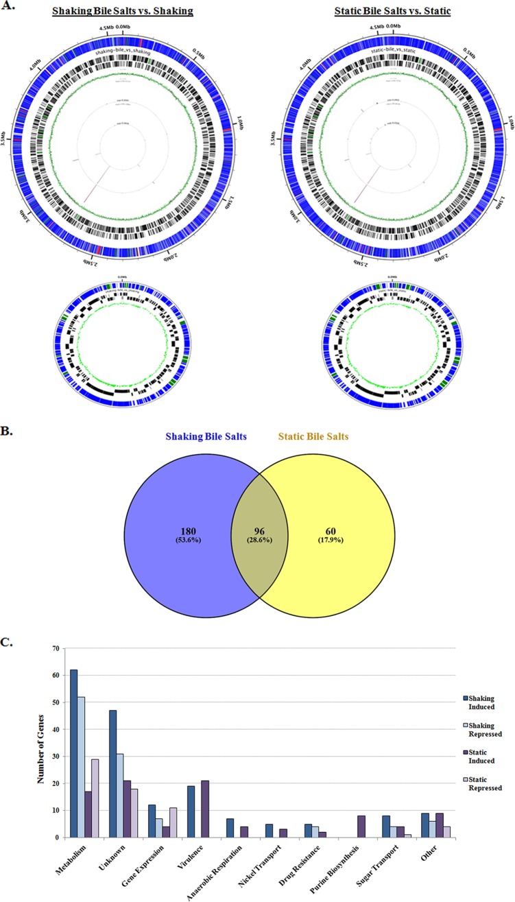
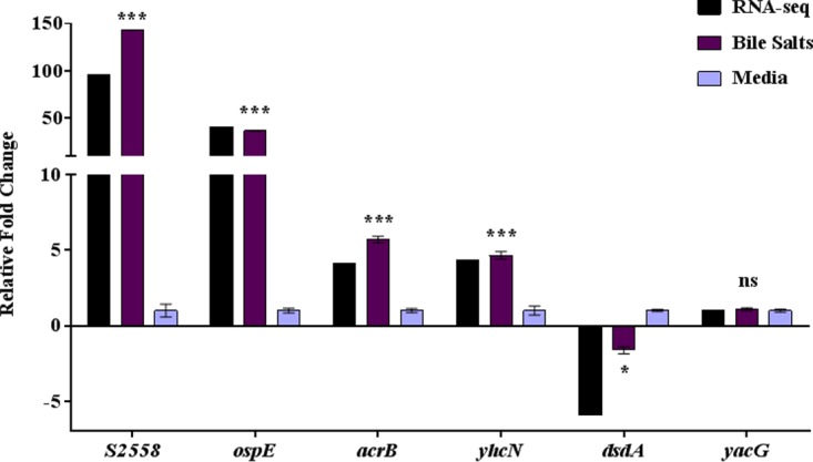
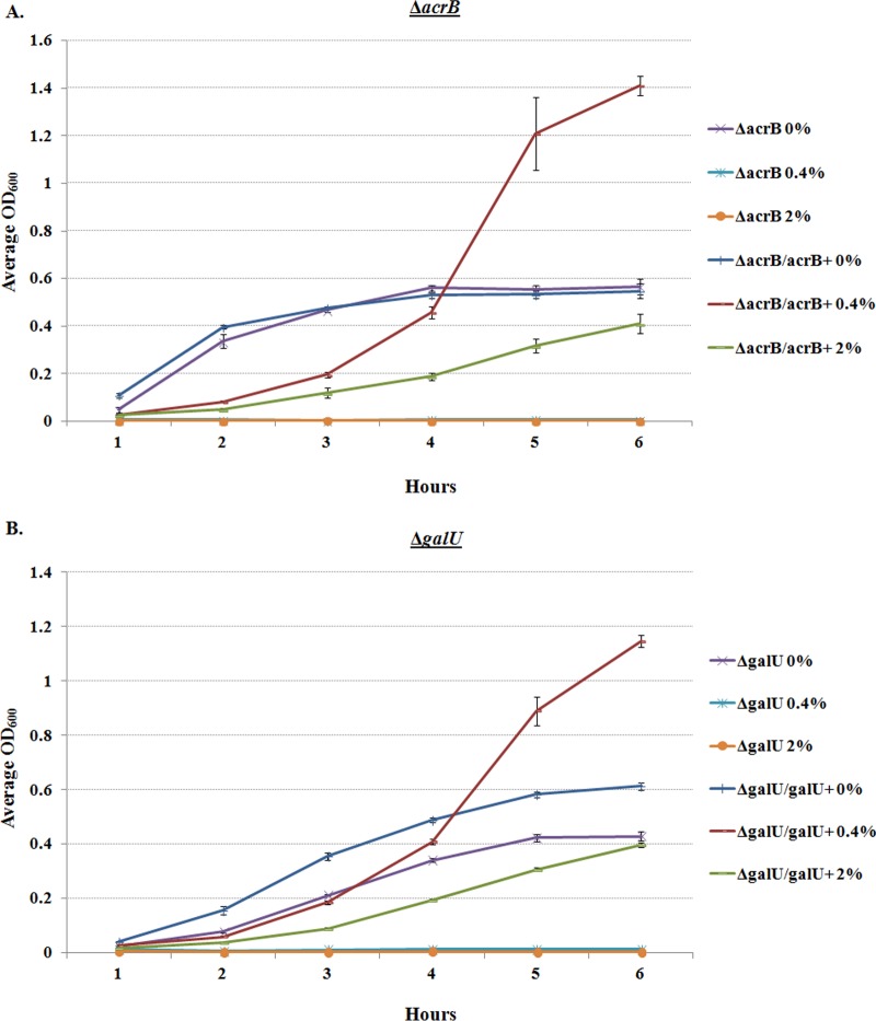
The Shigella species cause millions of cases of watery or bloody diarrhea each year, mostly in children in developing countries. While many aspects of Shigella colonic cell invasion are known, crucial gaps in knowledge regarding how the bacteria survive, transit, and regulate gene expression prior to infection remain. In this study, we define mechanisms of resistance to bile salts and build on previous research highlighting induced virulence in Shigella flexneri strain 2457T following exposure to bile salts. Typical growth patterns were observed within the physiological range of bile salts; however, growth was inhibited at higher concentrations. Interestingly, extended periods of exposure to bile salts led to biofilm formation, a conserved phenotype that we observed among members of the Enterobacteriaceae Characterization of S. flexneri 2457T biofilms determined that both bile salts and glucose were required for formation, dispersion was dependent upon bile salts depletion, and recovered bacteria displayed induced adherence to HT-29 cells. RNA-sequencing analysis verified an important bile salt transcriptional profile in S. flexneri 2457T, including induced drug resistance and virulence gene expression. Finally, functional mutagenesis identified the importance of the AcrAB efflux pump and lipopolysaccharide O-antigen synthesis for bile salt resistance. Our data demonstrate that S. flexneri 2457T employs multiple mechanisms to survive exposure to bile salts, which may have important implications for multidrug resistance. Furthermore, our work confirms that bile salts are important physiological signals to activate S. flexneri 2457T virulence. This work provides insights into how exposure to bile likely regulates Shigella survival and virulence during host transit and subsequent colonic infection.

Keywords: EPS matrix; Escherichia coli; Shigella; bile salts; biofilm; resistance; virulence; virulence genes.


Figures

FIG 1
FIG 1
Growth curve analysis of S. flexneri 2457T in bile salts. Bacterial growth was monitored in TSB medium or medium containing increasing concentrations of bile salts. (A) Average CFU/ml ± standard errors (SE) for three independent experiments are plotted. S. flexneri 2457T grew normally within the physiological range of 0.2% to 2.0% (wt/vol), but growth was slowed and inhibited at 5% and 10% (wt/vol), respectively. Statistical significance relative to the 0% control was detected at 5% and 10% (wt/vol) with P values of 0.001. (B) The corresponding average OD600 ± SE are plotted. In the physiological range of bile salts, sharp increases in optical density were detected. Statistical significance relative to the 0% control was detected at 0.4%, 5%, and 10% (all wt/vol) with P values ranging from <0.05 to 0.0001.
FIG 2
FIG 2
Prolonged exposure to bile salts leads to clumping of the bacteria. (A) Bacteria subcultured for 4 h under shaking conditions at 225 rpm (left) or single colonies inoculated into each well of a 24-well plate and grown statically overnight (right) in medium with or without bile salts. The bacteria exposed to 0.4% (wt/vol) bile salts clumped and formed an adhesive ring (white arrow) around the culture tube or to the bottom of the wells. Images are representative of three independent experiments. (B) Scanning electron microscopy analysis of bile salt-induced clumping. Bacteria were grown on coverslips in medium alone (top) or in the presence of 0.4% (wt/vol) bile salts (bottom), fixed, and processed for SEM analysis. In medium alone, the bacteria were dispersed on the coverslips. Following exposure to bile salts, the bacteria clumped and adhered better to the coverslips. Images are representative of three independent experiments. Lower magnification images (×4,000; scale bars, 10 μm) are on the left, and higher magnification images (×7,000 to ×9,000; scale bars, 5 μm) are in the middle and on the right.
FIG 3
FIG 3
Quantification of biofilm formation. Single colonies of the indicated bacterial strains were inoculated into TSB ± 0.4% (wt/vol) bile salts, grown overnight at 37°C, and subsequently processed with crystal violet staining. In medium without bile salts, biofilm formation was minimal; however, following bile salt exposure, biofilm formation was significantly induced in all strains of Shigella tested (*, P < 0.0001). The average OD540 ± SE values from three independent experiments are presented. There were no significant differences between bacterial strains.
FIG 4
FIG 4
Analysis of exopolysaccharide matrix production. (A) S. flexneri 2457T was grown on glass coverslips in TSB ± 0.4% (wt/vol) bile salts, fixed, and stained with DAPI (blue) and 25 μg/ml FITC-conjugated concanavalin A (green) to assess EPS production. Bacteria in medium alone were dispersed and had few areas staining positive for concanavalin A, while bacteria exposed to bile salts clumped and had more concanavalin A staining. The images are representative of three independent experiments. (B) To quantify the thickness of the biofilms, image stacks were taken every 0.25 μm of the biofilm, and quantification was determined from the full thickness of the stack. The image on the left represents 3D reconstructed stacks using ImageJ software. The average thickness (±SE) of each biofilm from three independent experiments is plotted on the right. There was a significant difference in biofilm thickness of S. flexneri 2457T following exposure to bile salts (***, P < 0.01).
FIG 5
FIG 5
Analysis of biofilm formation, dispersion, and subsequent adherence to HT-29 cells. (A) Analysis of glucose requirement in biofilm formation. Single colonies of S. flexneri 2457T were inoculated into medium (LB) with or without 0.4% (wt/vol) bile salts at the indicated percentage (wt/vol) of glucose. Crystal violet (left) and FITC-ConA (right) staining was performed, and the average OD540 and fluorescence at 488 nm, respectively, ± SE are presented. Bile salts increased bacterial aggregation, and the addition of glucose further increased this phenotype. However, EPS production was dependent on the presence of glucose. For both the crystal violet and FITC-ConA staining, all differences between medium alone and the corresponding condition with bile salts are indicated with a double dagger (‡, P < 0.001). Differences in biofilm formation between each of the glucose plus bile salts concentrations relative to 0.0% glucose plus bile salts concentration are indicated with an asterisk (*, P < 0.01). (B) Analysis of bile salt removal in biofilm dispersion. S. flexneri 2457T biofilms were gently washed with PBS and resuspended in prewarmed PBS or PBS with the indicated treatments for 30 min at 37°C. The supernatants were subsequently removed and dilution plated, and CFU were determined. Dispersion was minimal in the presence of bile salts (*, P < 0.01), regardless of the presence of glucose. The average dispersion ± SE relative to the PBS control is plotted for three independent experiments in which technical triplicates were performed for each assay. For reference, the average recovery for the PBS treatment was 7.6 × 10⁷ CFU while the average recovery for the PBS with bile salts treatment was 7.8 × 10⁵ CFU. (C) Analysis of S. flexneri 2457T adherence following dispersion from the biofilm. Bacteria were grown overnight in media (LB) without or with a combination of glucose and bile salts to initiate biofilm formation. On the next day, bacteria were collected, washed with 1× PBS, and analyzed for adherence to HT-29 cells. Strain 2457T consistently had significantly increased adherence following biofilm dispersion (*, P < 0.0001) compared to bacteria recovered from medium without glucose and bile salts. The average percent recovery of adherent bacteria ± SE is plotted for three independent experiments, each of which had technical triplicates.
FIG 6
FIG 6
RNA-sequencing analysis of S. flexneri 2457T grown in the presence and absence of 0.4% (wt/vol) bile salts. (A) Results of the RNA-seq analysis visualized with the Circleator program (71), in which blue represents unchanged genes, green represents induced genes (fold change ≥ 2, P < 0.05), and red represents repressed genes (fold change ≤ −2, P < 0.05) in the presence of bile salts for both the chromosome (top) and virulence plasmid (bottom). Barcodes represent forward and reverse genes, while the green circle represents the percent GC skew throughout the genome. (B) Venn diagram (72) of differentially expressed genes identified in the RNA-seq analysis. The numbers of induced and repressed genes for the “shaking + bile salts versus shaking” condition compared to the “static + bile salts versus static” condition are depicted. For both comparisons, there were 96 genes that were induced or repressed in the presence of bile salts. For the “shaking + bile salts versus shaking” analysis, there were an additional 180 genes induced or repressed, for a total of 276 differentially expressed genes in bile salts. For the “static + bile salts versus static” analysis, there were an additional 60 genes that were induced or repressed, for a total of 156 differentially expressed genes in bile salts. (C) Functional categories for differentially expressed genes in the presence of bile salts are plotted with the number of genes induced or repressed under the bile salts condition for both the shaking and static growth conditions. Please refer to Tables S2 and S3 in the supplemental material for details.
FIG 7
FIG 7
Quantitative reverse transcription-PCR analysis verifies the RNA-sequencing analysis. qRT-PCR was performed on three independent RNA samples isolated from S. flexneri 2457T grown with 0.4% (wt/vol) bile salts (Bile Salts) or without (Media). The relative fold changes ± SE from the ΔCT values are plotted for the 6 genes. The corresponding changes in gene expression from the RNA-seq analysis are provided as a reference in black. ospE indicates both ospE1 and ospE2. Significance symbols for the qRT-PCR data of expression of genes in bile salts compared to the expression of genes in media without bile salts: ***, P < 0.0001; *, P < 0.05; n.s., not significant.
FIG 8
FIG 8
Growth analysis of mutants in the presence of bile salts. The ΔacrB (A) and ΔgalU (B) mutants were analyzed for growth in TSB ± 0.4% and 2% (wt/vol) bile salts. The graphs depict the growth curve analysis of three independent experiments, with the average OD600 ± SE plotted. The mutants were unable to grow in either 0.4% or 2% bile salts (P < 0.001), and complementation restored growth in both 0.4% and 2% bile salts (P < 0.001).
FIG 9
FIG 9
Model of Shigella infection following bile exposure. As Shigella transits the small intestine, exposure to bile induces transcriptional changes, bile resistance mechanisms, and biofilm formation. In the terminal ileum, where the majority of bile is absorbed, Shigella disperses the biofilm and subsequently enters the colon. Transcriptional changes already induced in response to bile, such as increased anaerobic respiration and the induction of the osp and ipaH virulence genes, enable the bacteria to efficiently adapt to the colonic environment, attach and invade epithelial cells, and establish infection. The bacteria escape macrophages by inducing cell death, invade the basolateral pole of the colonic epithelium, and regulate interleukin-8 (IL-8) secretion to control polymorphonuclear (PMN) cell migration (3, 73–75).

Similar articles

Cited by

References

    1. Kotloff KL, Nataro JP, Blackwelder WC, Nasrin D, Farag TH, Panchalingam S, Wu Y, Sow SO, Sur D, Breiman RF, Faruque AS, Zaidi AK, Saha D, Alonso PL, Tamboura B, Sanogo D, Onwuchekwa U, Manna B, Ramamurthy T, Kanungo S, Ochieng JB, Omore R, Oundo JO, Hossain A, Das SK, Ahmed S, Qureshi S, Quadri F, Adegbola RA, Antonio M, Hossain MJ, Akinsola A, Mandomando I, Nhampossa T, Acacio S, Biswas K, O'Reilly CE, Mintz ED, Berkeley LY, Muhsen K, Sommerfelt H, Robins-Browne RM, Levine MM. 2013. Burden and aetiology of diarrhoeal disease in infants and young children in developing countries (the Global Enteric Multicenter Study, GEMS): a prospective, case-control study. Lancet 382:209–222. doi:10.1016/S0140-6736(13)60844-2. - DOI - PubMed
    1. Livio S, Strockbine NA, Panchalingam S, Tennant SM, Barry EM, Marohn ME, Antonio M, Hossain A, Mandomando I, Ochieng JB, Oundo JO, Qureshi S, Ramamurthy T, Tamboura B, Adegbola RA, Hossain MJ, Saha D, Sen S, Faruque AS, Alonso PL, Breiman RF, Zaidi AK, Sur D, Sow SO, Berkeley LY, O'Reilly CE, Mintz ED, Biswas K, Cohen D, Farag TH, Nasrin D, Wu Y, Blackwelder WC, Kotloff KL, Nataro JP, Levine MM. 2014. Shigella isolates from the global enteric multicenter study inform vaccine development. Clin Infect Dis 59:933–941. doi:10.1093/cid/ciu468. - DOI - PMC - PubMed
    1. Schroeder GN, Hilbi H. 2008. Molecular pathogenesis of Shigella spp.: controlling host cell signaling, invasion, and death by type III secretion. Clin Microbiol Rev 21:134–156. doi:10.1128/CMR.00032-07. - DOI - PMC - PubMed
    1. Bhattacharya D, Bhattacharya H, Sayi DS, Bharadwaj AP, Singhania M, Sugunan AP, Roy S. 2015. Changing patterns and widening of antibiotic resistance in Shigella spp. over a decade (2000-2011), Andaman Islands, India. Epidemiol Infect 143:470–477. doi:10.1017/S0950268814000958. - DOI - PMC - PubMed
    1. Reshetnyak VI. 2013. Physiological and molecular biochemical mechanisms of bile formation. World J Gastroenterol 19:7341–7360. doi:10.3748/wjg.v19.i42.7341. - DOI - PMC - PubMed

Publication types

LinkOut - more resources