. 2017 May 19;292(20):8244-8261.
doi: 10.1074/jbc.M117.775031. Epub 2017 Mar 27.

Structure of human promyeloperoxidase (proMPO) and the role of the propeptide in processing and maturation

Affiliations

Structure of human promyeloperoxidase (proMPO) and the role of the propeptide in processing and maturation

Irina Grishkovskaya et al. J Biol Chem. .

Abstract

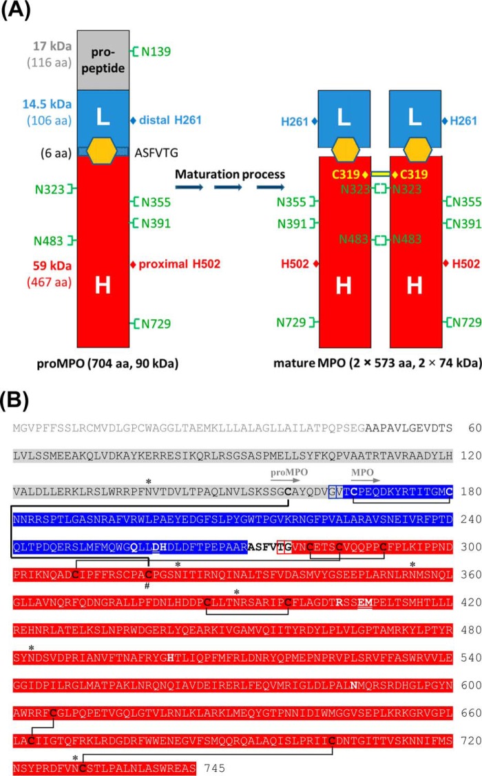
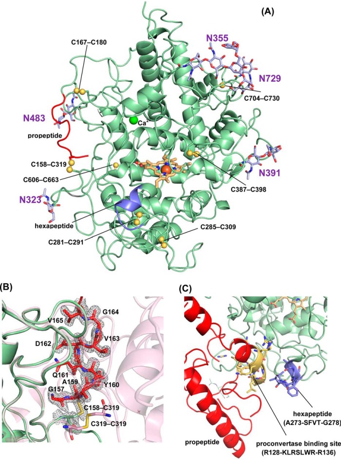
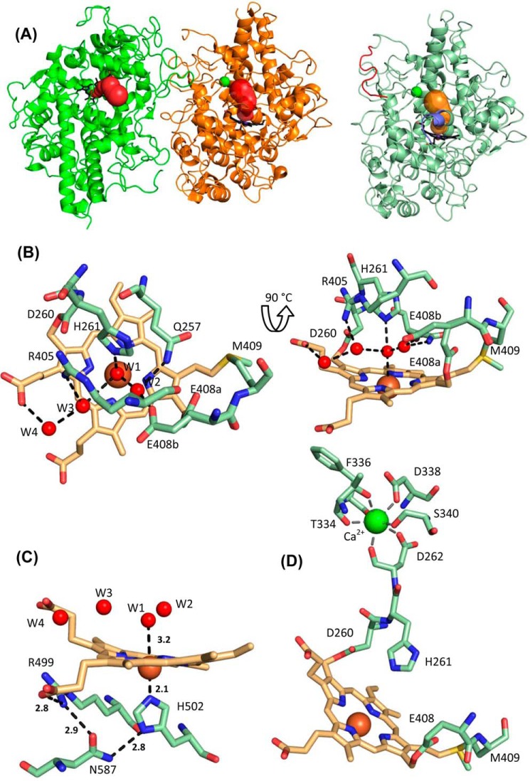
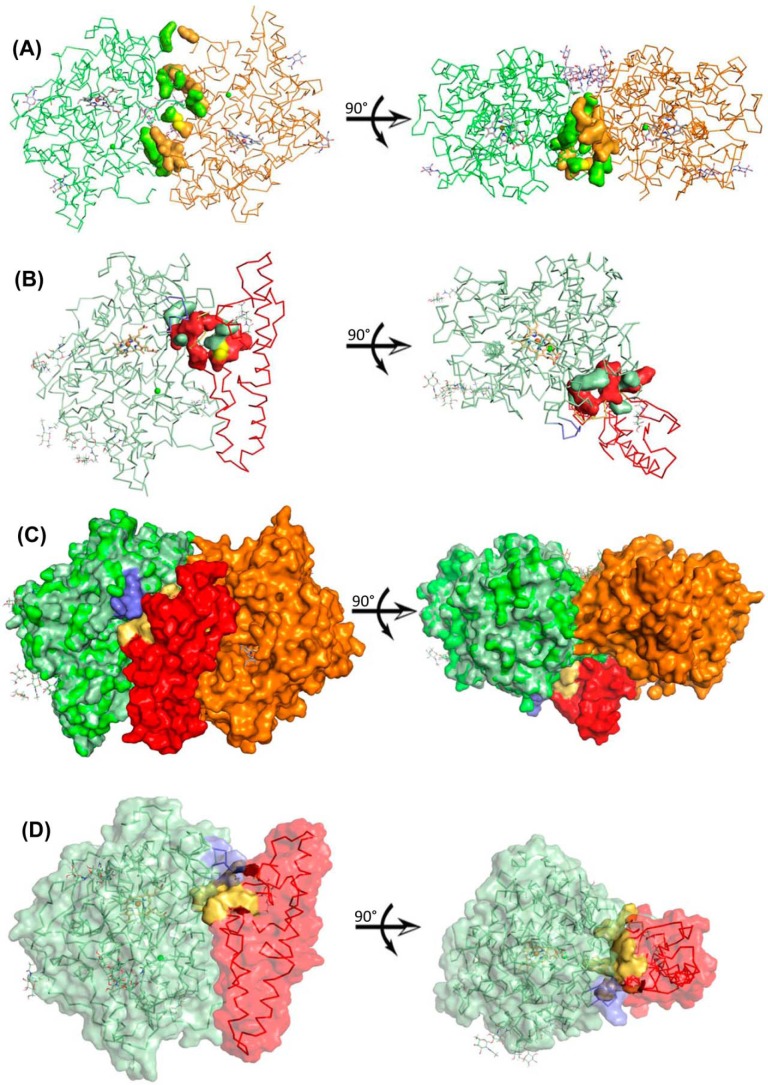
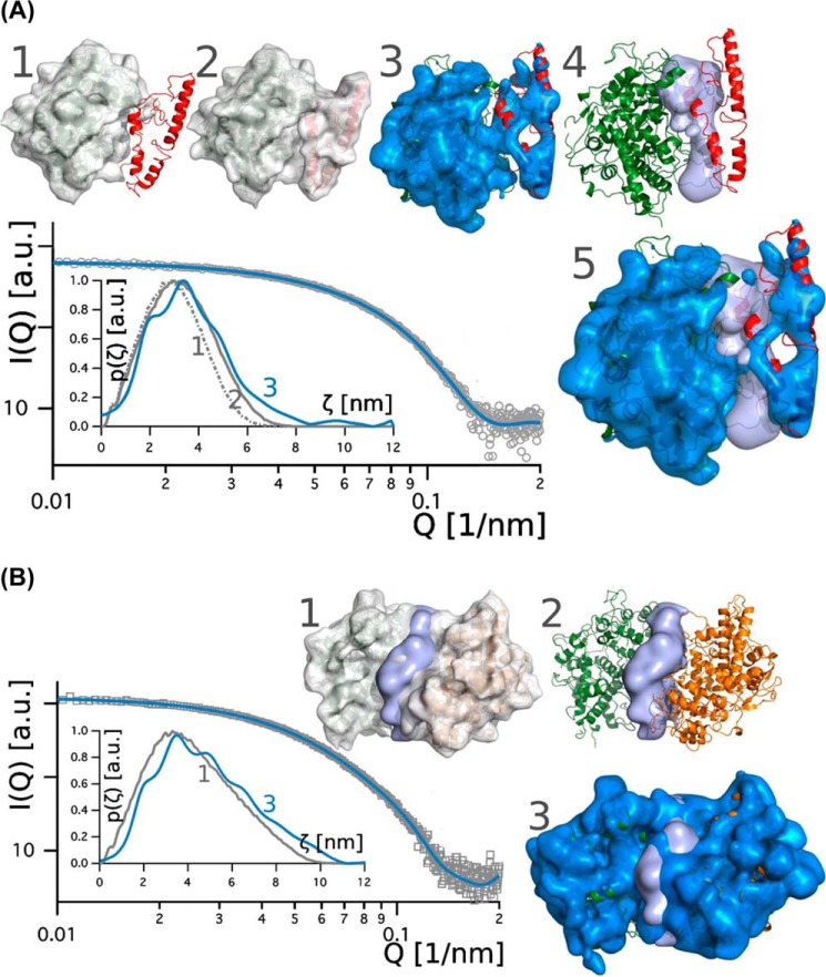
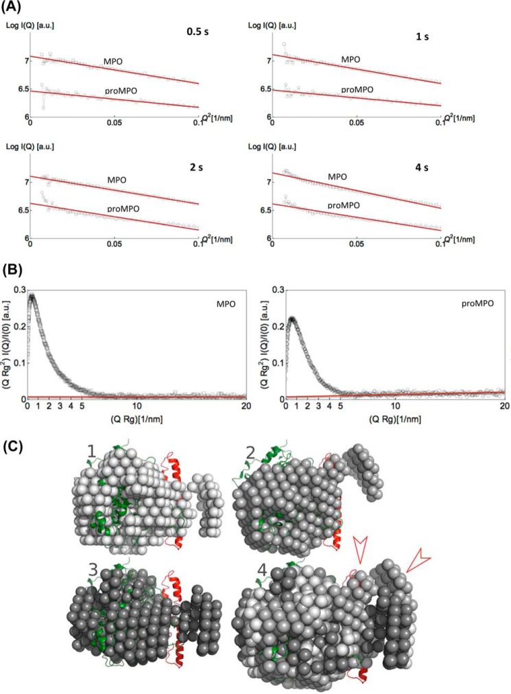
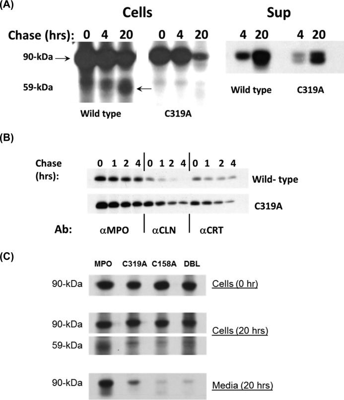
Myeloperoxidase (MPO) is synthesized by neutrophil and monocyte precursor cells and contributes to host defense by mediating microbial killing. Although several steps in MPO biosynthesis and processing have been elucidated, many questions remained, such as the structure-function relationship of monomeric unprocessed proMPO versus the mature dimeric MPO and the functional role of the propeptide. Here we have presented the first and high resolution (at 1.25 Å) crystal structure of proMPO and its solution structure obtained by small-angle X-ray scattering. Promyeloperoxidase hosts five occupied glycosylation sites and six intrachain cystine bridges with Cys-158 of the very flexible N-terminal propeptide being covalently linked to Cys-319 and thereby hindering homodimerization. Furthermore, the structure revealed (i) the binding site of proMPO-processing proconvertase, (ii) the structural motif for subsequent cleavage to the heavy and light chains of mature MPO protomers, and (iii) three covalent bonds between heme and the protein. Studies of the mutants C158A, C319A, and C158A/C319A demonstrated significant differences from the wild-type protein, including diminished enzymatic activity and prevention of export to the Golgi due to prolonged association with the chaperone calnexin. These structural and functional findings provide novel insights into MPO biosynthesis and processing.

Keywords: biosynthesis; crystallography; heme; innate immunity; myeloperoxidase.


Conflict of interest statement

The authors declare that they have no conflicts of interest with the contents of this article

Figures

Figure 1.
Figure 1.
Structure and sequence of proMPO and mature MPO. A, schematic presentation of the structure of monomeric unprocessed proMPO and dimeric mature MPO, including the locations of the distal and proximal catalytic histidines (His-261 and His-502) and the N-glycosylation sites. The propeptide of proMPO and the L- and H-chain of mature MPO are depicted in gray, blue, and red, respectively, together with the respective molar masses and number of amino acids. B, sequence of the primary translation product of human myeloperoxidase. Mature MPO is a homodimer with each monomer composed of a light (blue) and heavy chain (red). The signal peptide (light gray), the propeptide (gray), and a small peptide (depicted in bold black letters) are excised co- and posttranslationally. Boxed in blue and red are the alternative N termini of the light and heavy chains, respectively. Cysteine residues are depicted in bold black or white, and cysteine residue 319, which is responsible for dimer formation in mature MPO, is marked by the # symbol. Disulfides of MPO are shown by black lines. The cystine bridge in proMPO between Cys-158 and Cys-319 is shown as a bold black line. The N-glycosylation sites of both MPO forms are marked by an asterisk (*). ProMPO is a single peptide chain (Ala-49–Ser-745). The first resolved amino acid residues of the crystal structures of proMPO and MPO are depicted with gray arrows. Important catalytic residues are depicted in bold, and amino acid residues involved in the covalent heme to protein links are underlined and bold.
Figure 2.
Figure 2.
Biochemical properties of monomeric proMPO and dimeric MPO. A, UV-visible spectra of 8 μm (per heme) of proMPO (green) and MPO (gray) recorded in PBS. Spectra were shifted along the y axis for better visualization. B, thermal stability of proMPO and MPO evaluated by DSC. Thermal transitions of proMPO (green) and MPO (gray) were fitted to non-two-state equilibrium-unfolding models by the Levenberg-Marquardt nonlinear least squares method, and fits are depicted in dark green for proMPO and dark gray for MPO. C, SDS-PAGE of 2 μg of proMPO and MPO was resolved under reducing conditions on a 4–12% gradient gel.
Figure 3.
Figure 3.
Crystal packing. A, SDS-PAGE of proMPO crystals. B, crystal packing. The core protein of proMPO of the asymmetric unit is presented in green, and the propeptide derived from modeling is shown in red. Symmetry-related molecules are presented in gray. The crystallographic axis b is indicated to help identify the solvent channels parallel to it. Additionally, one solvent-channel-width dimension is given.
Figure 4.
Figure 4.
Overall structure of proMPO. A, crystal structure of recombinant proMPO. The disulfide bridges are depicted in yellow. In addition the N-glycosylation sites are shown in light violet. The segment of the propeptide visible in the electron density is shown in red, and the hexapeptide is shown in blue. B, close-up view of the C-terminal stretch of the propeptide (Gly-157–Val-165, shown in red) and the surrounding core-protein region, including the Cys-158–Cys-309 bridge, together with the 2 FoFc electron density map contoured at the 1σ level. The second protomer in dimeric MPO, including the Cys-319–Cys-319 disulfide bridge, is depicted in light pink for comparison. C, close-up view of the area around the hexapeptide 273ASFVTG278 (blue) and neighboring proposed proconvertase-binding site (128RKLRS132) (highlighted in yellow). The rest of the propeptide is depicted in red.
Figure 5.
Figure 5.
Heme cavity architecture of proMPO. A, substrate-access channels in mature MPO (left panel) and proMPO (right panel). The segment of the propeptide visible in the electron density map is shown in red. The channels were calculated using the tool CAVER 3.0 (57). For better orientation, the prosthetic group is highlighted in bold black. B, distal heme cavity and H-bonding network of proMPO. The catalytic residues His-261, Arg-405, and Gln-257 including W1–W4 and H-bondings are shown. The residues Asp-260, Glu-408, and Met-409 are involved in heme-to-protein linkages. The split conformation of Glu-408 is designated E408a and E408b. C, prosthetic group and proximal ligand His-502 together with the interaction network His-502–Asn-587–Arg-499 and the propionate of pyrrole ring D (distances are given in Å). D, calcium-binding site in proMPO.
Figure 6.
Figure 6.
Hybrid-model structure of proMPO. A, interface between the two identical protomers (chain A depicted as a green ribbon and chain B as an orange ribbon) of mature MPO (PDB accession code 3F9P). Residues involved in noncovalent interactions are presented as space-filling models (Arg-184–Glu-202, Ser-185–Ala-201, Thr-187–Gly-204, Arg-193–Asn-323, Arg-193–Ile-324, Ala-201–Ser-185, Glu-202–Arg-184, Gly-204–Thr-187, Lys-218–Glu-169, Cys-319–Cys-319, Asn-323–Arg-193, and Ile-324–Arg-193). B, interface between the propeptide (red) and the core protein of proMPO (pale green). Residues involved in noncovalent interactions are presented as space-filling amino acids (Glu-127–Cys-316, Arg-131–Arg-314, Arg-135–Gly-321, Lys-154–Thr-325, Ser-155–Cys-319, and Cys-158–Cys-319). Sulfur atoms in Cys-158–Cys-319 are shown in yellow. Residues belonging to the propeptide are shown in red. C, surface representation of the overlay of mature MPO and propeptide of proMPO in the colors described above. The hexapeptide is depicted in blue. A–C are represented from identical angles. D, proposed binding site (128RRKLRSLWR136) of proconvertase in proMPO (yellow surface). The propeptide is depicted in red and the core protein of proMPO in pale green.
Figure 7.
Figure 7.
Solution structures of proMPO and dimeric leukocyte MPO obtained by SAXS. A, promyeloperoxidase: plot of scattering intensity I(Q) (gray circles) and fit (blue line) versus scattering vector. The inset shows pair-density distributions p(ζ) computed for: the proMPO crystal structure (dotted gray line 1); the hybrid model, which contains the propeptide (gray line 2); and p(ζ) computed from the scattering intensity (solid blue line 3). The corresponding surface models (depicted in gray) are superimposed onto the schematic representation of the crystal structure (model 1) and the hybrid model structure (model 2) of proMPO. The blue surfaces in models 3 and 5 comprise scattering sites that contribute to I(Q); χ2 of the model is 0.91. The light blue surface comprises residues that potentially contribute to the interface between the propeptide and the compact rest of the protein. In model 4 the secondary structural elements of the core protein (green) and the propeptide (red) are shown. B, mature dimeric leukocyte MPO: plot of scattering intensity I(Q) (gray circles) versus scattering vector including fit (blue line). In the inset we compared the pair-density distribution p(ζ) computed for the background-corrected data (blue line) and the MPO crystal structure (gray line). The corresponding surface models are depicted in gray (model 1) and blue (model 3); χ2 of the model is 0.90. The light blue surface comprises residues that potentially contribute to the interface of the dimer. In model 2 the secondary structural elements of the two protomers of MPO are depicted in green and orange. a. u., arbitrary units.
Figure 8.
Figure 8.
Guinier and normalized Kratky plots of dimeric MPO and proMPO and bead models of proMPO. A, Guinier plots. The logarithm of scattering intensity (lnI(Q)) of dimeric mature MPO (gray squares) and proMPO (gray circles) are given as a function of Q2. Four different times of data acquisition (0.5, 1, 2, and 4 s) are compared. For leukocyte MPO, nonlinearity is seen after 4 s, whereas for proMPO, nonlinear effects are seen already after 2 s of beam exposure. Nonlinearity is taken as an indication of the onset of radiation damage. B, normalized Kratky plot given as a function of the radii of gyration Rg and extrapolated scattering intensity I(Q) for dimeric mature MPO (gray squares) and proMPO (gray circles). Note the difference in slope of the baseline (red line). Although the baseline for leukocyte MPO is constant, the baseline for proMPO shows a positive slope, which is an indication of the soft binding of the propeptide to the core of proMPO. a. u., arbitrary units. C, bead models of proMPO derived from data analysis using the software ATSAS 2.7.2. We computed three different models using the program package ATSAS 2.7.2. For computation of the bead models, we used the pair-density distributions depicted in Fig. 6A. These bead models were aligned by PyMOL and superimposed in model 4.
Figure 9.
Figure 9.
Biosynthesis of mutants C319A and C158A and double mutant C158A/C319A. A, transfectants expressing wild-type MPO or C319A were radiolabeled biosynthetically and chased for 0–20 h. Cell lysates (Cells) and supernatants (Sup) were immunoprecipitated for MPO-related proteins, including the 90-kDa precursors (apo- and proMPO) and the 59-kDa heavy subunit of mature MPO. B, stable transfectants expressing wild-type MPO or C319A were radiolabeled biosynthetically and chased for 0–4 h. Lysates were immunoprecipitated for MPO (αMPO) or sequentially immunoprecipitated with αCLN → αMPO to recover CLN-associated MPO (αCLN) or with αCRT → αMPO to recover CRT-associated MPO (αCRT). A representative of three to five independent experiments is shown. C, transfectants expressing wild-type MPO, C319A, C158A, or C319A/C158A (DBL) were radiolabeled biosynthetically and chased for 0–20 h. Cell lysates (Cells) and supernatants (Media) were immunoprecipitated for MPO-related proteins, including the 90-kDa precursors (apo- and proMPO) and the 59-kDa heavy subunit of mature MPO. A representative of four to six independent experiments is shown.

References

    1. Nauseef W. M. (2014) Myeloperoxidase in human neutrophil host defence. Cell. Microbiol. 16, 1146–1155 - PMC - PubMed
    1. Olsson I., Persson A. M., and Strömberg K. (1984) Biosynthesis, transport and processing of myeloperoxidase in the human leukaemic promyelocytic cell line HL-60 and normal marrow cells. Biochem. J. 223, 911–920 - PMC - PubMed
    1. Zamocky M., Jakopitsch C., Furtmüller P. G., Dunand C., and Obinger C. (2008) The peroxidase-cyclooxygenase superfamily: reconstructed evolution of critical enzymes of the innate immune system. Proteins 72, 589–605 - PubMed
    1. Zámocký M., Hofbauer S., Schaffner I., Gasselhuber B., Nicolussi A., Soudi M., Pirker K. F., Furtmüller P. G., and Obinger C. (2015) Independent evolution of four heme peroxidase superfamilies. Arch. Biochem. Biophys. 574, 108–119 - PMC - PubMed
    1. Fiedler T. J., Davey C. A., and Fenna R. E. (2000) X-ray crystal structure and characterization of halide binding sites of human myeloperoxidase at 1.8 Å resolution. J. Biol. Chem. 275, 11964–11971 - PubMed

Publication types

LinkOut - more resources