. 2017 May 19;292(20):8472-8483.
doi: 10.1074/jbc.M117.778076. Epub 2017 Mar 27.

The herpes simplex virus 1 UL36USP deubiquitinase suppresses DNA repair in host cells via deubiquitination of proliferating cell nuclear antigen

Affiliations

The herpes simplex virus 1 UL36USP deubiquitinase suppresses DNA repair in host cells via deubiquitination of proliferating cell nuclear antigen

Xiaodong Dong et al. J Biol Chem. .

Abstract

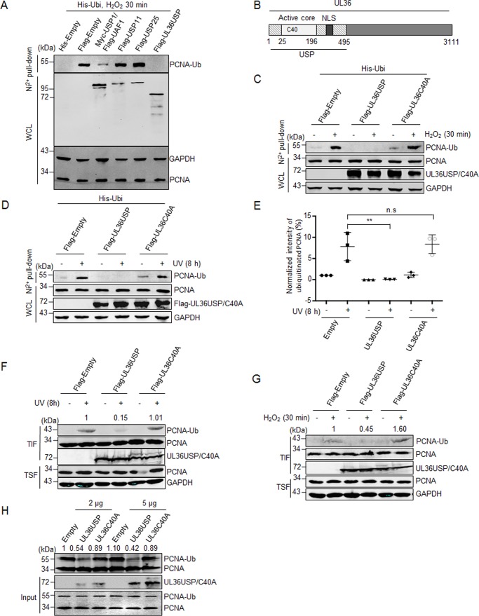
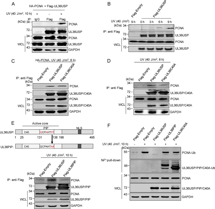
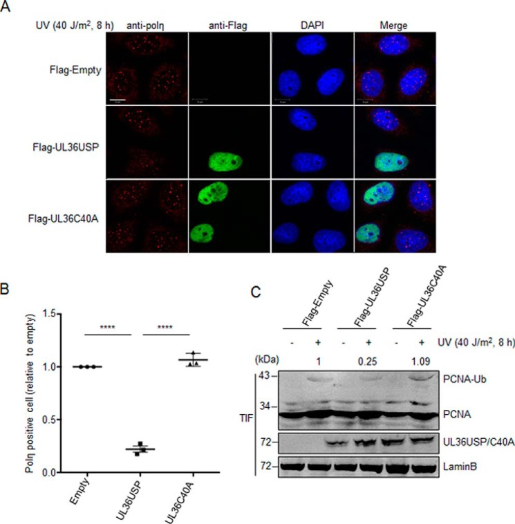
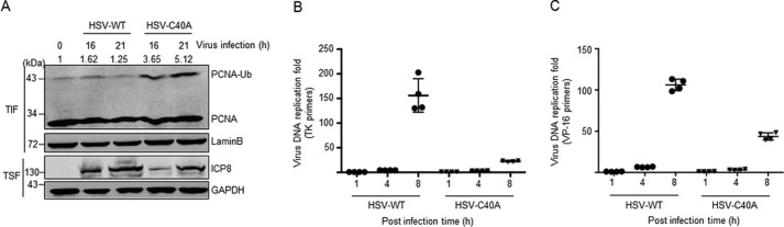
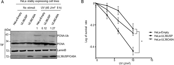
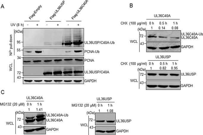
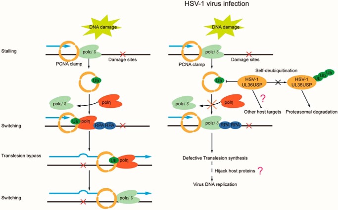
Herpes simplex virus 1 (HSV-1) infection manipulates distinct host DNA-damage responses to facilitate virus proliferation, but the molecular mechanisms remain to be elucidated. One possible HSV-1 target might be DNA damage-tolerance mechanisms, such as the translesion synthesis (TLS) pathway. In TLS, proliferating cell nuclear antigen (PCNA) is monoubiquitinated in response to DNA damage-caused replication fork stalling. Ubiquitinated PCNA then facilitates the error-prone DNA polymerase η (polη)-mediated TLS, allowing the fork to bypass damaged sites. Because of the involvement of PCNA ubiquitination in DNA-damage repair, we hypothesized that the function of PCNA might be altered by HSV-1. Here we show that PCNA is a substrate of the HSV-1 deubiquitinase UL36USP, which has previously been shown to be involved mainly in virus uptake and maturation. In HSV-1-infected cells, viral infection-associated UL36USP consistently reduced PCNA ubiquitination. The deubiquitination of PCNA inhibited the formation of polη foci and also increased cell sensitivity to DNA-damage agents. Moreover, the catalytically inactive mutant UL36C40A failed to deubiquitinate PCNA. Of note, the levels of virus marker genes increased strikingly in cells infected with wild-type HSV-1, but only moderately in UL36C40A mutant virus-infected cells, indicating that the UL36USP deubiquitinating activity supports HSV-1 virus replication during infection. These findings suggest a role of UL36USP in the DNA damage-response pathway.

Keywords: DNA damage response; DNA polymerase; UL36USP; deubiquitylation (deubiquitination); herpesvirus; proliferating cell nuclear antigen (PCNA); translesion synthesis.


Conflict of interest statement

The authors declare that they have no conflicts of interest with the contents of this article

Figures

Figure 1.
Figure 1.
UL36USP deubiquitinates PCNA in vivo and in vitro. A, UL36USP reduces PCNA ubiquitination more efficiently than USP1/UAF1. HEK293T cells were cotransfected with His-ubiquitin and the indicated deubiquitinase plasmids, at 48 h after transfection, cells were treated with 100 μm H2O2 for 30 min and lysed. Cell lysates were subjected to His pulldown analysis, and the ubiquitinated PCNA was detected by Western blotting using anti-PCNA antibody. B, UL36USP is the N-terminal region of the HSV large tegument protein UL36. The cysteine at position 40 is the key amino acid of deubiquitinating activity. C–G, wild-type UL36USP, but not the C40A mutant, deubiquitinates PCNA in response to DNA damage induced by H2O2 or UV treatment. HEK293T cells were transfected with UL36USP or mutant UL36C40A that were defective in deubiquitination. At 40 h after transfection, cells were stimulated with or without H2O2 (100 μm, C) or UV (40 J/m2, D) and subjected to a His pulldown assay as described above. E, quantification of ubiquitinated PCNA normalized to the empty vector expressing group. Three independent His pulldown experiments were performed at the same conditions of D, the normalized intensities of PCNA ubiquitination was shown in scatter plots with standard deviation to represent error. F and G, HEK293T cells were transfected with UL36USP or UL36C40A for 40 h, and then treated with or without UV (F) or H2O2 (G). Cells were harvested, incubated with Triton X-100 buffer, and fractionated into chromatin-containing insoluble (TIF) and soluble (TSF) fractions. PCNA were concentrated in the insoluble fraction and probed by anti-PCNA antibody. The intensity of ubiquitinated PCNA was normalized to the total quantity of PCNA in TIF. H, in vitro deubiquitination assay shows UL36USP but not C40A deubiquitinates PCNA. HEK293T cells were transfected with His-ubiquitin and HA-PCNA plasmids. At 48 h after transfection, UV was used to promote PCNA ubiquitination. Ubiquitinated PCNA was precipitated by using Ni2+ beads and incubated with purified UL36USP or UL36C40A recombinant protein, as indicated, at 30 °C overnight, and then detected by Western blotting with antibody targeting PCNA. The intensity of ubiquitinated PCNA was normalized to the total quantity of PCNA calculated by adding ubiquitinated and unmodified PCNA. UL36USP and UL36C40A proteins were purified from E. coli. All the experiments described have been repeated more than 3 times. WCL, whole cell lysates; **, p value < 0.01; n.s., not significant.
Figure 2.
Figure 2.
UL36USP and UL36C40A interact with PCNA in response to DNA damage. A and B, UV treatment enhances the interaction between PCNA and UL36USP. HEK293T cells transfected with FLAG-UL36USP and HA-PCNA plasmids were treated with or without UV for the indicated time. After stimulation, a co-IP assay was performed to assess the interaction between PCNA with UL36USP or -C40A using the indicated antibodies. C, PCNA interacts with both UL36USP and UL36C40A. HEK293T cells transfected with HA-PCNA, FLAG-Empty, FLAG-UL36USP, or UL36C40A plasmids were treated with 40 J/m2 UV. At 8 h post-stimulation, cells were harvested and subjected to co-IP experiment. D, endogenous PCNA interacts with UL36USP and UL36C40A. HEK293T cells were transfected with FLAG-empty, FLAG-UL36USP, or UL36C40A plasmids and harvested as described in B. E, a schematic diagram of the UL36USP PIP domain and the UL36PIP mutation construct. The conserved amino acids of PIP domain are indicated by red, and the contiguous phenylalanines were mutated to alanines. The interaction between PCNA and UL36USP or UL36PIP was assessed by co-IP analysis using anti-FLAG® M2 magnetic beads, followed by Western blotting with anti-PCNA antibody. F, UL36PIP does not inhibit PCNA ubiquitination. HEK293T cells transfected with the indicated plasmids were subjected to 40 J/m2 UV stimuli for 10 h. And then a His pulldown assay was performed to examine ubiquitinated PCNA.
Figure 3.
Figure 3.
UL36USP inhibits polη foci formation in response to oxidative DNA damage. A, the number of polη foci was significantly decreases by UL36USP but not C40A. HeLa cells were transfected with UL36USP or -C40A plasmids and subjected to UV treatment. At 30 min after DNA-damage stimuli cells were fixed by cold methyl alcohol, immunofluorescence staining was performed using anti-polη and anti-FLAG antibodies. Endogenous polη were indicated by red fluorescence and UL36USP or UL36C40A by green. The scale bar represents 10 μm. B, quantification of polη foci-positive cells. Percentages of polη foci-positive cells (≥10 polη foci) were counted separately by Bitplane Imaris software. Three independent experiments were performed and ∼1000 cells were counted in each group. Error bars shows S.D. ****, p value < 0.0001. C, PCNA ubiquitination levels decreased in UL36USP-expressing cells compared with the control or UL36C40A group. HeLa cells transfected with UL36USP or -C40A plasmids were treated with or without UV. At 30 min after stimuli, cells were incubated with Triton X-100 buffer. Ubiquitinated PCNA in Triton X-100-soluble and -insoluble fractions was detected separately by Western blotting. The amount of ubiquitinated PCNA was normalized to Lamin B and indicated above the blot.
Figure 4.
Figure 4.
The deubiquitination activity of UL36USP is essential for viral DNA replication during HSV-1 infection. A, HSV-1 C40A mutant virus infection induced ubiquitination of PCNA, whereas wild-type virus exhibited an equivalent level compared with the control group. HEK293T cells were mock infected or infected with HSV-1 wild-type or UL36C40A mutant virus at 0.1 m.o.i. At 16 or 21 h post-infection, cells were fractionated into chromatin-containing fractions by incubating with Triton X-100 buffer. Western blotting was performed using the indicated antibodies. The intensity of ubiquitinated PCNA was normalized to Lamin B. B and C, HSV-1 wild-type virus DNA replication rate is higher than the UL36C40A mutant virus. Vero cells were infected with HSV-1 wild-type or UL36C40A mutant virus. Cells were harvested at the indicated time for quantitative real-time PCR analysis. The quantity of virus DNA was indicated by virus genes tk (B) and vp16 (C), respectively.
Figure 5.
Figure 5.
UL36USP stably expressing HeLa cells exhibited lower viability than that of wild-type or UL36C40A stably expressing cells after DNA damage. A, PCNA ubiquitination level decreased in UL36USP stably expressing HeLa cells compared with the control or UL36C40A group. HeLa cells stably expressing UL36USP or -C40A were treated with or without 40 J/m2 UV for 10 h and then incubated with Triton X-100. Triton-insoluble fractions were subjected to Western blotting to detect PCNA ubiquitination and UL36USP or -C40A expression. The intensity of ubiquitinated PCNA was normalized to Lamin B. B, UL36USP stably expressing HeLa cells showed decreased viability after DNA damage. UL36USP or UL36C40A stably expressing HeLa cells were treated with various ultraviolet radiation conditions as indicated. After 12 days, cell colonies were stained with crystal violet and counted; *, p value < 0.05.
Figure 6.
Figure 6.
UL36USP and UL36C40A are ubiquitinated and degraded by proteasome, whereas UL36USP deubiquitinates itself to stabilize itself. A, UL36C40A had a stronger ubiquitination modification compared with UL36USP. HEK293T cells were transfected with His-ubiquitin and UL36USP or -C40A plasmids and treated with or without ultraviolet radiation. His pulldown assay was performed to examine the ubiquitination of UL36USP and UL36C40A using anti-FLAG antibody. B, UL36C40A degraded after CHX treatment but UL36USP does not. HEK293T cells stably expressing UL36USP or UL36C40A were treated with CHX to block protein synthesis for the indicated times. Whole cell lysates were detected by Western blotting. C, UL36C40A degradation was blocked by MG132 treatment. HEK293T cells stably expressing UL36USP or -C40A were treated with MG132 to block proteasome degradation. Whole cell lysates (WCL) were detected by Western blotting. The intensity of UL36USP and UL36C40A were normalized against those of GAPDH.
Figure 7.
Figure 7.
HSV-1 UL36USP suppresses TLS pathway by deubiquitinating PCNA. DNA damage caused by UV and H2O2 induces PCNA monoubiquitination, which promotes polη foci formation and facilitates cell survival after DNA damage. HSV-1 inhibits the TLS pathway by deubiquiting PCNA through the deubiquitinase activity of the UL36USP protein, which results in a reduced number of polη foci, thus decreasing cell viability. Meanwhile, UL36USP deubiquitinates itself to increase protein stability.

Similar articles

Cited by

References

    1. Luftig M. A. (2014) Viruses and the DNA damage response: activation and antagonism. Annu. Rev. Virol. 1, 605–625 - PubMed
    1. Kannouche P. L., Wing J., and Lehmann A. R. (2004) Interaction of human DNA polymerase η with monoubiquitinated PCNA: a possible mechanism for the polymerase switch in response to DNA damage. Mol. Cell 14, 491–500 - PubMed
    1. Chapman J. R., Taylor M. R., and Boulton S. J. (2012) Playing the end game: DNA double-strand break repair pathway choice. Mol. Cell 47, 497–510 - PubMed
    1. Mailand N., Gibbs-Seymour I., and Bekker-Jensen S. (2013) Regulation of PCNA-protein interactions for genome stability. Nat. Rev. Mol. Cell Biol. 14, 269–282 - PubMed
    1. Davies A. A., Huttner D., Daigaku Y., Chen S., and Ulrich H. D. (2008) Activation of ubiquitin-dependent DNA damage bypass is mediated by replication protein A. Mol. Cell 29, 625–636 - PMC - PubMed

Substances

LinkOut - more resources