. 2017 May 26;292(21):8667-8682.
doi: 10.1074/jbc.M116.771675. Epub 2017 Mar 27.

Delineating distinct heme-scavenging and -binding functions of domains in MF6p/helminth defense molecule (HDM) proteins from parasitic flatworms

Affiliations

Delineating distinct heme-scavenging and -binding functions of domains in MF6p/helminth defense molecule (HDM) proteins from parasitic flatworms

Victoria Martínez-Sernández et al. J Biol Chem. .

Abstract

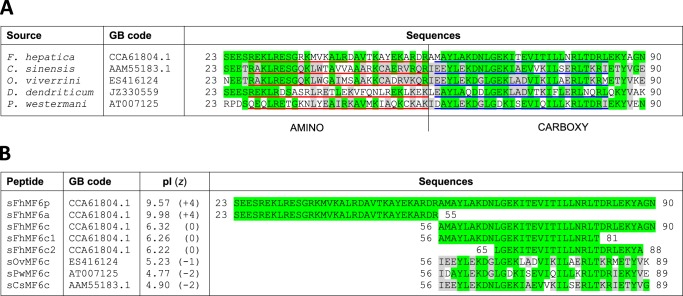
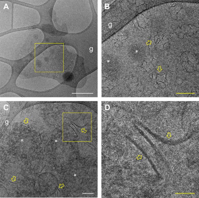
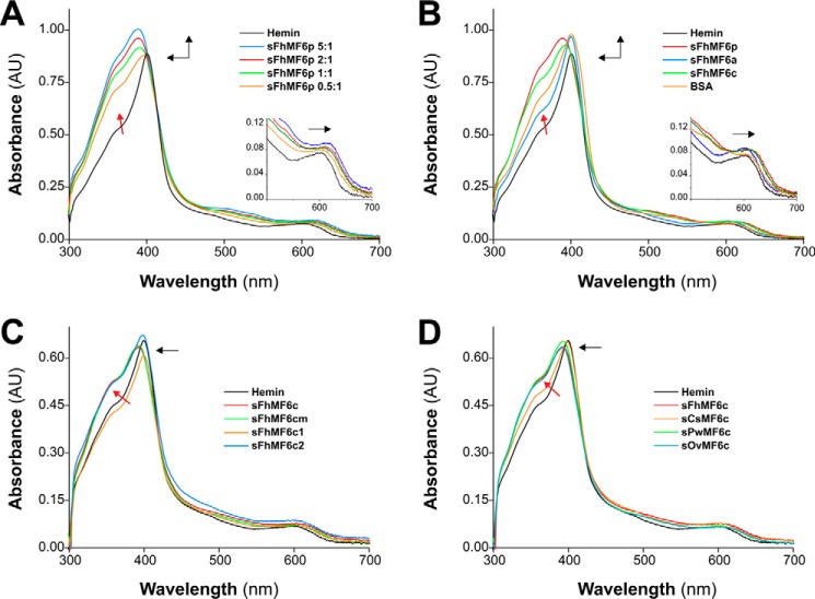
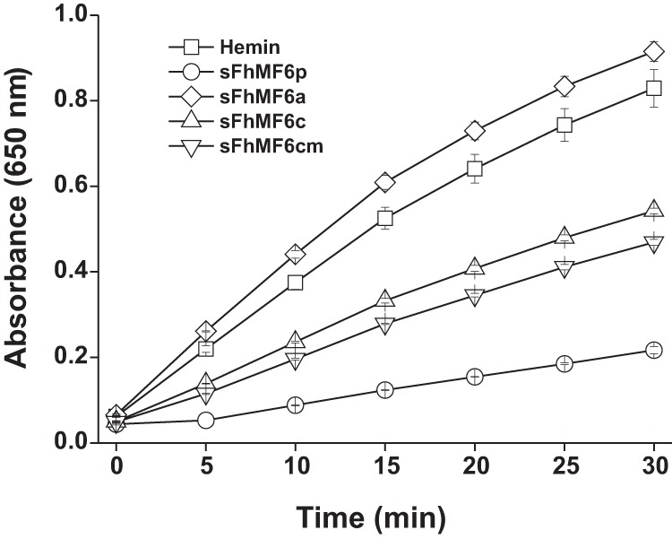
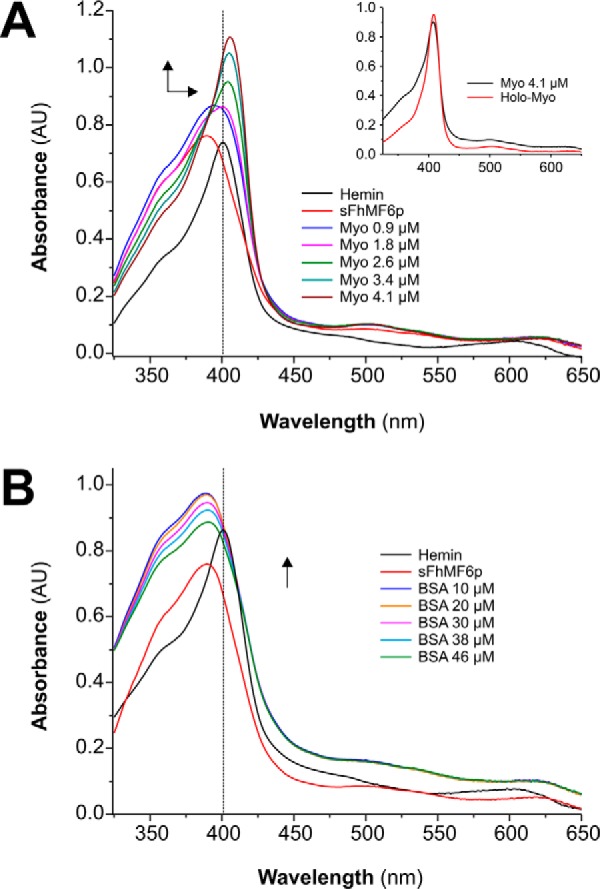
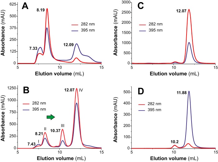
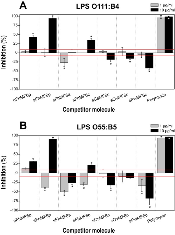
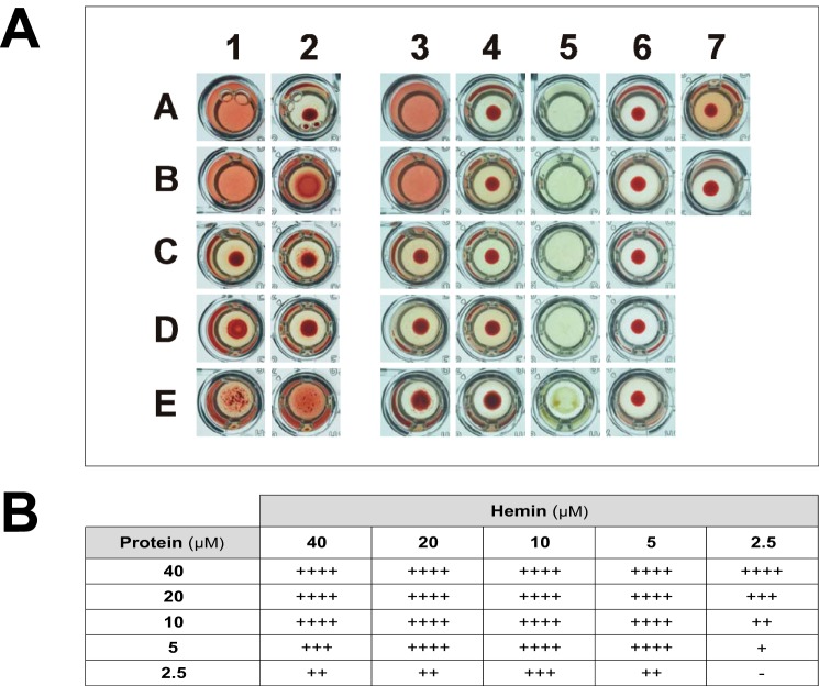
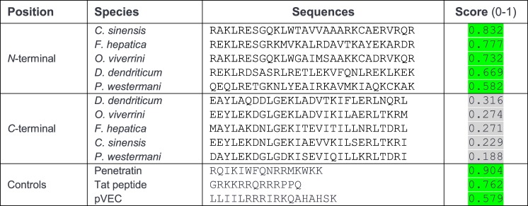
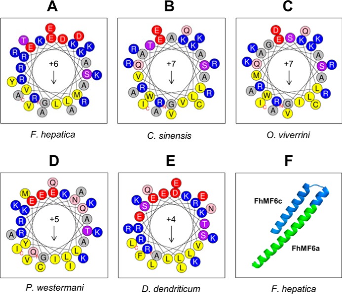
MF6p/FhHDM-1 is a small protein secreted by the parasitic flatworm (trematode) Fasciola hepatica that belongs to a broad family of heme-binding proteins (MF6p/helminth defense molecules (HDMs)). MF6p/HDMs are of interest for understanding heme homeostasis in trematodes and as potential targets for the development of new flukicides. Moreover, interest in these molecules has also increased because of their immunomodulatory properties. Here we have extended our previous findings on the mechanism of MF6p/HDM-heme interactions and mapped the protein regions required for heme binding and for other biological functions. Our data revealed that MF6p/FhHDM-1 forms high-molecular-weight complexes when associated with heme and that these complexes are reorganized by a stacking procedure to form fibril-like and granular nanostructures. Furthermore, we showed that MF6p/FhHDM-1 is a transitory heme-binding protein as protein·heme complexes can be disrupted by contact with an apoprotein (e.g. apomyoglobin) with higher affinity for heme. We also demonstrated that (i) the heme-binding region is located in the MF6p/FhHDM-1 C-terminal moiety, which also inhibits the peroxidase-like activity of heme, and (ii) MF6p/HDMs from other trematodes, such as Opisthorchis viverrini and Paragonimus westermani, also bind heme. Finally, we observed that the N-terminal, but not the C-terminal, moiety of MF6p/HDMs has a predicted structural analogy with cell-penetrating peptides and that both the entire protein and the peptide corresponding to the N-terminal moiety of MF6p/FhHDM-1 interact in vitro with cell membranes in hemin-preconditioned erythrocytes. Our findings suggest that MF6p/HDMs can transport heme in trematodes and thereby shield the parasite from the harmful effects of heme.

Keywords: Fasciola; MF6p/FhHDM-1; MF6p/HDM; cell-penetrating peptide (CPP); fluke; heme; homeostasis; oligomerization; parasite; trematode.


Conflict of interest statement

The authors declare that they have no conflicts of interest with the contents of this article

Figures

Figure 1.
Figure 1.
Sequence alignment of the MF6p/HDM family of proteins. A, aligned sequences of complete mature orthologous proteins present in F. hepatica and related trematodes. The 28–29 regions considered for bioinformatics analyses are underlined in red (N-terminal fragment) and blue (C-terminal fragment). B, aligned sequences of the synthetic peptides used in the present study. Amino acid residues in common with F. hepatica are shaded in green, and residues shared among trematodes other than F. hepatica are shaded in gray. GB, GenBankTM.
Figure 2.
Figure 2.
Fractionation of sMF6p/FhHDM-1·hemin complexes by SEC. Elution profiles obtained by SEC (Superdex 75 HR 10/30) of 0.06 mm hemin samples preincubated with sMF6p/FhHDM-1 at protein to hemin molar ratios of 0.5:1 (blue), 1:1 (black), and 2:1 (green) in TBS-C. A sample of F. hepatica SAs diluted in TBS-C (red) was also analyzed for comparison (red vertical axis). Absorbance at a wavelength of 395 nm is shown for heme, and the elution volume of the major peaks is indicated above each peak. The following internal standards are indicated (vertical bars): albumin (67 kDa), ovalbumin (43 kDa), and chymotrypsin (25 kDa). Arrows indicate the tendency of sMF6p/FhHDM-1·hemin to form higher-molecular-mass complexes as the molar ratio of protein to hemin increased. Some of these complexes were eluted at a retention volume equivalent to nMF6p/FhHDM-1·heme complexes present in SAs (8.36 ml). mAU, milliabsorbance units.
Figure 3.
Figure 3.
The sMF6p/FhHDM-1 protein tends to oligomerize and form high-molecular-mass complexes with hemin. Shown are elution profiles obtained by SEC (Superdex 75 HR 10/30) of sMF6p/FhHDM-1 (0.2-ml sample at 0.12 mm in PBS) applied to a column to which hemin (0.2-ml sample at 0.06 mm in PBS) was previously adsorbed (A), F. hepatica SAs diluted in PBS (B), and a sample of sMF6p/FhHDM-1 applied to a clean column in the absence of hemin (C). Absorbance at wavelengths of 280 (black) and 395 nm (red) are shown for protein and hemin, respectively. The elution volume of the major peaks is indicated above each peak. Hemin trapped by the column beads in A was eluted with sMF6p/FhHDM-1, forming complexes of several molecular masses between that of nMF6p/FhHDM-1·heme complexes (elution volume, 8.98 ml; B), and sMF6p/FhHDM-1 (elution volume, 11.23 ml; C), which forms oligomers of 35–40 kDa. mAU, milliabsorbance units.
Figure 4.
Figure 4.
Cryo-TEM analysis of the structure of nMF6p/FhHDM-1·hemin complexes present in F. hepatica SAs. A sample of a solution of immunopurified nMF6p/FhHDM-1 prepared in distilled water was analyzed by cryo-TEM. A, Grid 1, low-magnification cryo-TEM micrograph showing an overall view of nMF6p/FhHDM-1·hemin complexes extended over the lacey carbon grid (g). White scale bar, 400 nm. B, a higher magnification of the square region marked in yellow in A showing the presence of fibril-like nanostructures (yellow arrows) and electron-dense granular structures of 5–20 nm in diameter (white asterisks). Yellow scale bar, 100 nm. C, Grid 2, intermediate-magnification micrograph from another grid showing abundant fibril-like (yellow arrows) and granular nanostructures (white asterisks). White scale bar, 100 nm. D, a higher magnification of the square region marked in yellow in B showing the fibril-like nanostructures in greater detail. Yellow scale bar, 50 nm.
Figure 5.
Figure 5.
Comparison of hemin spectra in several solvent systems. A, UV/visible absorption spectra of hemin (12 μm) in TBS (pH 7.3), TBS-T, or TBS-C. B, UV/visible absorption spectra of hemin (12 μm) alone, mixed with LSZ, or with PMX at protein to hemin molar ratios of 2:1 and 1:1 in TBS-C. Addition of LSZ or PMX did not shift the absorbance maxima of hemin spectra. AU, absorbance units.
Figure 6.
Figure 6.
The C-terminal region of the MF6p/HDM family of proteins is mainly responsible for the alterations observed in the UV/visible absorption spectra of hemin. Shown are UV/visible absorption spectra of hemin (12 μm) prepared in TBS-C alone (black) or incubated with one of the following: sMF6p/FhHDM-1 (sFhMF6p) at protein to hemin molar ratios of 0.5:1, 1:1, 2:1 and 5:1 (A); sMF6p/FhHDM-1, sFhMF6a, sFhMF6c, or BSA at a protein to hemin molar ratio of 2:1 (B); sFhMF6c, two shorter derivatives (sFhMF6c1 and sFhMF6c2), or an N-acetylated and C-amidated derivative of sFhMF6c (sFhMF6cm) at a protein to hemin molar ratio of 2:1 (C); sFhMF6c or the C-terminal region of orthologous proteins sCsMF6c, sOvMF6c, and sPwMF6c (D). The direction of spectral changes of protein plus hemin samples relative to hemin alone is indicated by black arrows. Insets in A and B depict a magnification of the Q bands. A blue shift of the Soret band (λmax, 401–389 nm), a slight red shift of the Q bands, and the emergence of an apparent shoulder close to 350 nm (red arrow) were observed in A as the molar ratio of sMF6p/FhHDM-1 to hemin increased. Although less intense (see B–D), these spectral changes were also reproduced by F. hepatica sFhMF6c (λmax, 393 nm), by the orthologous peptides sOvMF6c (λmax, 392 nm) and sPwMF6c (λmax, 393 nm), and to a lesser extent by sFhMF6c2 (λmax, 398 nm) and sCsMF6c (λmax, 398 nm). Incubation of hemin with sFhMF6a, sFhMF6c1, or BSA did not shift the absorbance maxima of hemin spectra. AU, absorbance units.
Figure 7.
Figure 7.
The C-terminal region of sMF6p/FhHDM-1 is responsible for the inhibitory effect on hemin peroxidase-like activity. Hemin alone or preincubated with sMF6p/FhHDM-1 (sFhMF6p), sFhMF6a, sFhMF6c, and sFhMF6cm (N-acetylated and C-amidated sFhMF6c) at a protein to hemin ratio of 5:1 were mixed with a commercial solution of TMB (1 mm TMB, 3 mm H2O2) adjusted to pH 6, and TMB oxidation was monitored at 650 nm for 30 min. The sMF6p/FhHDM-1 protein showed the highest level of inhibitory activity followed by both sFhMF6c and sFhMF6cm, whereas sFhMF6a had no effect on the peroxidase-like activity of hemin. The data points show the mean value ±S.D. (error bars) for duplicate wells.
Figure 8.
Figure 8.
sMF6p/FhHDM-1 transfers hemin to apoMYO but not to BSA. A, 20 μm hemin was incubated with 40 μm sMF6p/FhHDM-1 in TBS containing 25 mm caffeine. Both UV/visible absorption spectra prior to (black) and after (red) addition of sMF6p/FhHDM-1 (sFhMF6p) were measured. The sample was then titrated with increasing amounts of apoMYO (0.9, 1.8, 2.6, 3.4, and 4.1 μm final concentrations), and hemin transfer to apoMYO was followed by the increase in the absorbance at 407 nm. The inset shows the UV/visible spectra of MYO prior to hemin extraction (holoMYO) and after hemin reconstitution (MYO). B, another sample of hemin preincubated with sMF6p/FhHDM-1 was titrated with increasing amounts of BSA (10, 20, 30, 38, and 46 μm final concentrations), and the UV/visible absorption spectra were measured. Arrows represent the direction of spectral changes upon addition of apoMYO or BSA. Spectra were not corrected for dilution. AU, absorbance units.
Figure 9.
Figure 9.
Disruption of Fasciola nMF6p/FhHDM-1·hemin complexes of high molecular mass by apoMYO. Size-exclusion chromatography analyses were performed on an FPLC system equipped with a Superdex 75 HR 10/30 column and continuous monitoring at 282 (blue line) and 395 nm (red line). Elution volumes are indicated above each peak fraction. A and B, elution profiles of F. hepatica SAs subjected to SEC alone (A) or mixed with an excess of apoMYO (B). After mixing the SAs with apoMYO, most of the heme signal observed at 8.11 ml in A was eluted at 12.07 ml (B). MALDI-TOF MS confirmed the presence of nMF6p/FhHDM-1 in peak II, MYO in peak III, and a mixture of nMF6p/FhHDM-1 and MYO in peak IV. C and D, elution profiles of myoglobin prior to (C) and after (D) acidification and MEK extraction to obtain apoMYO. Most heme signal (red line) disappeared after MEK treatment, and the protein (black line) eluted as a main peak in both chromatograms (elution volume, 12.7 and 11.88 ml, respectively). A small peak (elution volume, 10.2 ml), probably corresponding to apoMYO aggregates, is also observed in D. mAU, milliabsorbance units.
Figure 10.
Figure 10.
Competitive displacement of LPS binding to PMX. Samples of FITC-LPS from E. coli serotypes O111:B4 (A) and O55:B5 (B) were preincubated at concentrations of 1.25 and 2.5 μg/ml, respectively, in PBS-EDTA-BSA with 10 and 1 μg/ml concentrations of the following proteins/peptides: whole native (nFhMF6p) and synthetic (sFhMF6p); the N- (sFhMF6a) and C-terminal (sFhMF6c) regions of Fasciola MF6p/FhHDM-1; the C-terminal region of orthologous proteins sCsMF6c, sOvMF6c, and sPwMF6c; and PMX. A 100-μl volume of each sample was then added to the wells containing biotinylated PMX captured by deglycosylated avidin. The results are expressed as percentage of inhibition of LPS binding to PMX by the target protein/peptide and are the mean values ±S.D. (error bars) for duplicate wells. The average A (492 nm) obtained for control wells (without inhibitor) was 1.06 ± 0.09 and 0.81 ± 0.05 for serotypes O111:B4 and O55:B5, respectively. The red line indicates the ±S.D. percentage values of control wells. Differences in bars marked with an asterisk were significant at p < 0.05.
Figure 11.
Figure 11.
The sMF6p/FhHDM-1 protein and the peptide sFhMF6a derived from its N-terminal region are able to hemolyze RBCs preconditioned with hemin. A, hemolysis assays showing the effect of the sMF6p/FhHDM-1 protein or derived peptides and hemin on RBCs. The assay was carried out in U-shaped wells of microtiter plates and photographed after 6 h of incubation at room temperature. Wells in column 1 contain protein/peptides incubated with RBCs preconditioned with hemin, wells in column 2 contain protein/peptides preincubated with hemin and subsequently added to RBCs, wells in columns 3 and 4 are the same as in columns 1 and 2 after centrifuging at 800 × g, wells in column 5 contain protein/peptides incubated with hemin alone, and wells in column 6 contain protein/peptides incubated with RBCs alone. The proteins/peptides tested by rows were as follows: A, sMF6p/FhHDM-1; B, sFhMF6a; C, sFhMF6c. OVA (D) and LSZ (E) were used as controls. Proteins/peptides were incubated at 40 μm, hemin was incubated at 20 μm, and RBCs were incubated at 0.5% in TBS. Control wells containing RBCs plus hemin and RBCs only were placed in wells 7A and 7B, respectively. B, table depicting the degree of hemolysis of hemin-preconditioned RBCs produced by different combinations of sMF6p/FhHDM-1 protein and hemin concentrations. The values depict the degree of hemolysis, ranging from no hemolysis (−) to intermediate (++ and +++) and complete hemolysis (++++) as determined by macroscopic observation of the turbidity of RBCs (0.5%) preconditioned with 2.5–40 μm hemin and subsequently incubated with different concentrations of the sMF6p/FhHDM-1 protein (2.5–40 μm).
Figure 12.
Figure 12.
Predictive analysis for the presence of CPPs within the N- and C-terminal sequences of the MF6p/HDM family of proteins. The presence of CPP regions was predicted with the CPPpred bioinformatics tool as indicated under “Experimental procedures.” In addition to the N- and C-terminal sequences derived from the trematode MF6p/HDM family of proteins, three well known CPPs were analyzed as controls with the same tool. The score indicates the probability (p range, 0–1) of the peptides being CPPs. Scores of peptides being CPPs with a probability of ≥50% are shaded in green. The positive CPP control sequences derived from the proteins Antennapedia (Penetratin; residues 43–58), human immunodeficiency virus type 1 Tat (residues 48–60), and murine vascular endothelium cadherin (pVEC; residues 615–632), respectively (25).
Figure 13.
Figure 13.
Helical wheel projections of N-terminal sequences derived from the MF6p/HDM family of proteins and predicted 3D structure of the Fasciola MF6p/FhHDM-1 protein. A–E, helical wheel projections corresponding to peptide FhMF6a (33 residues; see Fig. 1) and related orthologs from the trematodes C. sinensis, O. viverrini, P. westermani, and D. dendriticum. The projections were performed with the HeliQuest v2 tool covering the full sequence. The data indicate that all peptides are amphiphilic with moderate hydrophobic moments (arrows; μH range, 0.338–0.434) and have a net positive charge (z values ranging from +4 to +7). F, ab initio calculation of the 3D structure of the full-length mature sMF6p/FhHDM-1 protein with the QUARK bioinformatics tool predicted that it is composed of two α-helices of different length connected by a small random coil. The portion of the sequence corresponding to the peptides FhMF6c and FhMF6a is marked in blue and green, respectively.

Similar articles

Cited by

References

    1. Keiser J., and Utzinger J. (2005) Emerging foodborne trematodiasis. Emerg. Infect. Dis. 11, 1507–1514 - PMC - PubMed
    1. Ashrafi K., Bargues M. D., O'Neill S., and Mas-Coma S. (2014) Fascioliasis: a worldwide parasitic disease of importance in travel medicine. Travel Med. Infect. Dis. 12, 636–649 - PubMed
    1. Haçarız O., Sayers G., and Baykal A. T. (2012) A proteomic approach to investigate the distribution and abundance of surface and internal Fasciola hepatica proteins during the chronic stage of natural liver fluke infection in cattle. J. Proteome Res. 11, 3592–3604 - PubMed
    1. Mulvenna J., Sripa B., Brindley P. J., Gorman J., Jones M. K., Colgrave M. L., Jones A., Nawaratna S., Laha T., Suttiprapa S., Smout M. J., and Loukas A. (2010) The secreted and surface proteomes of the adult stage of the carcinogenic human liver fluke Opisthorchis viverrini. Proteomics 10, 1063–1078 - PMC - PubMed
    1. Cao X., Fu Z., Zhang M., Han Y., Han Q., Lu K., Li H., Zhu C., Hong Y., and Lin J. (2016) Excretory/secretory proteome of 14-day schistosomula, Schistosoma japonicum. J. Proteomics 130, 221–230 - PubMed

Publication types

MeSH terms

LinkOut - more resources