. 2017 May 1;144(9):1600-1606.
doi: 10.1242/dev.135681. Epub 2017 Mar 27.

FGFR2 is required for airway basal cell self-renewal and terminal differentiation

Affiliations

FGFR2 is required for airway basal cell self-renewal and terminal differentiation

Gayan I Balasooriya et al. Development. .

Abstract

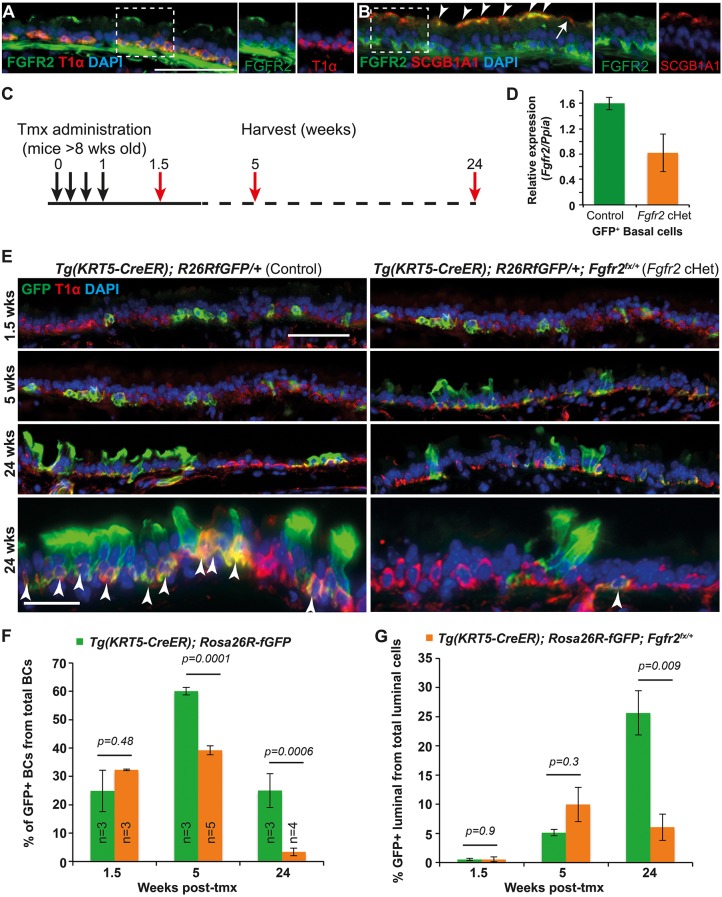
Airway stem cells slowly self-renew and produce differentiated progeny to maintain homeostasis throughout the lifespan of an individual. Mutations in the molecular regulators of these processes may drive cancer or degenerative disease, but are also potential therapeutic targets. Conditionally deleting one copy of FGF receptor 2 (FGFR2) in adult mouse airway basal cells results in self-renewal and differentiation phenotypes. We show that FGFR2 signalling correlates with maintenance of expression of a key transcription factor for basal cell self-renewal and differentiation: SOX2. This heterozygous phenotype illustrates that subtle changes in receptor tyrosine kinase signalling can have significant effects, perhaps providing an explanation for the numerous changes seen in cancer.

Keywords: Cre-Lox; Lung; Mouse; Progenitor; Trachea.


Conflict of interest statement

Competing interestsThe authors declare no competing or financial interests.

Figures

Fig. 1.
Fig. 1.
Decreasing Fgfr2 levels in basal cells results in altered tracheal homeostasis. (A,B) Adult tracheal sections. (A) Green, FGFR2; red, T1α (basal cells). (B) Green, FGFR2; red, SCGB1A1 (secretory cells). FGFR2+ secretory cells (arrowheads); rare SCGB1A1+, FGFR2 cells (arrow). (C) Experimental schematic. (D) Relative expression of Fgfr2 mRNA in GFP+ basal cells from control and Fgfr2cHet mice 3 weeks post-tmx. (E) Representative sections from control Tg(KRT5-CreER); Rosa26RfGFP/+ and cHet Tg(KRT5-CreER); Rosa26RfGFP/+; Fgfr2fx/+ tracheae. Green, GFP (Rosa reporter); red, T1α (basal cells). Arrowheads indicate GFP+ basal cells. (F,G) Percentage of the total T1α+ BCs that are also GFP+ (F) and percentage of the total T1α luminal cells that are also GFP+ (G). Blue, DAPI. Error bars indicate s.e.m. Scale bars: 50 μm.
Fig. 2.
Fig. 2.
Fgfr2 conditional heterozygous basal cells do not produce terminally differentiated luminal cells. (A) Confocal projections from control and Fgfr2cHet tracheae 5 weeks post-tmx. Green, GFP (Rosa reporter); red, KRT5 (basal cells); white, KRT8 (luminal cells); blue, DAPI (nuclei). Arrowheads indicate GFP+ luminal cells. Arrows indicate GFP+ basal cells. (B) Percentage of all GFP+ cells 5 weeks post-tmx that are GFP+, T1 α (see Fig. 1D) or GFP+, KRT8+ (see A). (C) Sections from control and Fgfr2cHet tracheae 5 weeks post-tmx. Green, GFP (Rosa reporter); red, SCGB1A1 (club cells); white, MUC5AC (mucous). Arrows indicate club cells containing a low level of MUC5AC protein. (D) Percentage of all GFP+ cells 5 weeks post-tmx that are GFP+, SCGB1A1+. (E) Confocal sections from control and Fgfr2 cHet tracheae at 24 weeks post-tmx. Green, GFP (Rosa reporter); red, acetylated tubulin (cilia). Error bars indicate s.e.m. Scale bars: 20 μm in A,C; 25 μm in E.
Fig. 3.
Fig. 3.
Fgfr2 conditional heterozygous basal cells have high levels of β-galactosidase and low levels of SOX2. (A) Experimental schematic for B-G. (B) Percentage tracheal epithelial cells at day 6 post-seeding expressing KRT5 and/or KRT8. (C,D) Control and Fgfr2cHet tracheal cells day 6 post-seeding. (C) Green, KRT5 (basal cells); red, KRT8 (luminal cells). (D) X-gal assay for β-galactosidase activity (blue pigment). (E) Representative western blots from control and Fgfr2cHet BCs. (F) Quantification of protein levels in E. (G) SOX2 in cHet BCs day 6 post-seeding. Green, E-cadherin (lateral cell membranes); red, SOX2. (H,I) Confocal images of control and Fgfr2cHet tracheal sections 5 weeks post-tmx. Green, GFP (Rosa reporter); red, SOX2; magenta, FGFR2. White arrows indicate lineage-labelled cells with decreased levels of SOX2. Arrowheads indicate lineage-labelled cells with no change in SOX2. Yellow arrows indicate unlabelled cells with decreased SOX2. Brackets in I indicate a patch of GFP+ cells that have decreased FGFR2 and no SOX2. Blue, DAPI. Error bars indicate s.e.m. Scale bars: 100 μm in C; 250 μm in D; 50 μm in G; 25 μm in H,I.
Fig. 4.
Fig. 4.
FGF7 and FGF10 increase colony size of wild-type basal cells. (A) Experimental schematic. Epithelial cells plated at low density, 3×10⁴ cells/insert. (B) Colonies formed by control, FGF7- or FGF10-treated wild-type cells. Red, E-cadherin; blue, DAPI. Scale bar: 100 μm. (C) Number of cells per colony in B. Data are mean±s.e.m. (D) Level of Sox2 mRNA relative to control (normalized to 1) in cells treated with FGF7 or FGF10 for 1 or 2 days. Error bars indicate s.e.m. (E) Fgfr2cHet BCs rarely make self-renewing divisions in which a new BC is produced. Mutant BCs are more likely to produce descendants with luminal morphology/markers that are unable to completely differentiate, possibly because they senesce. The result is that GFP+ Fgfr2cHet cells are gradually diluted out from both the basal and luminal populations, and the epithelium is sustained by GFP wild-type BCs.

References

    1. Abler L. L., Mansour S. L. and Sun X. (2009). Conditional gene inactivation reveals roles for Fgf10 and Fgfr2 in establishing a normal pattern of epithelial branching in the mouse lung. Dev. Dyn. 238, 1999-2013. 10.1002/dvdy.22032 - DOI - PMC - PubMed
    1. Balasooriya G. I., Johnson J.-A., Basson M. A. and Rawlins E. L. (2016). An FGFR1-SPRY2 signaling axis limits basal cell proliferation in the steady-state airway epithelium. Dev. Cell 37, 85-97. 10.1016/j.devcel.2016.03.001 - DOI - PMC - PubMed
    1. Correia L. L., Johnson J. A., McErlean P., Bauer J., Farah H., Rassl D. M., Rintoul R. C., Sethi T., Lavender P., Rawlins E. L. et al. (2017). SOX2 drives bronchial dysplasia in a novel organotypic model of early human squamous lung cancer. Am. J. Respir. Crit. Care. Med. 10.1164/rccm.201510-2084OC - DOI - PMC - PubMed
    1. de Vries W. N., Binns L. T., Fancher K. S., Dean J., Moore R., Kemler R. and Knowles B. B. (2000). Expression of Cre recombinase in mouse oocytes: a means to study maternal effect genes. Genesis 26, 110-112. 10.1002/(SICI)1526-968X(200002)26:2<110::AID-GENE2>3.0.CO;2-8 - DOI - PubMed
    1. Ferone G., Song J.-Y., Sutherland K. D., Bhaskaran R., Monkhorst K., Lambooij J.-P., Proost N., Gargiulo G. and Berns A. (2016). SOX2 is the determining oncogenic switch in promoting lung squamous cell carcinoma from different cells of origin. Cancer Cell 30, 519-532. 10.1016/j.ccell.2016.09.001 - DOI - PMC - PubMed

Publication types

Substances