Review
. 2017 May 19;372(1720):20150516.
doi: 10.1098/rstb.2015.0516.

Mechanical design in embryos: mechanical signalling, robustness and developmental defects

Affiliations
Review

Mechanical design in embryos: mechanical signalling, robustness and developmental defects

Lance A Davidson. Philos Trans R Soc Lond B Biol Sci. .

Abstract

Embryos are shaped by the precise application of force against the resistant structures of multicellular tissues. Forces may be generated, guided and resisted by cells, extracellular matrix, interstitial fluids, and how they are organized and bound within the tissue's architecture. In this review, we summarize our current thoughts on the multiple roles of mechanics in direct shaping, mechanical signalling and robustness of development. Genetic programmes of development interact with environmental cues to direct the composition of the early embryo and endow cells with active force production. Biophysical advances now provide experimental tools to measure mechanical resistance and collective forces during morphogenesis and are allowing integration of this field with studies of signalling and patterning during development. We focus this review on concepts that highlight this integration, and how the unique contributions of mechanical cues and gradients might be tested side by side with conventional signalling systems. We conclude with speculation on the integration of large-scale programmes of development, and how mechanical responses may ensure robust development and serve as constraints on programmes of tissue self-assembly.This article is part of the themed issue 'Systems morphodynamics: understanding the development of tissue hardware'.

Keywords: biomechanics; developmental defect; mechanobiology; morphogenesis; organogenesis; self-assembly.


Figures

Figure 1.
Figure 1.
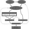
The roles of physical mechanics during development. The direct role of mechanics under genetic control (solid-line arrows). Genetic factors provided maternally and through pattern formation processes that establish the germ layers generate patterns of expression of morphogenetically ‘active’ proteins that regulate adhesion, the cytoskeleton, motors, ECM, etc. These protein complexes are responsible for establishing patterns of cell behaviours such as directed migration and apical contraction. Cell behaviours and their orientation are responsible for both the direction and magnitude of force generation in the embryo. These active proteins also establish the material properties of the embryo responsible for elastic, fluid and viscoelastic responses to internally or externally generated forces. Tissue movements, or deformations, are the net result of forces acting against the mechanical structures of the embryo. The rate and direction of these movements are controlled by the magnitude and direction of forces generated by multiple tissues and also by the anisotropic mechanical response of the tissue, including not only bulk elastic properties but how they adhere to one another and the geometry of their assembly. Mechanical signalling systems connect with conventional biochemical signalling systems through feedback to control molecular complexes and cell behaviours and to control gene regulatory networks and cell identity (dashed-line arrows). Feedback systems may involve mechanosensors that are integrated into the active protein complexes that regulate force production and material properties but may also reside in separate complexes that also detect environmental conditions.
Figure 2.
Figure 2.
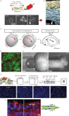
Quantitative biomechanical analysis of sea urchin embryo mechanical design reveals the contribution of stiff ECM to epithelial folding, ruling out a role for actomyosin-driven apical constriction. (a) The start of sea urchin gastrulation involves the folding of a single cell–layered epithelium to form the primitive gut tube. The basal surface of the epithelium of the near spherical embryo faces the fluidic-filled blastocoel while the apical surface is attached to a complex ECM. Folding of the columnar epithelium of the vegetal plate produces a short cylindrical archenteron composed of endoderm and secondary mesenchyme (primary mesenchyme cells have already ingressed to begin spiculogenesis within the blastocoel). (δ, depth of invagination) (b) Fully three-dimensional computational models of sea urchin gastrulation were constructed based on the sampled geometry of a living embryo with layers representing two layers of ECM and a single cell layer. These models demonstrated that several hypotheses were able to generate plausible invaginations [19] but that each placed specific constraints on the embryo's mechanical design (‘topo-map’ shows dependence of simulated depth on modulus of two ECM layers with a fixed cell modulus). Apical constriction, shown here, required a relatively soft apical ECM in order to produce a successful invagination (more than 12 µm). (c) In order to identify the mechanical regime of the living embryo, biomaterials testing with parallel plate compression were combined with ECM and cytoskeleton disruption. The measured modulus under different disruptions revealed the apical ECM was considerably stiffer (more than 4.5 kPa) than cells (0.7 kPa). (d) The measured mechanical properties of the embryo ruled out the role of apical constriction in the physical mechanics of invagination [20].
Figure 3.
Figure 3.
Biomechanical analysis of Xenopus convergent extension reveals cryptic features of mechanical design and function of Rho kinase. Dorsal convergence and extension (CE) is one of the central tissue-scale movements driving gastrulation and anterior–posterior elongation of the vertebrate body plan. The process of CE can be separated from other tissue movements and studied within explants (dorsal isolates) composed of the dorsal-most tissues of early gastrula stage embryos. The rate of extension of dorsal isolates is the same as the extension of the same tissues within intact embryos. Isolates, by contrast to whole embryos, allow more experimental control over their biochemical and mechanical microenvironment (e.g. boundary conditions, surrounding materials) and provide a more regular geometry that allows easily interpretable biomechanical testing. (a) Dorsal isolates incubated in Y-27632 extend at the same rate (strain per hour) as control tissues [22] but exhibit reduced stiffness [23], which is also reflected in reduced actomyosin contractility and formation of punctuated actin ‘asters’ (b; [24]). (c) The reduced contractility without observable changes in deformation rates was paradoxical. However, a novel force-sensing gel (c) revealed that force production after Rho kinase inhibition was reduced by approximately 70% (d; [22]). Biomechanical analysis demonstrated Rho kinase controlled both stiffness and force production to a similar degree that had minimal impact on morphogenetic movements.
Figure 4.
Figure 4.
Mechanical cues can instruct cell behaviours and guide cell fate choices. (a,b) Internal collective movements of mesendodermal cell precursors in the Xenopus embryo migrate from the marginal zone (an annulus centered at the ‘equator’ of the early gastrula) to the future ventral regions of the embryo that will give rise to ventral organs such as the heart. Collective migration of these cells, which exhibit a mesenchymal phenotype is coordinated and highly directed. Both biochemical and biomechanical cues for this directed movement have been proposed. (a) In order to test the role of mechanical cues, single cells isolated from mesendoderm can be cultured on a two-dimensional ECM substrate and subjected to externally applied force. Tension from magnetic beads transmitted through cadherin and the intermediate filament keratin can redirect cell protrusions in Xenopus embryonic mesendoderm cells and (b) may guide in vivo collective migration during ‘free-edge’ spreading of embryonic mesendoderm in Xenopus. (Figure adapted from [31].) (ce) Early patterns of strain generated by gastrulation movement of epiboly, the spreading of prospective epidermis to cover the entire surface of the embryo (c), were proposed to play a role in establishing the planar polarity of cilia-generated fluid flow over the tadpole skin. Directional fluid flow, from anterior-dorsal to posterior-ventral, is driven by the coordinated beating of multiciliated cells distributed over the epidermis. Early anisotropic tissue strain during gastrulation gives rise to polarized microtubules in the ectoderm that presage the planar polarity of multiciliated cells. (e) The patterning effect of strain on cilia and flow patterns can be recreated within naive tissues using micropipette aspiration to generate strain. (Figure adapted from [32].) (f,g) Extensive non-uniform growth of epithelial tissues such as the skin and in endothelial cells such as the kidney suggested mechanical cues could drive both addition and removal of cells to maintain homeostatic balance in cell density. (f) Experimental stretching of cultured epithelial cells revealed tissues could maintain homeostatic control over cell density by extruding cells when surfaces were under compression. (g) This same mechanism appears to control epidermal cell extrusion in the forming tail and fin of zebrafish larva. Density appears to be sensed through the tension-gated membrane channel Piezo. (Figure adapted from [33].) Permissions for re-use of figures provided courtesy of their respective authors.

References

    1. His W. 1888. On the principles of animal morphology. Proc. R. Soc. Edinb. 15, 287–298.
    1. Morgan TH. 1898. Developmental mechanics. (from ‘Biological Problems of To-Day’). Science 7, 156–158. (10.1126/science.7.162.156) - DOI - PubMed
    1. Heisenberg CP, Bellaiche Y. 2013. Forces in tissue morphogenesis and patterning. Cell 153, 948–962. (10.1016/j.cell.2013.05.008) - DOI - PubMed
    1. Eaton S, Jülicher F. 2011. Cell flow and tissue polarity patterns. Curr. Opin Genet. Dev. 21, 747–752. (10.1016/j.gde.2011.08.010) - DOI - PubMed
    1. Daley WP, Yamada KM. 2013. ECM-modulated cellular dynamics as a driving force for tissue morphogenesis. Curr. Opin Genet. Dev. 23, 408–414. (10.1016/j.gde.2013.05.005) - DOI - PMC - PubMed