Review
. 2017 May 19;372(1720):20150519.
doi: 10.1098/rstb.2015.0519.

Mechanocellular models of epithelial morphogenesis

Affiliations
Review

Mechanocellular models of epithelial morphogenesis

Alexander G Fletcher et al. Philos Trans R Soc Lond B Biol Sci. .

Abstract

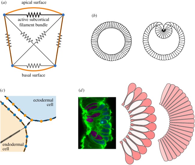
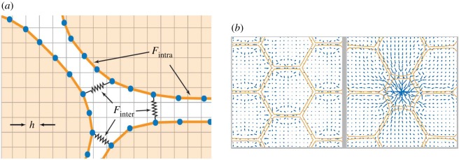
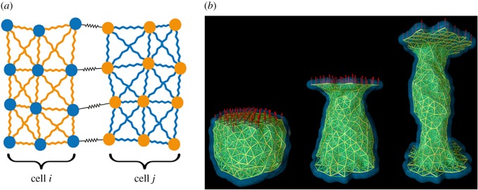
Embryonic epithelia achieve complex morphogenetic movements, including in-plane reshaping, bending and folding, through the coordinated action and rearrangement of individual cells. Technical advances in molecular and live-imaging studies of epithelial dynamics provide a very real opportunity to understand how cell-level processes facilitate these large-scale tissue rearrangements. However, the large datasets that we are now able to generate require careful interpretation. In combination with experimental approaches, computational modelling allows us to challenge and refine our current understanding of epithelial morphogenesis and to explore experimentally intractable questions. To this end, a variety of cell-based modelling approaches have been developed to describe cell-cell mechanical interactions, ranging from vertex and 'finite-element' models that approximate each cell geometrically by a polygon representing the cell's membrane, to immersed boundary and subcellular element models that allow for more arbitrary cell shapes. Here, we review how these models have been used to provide insights into epithelial morphogenesis and describe how such models could help future efforts to decipher the forces and mechanical and biochemical feedbacks that guide cell and tissue-level behaviour. In addition, we discuss current challenges associated with using computational models of morphogenetic processes in a quantitative and predictive way.This article is part of the themed issue 'Systems morphodynamics: understanding the development of tissue hardware'.

Keywords: computational modelling; epithelial morphogenesis; finite-element model; immersed boundary method; subcellular element model.


Figures

Figure 1.
Figure 1.
Polygon-based models of gastrulation. (a) Model of an epithelial cell cross section, incorporating viscoelastic cytoskeletal elements and active apical contractility. Adapted from [35]. (b) Simulating this model from a cylindrically symmetric configuration, while imposing a constant inner volume representing a yolk-filled lumen, leads to behaviour redolent of Drosophila ventral furrow formation. Reproduced from [35]. (c) Model of endodermal (yellow) and ectodermal (blue) cells in the Nematostella vectensis blastula, showing contractile elements (black) within and between cells. Adapted from [39]. (d) Simulation of bottle cell formation with this model. Left: In vivo image showing bottle (red) and squat (blue) cells. Middle/right: two model configurations, where endodermal cells are bound apically only (middle) or also basally (right), resulting in distinct cell shapes during apical constriction. Reproduced from [39].
Figure 2.
Figure 2.
The immersed boundary method. (a) Model schematic shows an off-lattice discretization (blue nodes) of the immersed boundaries representing individual cells (orange) and the regular grid use to discretize the fluid flow problem. Adhesion links exist between blue nodes within each immersed boundary, as well as between neighbouring boundaries. (b) A simulation viewed at two time points shows the computed fluid velocity (blue arrows) and immersed boundary geometry (orange lines). Immersed boundaries are initially at rest in a honeycomb pattern before reacting to the central cell reducing its surface area.
Figure 3.
Figure 3.
The subcellular element model. (a) Model schematic diagram shows two cells and a subset of the intra- and intercellular interactions between their elements. (b) SEM simulation under a creep-stress protocol. Reproduced from [70]. © IOP Publishing. Reproduced with permission. All rights reserved.

References

    1. Oates AC, Gorfinkiel N, González-Gaitán M, Heisenberg C-P. 2009. Quantitative approaches in developmental biology. Nat. Rev. Genet. 10, 517–530. (10.1038/nrg2548) - DOI - PubMed
    1. Gorfinkiel N, Blanchard GB. 2011. Dynamics of actomyosin contractile activity during epithelial morphogenesis. Curr. Opin. Cell Biol. 23, 531–539. (10.1016/j.ceb.2011.06.002) - DOI - PubMed
    1. Martin AC, Kaschube M, Wieschaus EF. 2009. Pulsed contractions of an actin–myosin network drive apical constriction. Nature 457, 495–499. (10.1038/nature07522) - DOI - PMC - PubMed
    1. Blanchard GB, Kabla AJ, Schultz NL, Butler LC, Sanson B, Gorfinkiel N, Mahadevan L, Adams RJ. 2009. Tissue tectonics: morphogenetic strain rates, cell shape change and intercalation. Nat. Methods 6, 458–464. (10.1038/nmeth.1327) - DOI - PMC - PubMed
    1. Miller CJ, Davidson LA. 2013. The interplay between cell signalling and mechanics in developmental processes. Nat. Rev. Genet. 14, 733–744. (10.1038/nrg3513) - DOI - PMC - PubMed