. 2017 May 1;198(9):3494-3506.
doi: 10.4049/jimmunol.1600965. Epub 2017 Mar 27.

Low Antigen Dose in Adjuvant-Based Vaccination Selectively Induces CD4 T Cells with Enhanced Functional Avidity and Protective Efficacy

Affiliations

Low Antigen Dose in Adjuvant-Based Vaccination Selectively Induces CD4 T Cells with Enhanced Functional Avidity and Protective Efficacy

Rolf Billeskov et al. J Immunol. .

Abstract

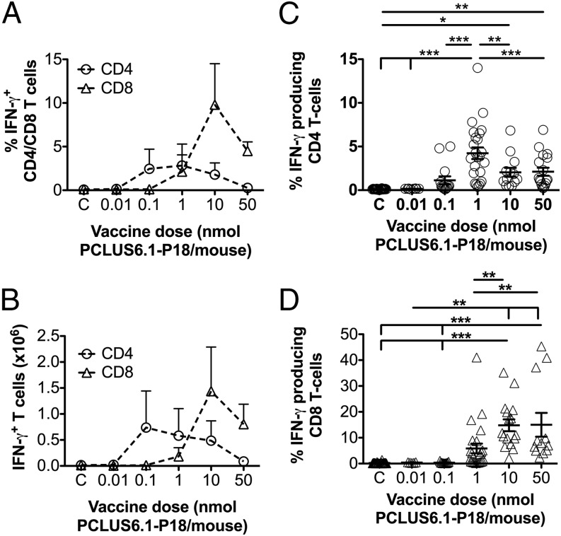
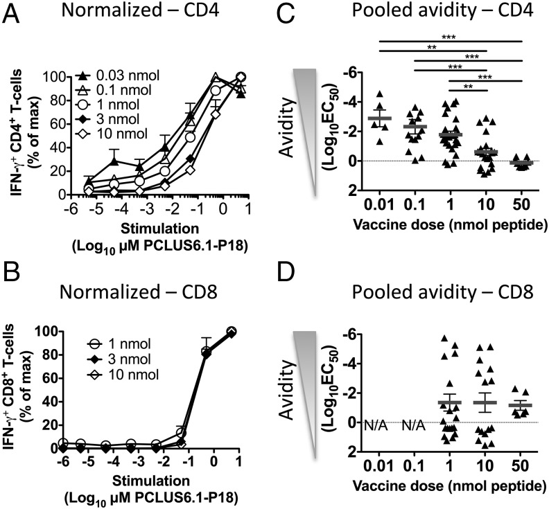
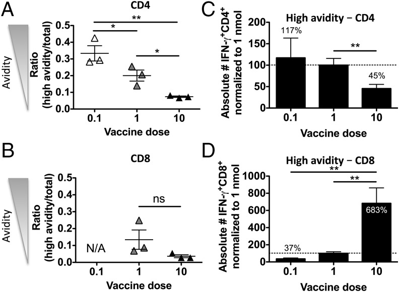
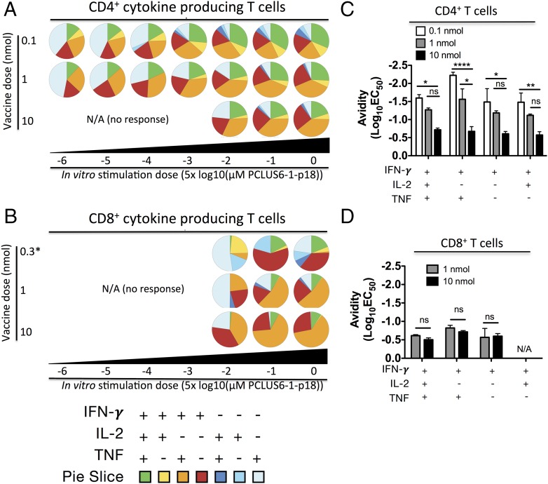
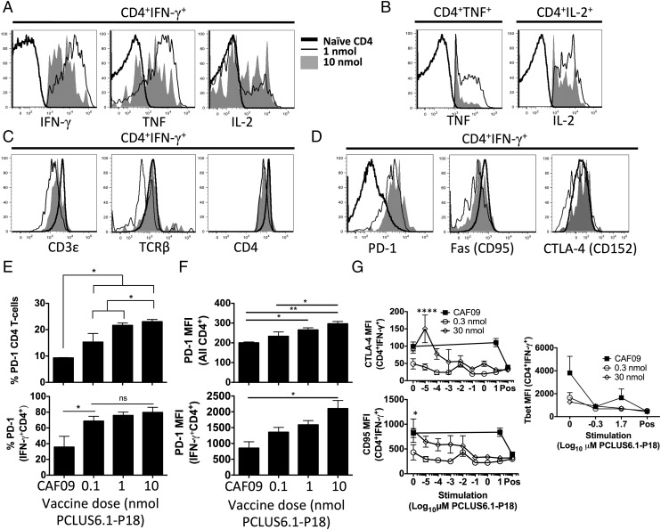
T cells with high functional avidity can sense and respond to low levels of cognate Ag, a characteristic that is associated with more potent responses against tumors and many infections, including HIV. Although an important determinant of T cell efficacy, it has proven difficult to selectively induce T cells of high functional avidity through vaccination. Attempts to induce high-avidity T cells by low-dose in vivo vaccination failed because this strategy simply gave no response. Instead, selective induction of high-avidity T cells has required in vitro culturing of specific T cells with low Ag concentrations. In this study, we combined low vaccine Ag doses with a novel potent cationic liposomal adjuvant, cationic adjuvant formulation 09, consisting of dimethyldioctadecylammonium liposomes incorporating two immunomodulators (monomycolyl glycerol analog and polyinosinic-polycytidylic acid) that efficiently induces CD4 Th cells, as well as cross-primes CD8 CTL responses. We show that vaccination with low Ag dose selectively primes CD4 T cells of higher functional avidity, whereas CD8 T cell functional avidity was unrelated to vaccine dose in mice. Importantly, CD4 T cells of higher functional avidity induced by low-dose vaccinations showed higher cytokine release per cell and lower inhibitory receptor expression (PD-1, CTLA-4, and the apoptosis-inducing Fas death receptor) compared with their lower-avidity CD4 counterparts. Notably, increased functional CD4 T cell avidity improved antiviral efficacy of CD8 T cells. These data suggest that potent adjuvants, such as cationic adjuvant formulation 09, render low-dose vaccination a feasible and promising approach for generating high-avidity T cells through vaccination.


Figures

FIGURE 1.
FIGURE 1.
Low-dose immunizations favor induction of CD4 T cells over CD8 T cells. (A and B) BALB/c mice were immunized i.p. three times at 2-wk intervals with different doses of PCLUS6.1-P18 in CAF09, as indicated on the x-axis (C, control group receiving CAF09 only). One week after the third immunization, splenocytes were restimulated in vitro with 5 μM PCLUS6.1-P18 in the presence of brefeldin A and assessed for intracellular IFN-γ production by flow cytometry. The graphs depict the mean (+ SEM) percentages (A) and absolute numbers (B) of CD4 T cells and CD8 T cells producing IFN-γ after stimulation in each vaccine dose group (n = 3 per group). These results are representative of nine experiments with similar results. (C and D) Pooled analysis of eight repeated immunization experiments. Not all experiments included all vaccine doses, and one repeated experiment used different doses and could not be pooled. Mice were immunized, and IFN-γ production was assessed by flow cytometry, as described for (A) and (B). The graphs depict the mean percentages (± SEM) of CD4 T cells (○) and CD8 T cells (△) producing IFN-γ following stimulation with 5 μM PCLUS6.1-P18. n = 28 for CAF09; n = 5, 11, 24, 16, and 12 for vaccine groups dosed at 0.01, 0.1, 1, 10, and 50 nmol PCLUS6.1-P18, respectively. *p < 0.05, **p < 0.01, ***p < 0.001, one-way ANOVA and Newman–Keul posttest.
FIGURE 2.
FIGURE 2.
Low-dose immunization selectively enhances functional avidity of CD4, but not CD8, T cells. BALB/c mice were immunized i.p. three times at 2-wk intervals with different doses of PCLUS6.1-P18 in CAF09 and euthanized 1 wk later, when functional avidity of splenocytes was assessed by ICS and flow cytometry. Relative percentage of CD4 (A) and CD8 (B) T cells producing IFN-γ after in vitro stimulation with increasing concentrations (5 × 10⁻⁶ to 5 μM) of Ag, as indicated on the x-axis. Responses were normalized to the maximum response for each mouse (set to 100%; magnitudes of responses are shown in Supplemental Fig. 1C) and plotted as a function of peptide concentration used for stimulation. Data points represent mean and SEM of n = 3 mice per group from mice immunized with the PCLUS6.1-P18 doses shown. Statistical analyses were performed using two-way repeated-measures ANOVA and the Bonferroni correction for multiple comparisons. Only groups with a response significantly different from the control group are shown. Pooled avidity [log10(EC50)] for CD4 (C) and CD8 (D) T cells calculated based on normalized curves, such as the ones shown in (A) and (B), from repeated experiments. n = 14–26 for 0.1, 1, and 10 nmol vaccine groups; n = 5–9 for the remaining groups. Data points represent functional T cell avidity [log10(EC50)] from individual mice; mean and SEM are shown. The data are representative of 10 experiments with similar results. **p < 0.01, ***p < 0.001, one-way ANOVA and Newman–Keul posttest for multiple comparisons.
FIGURE 3.
FIGURE 3.
Low-dose immunizations favor increased relative, as well as absolute, numbers of high-avidity CD4 T cells but not CD8 T cells. BALB/c mice were immunized i.p. three times at 2-wk intervals, as described, with a low (0.1 nmol), medium (1 nmol), or high (10 nmol) dose of PCLUS6.1-P18 in CAF09. One week later, functional avidity was assessed by ICS and by flow cytometry. (A) CD4 T cells of high functional avidity were defined as cells producing IFN-γ after stimulation with 5 × 10⁻³ μM PCLUS6.1-P18 (below the EC50 in all groups to assure cells were of high avidity), and the total response was determined as the highest response observed in all groups over all stimulation concentrations. Data points depict the ratio of high avidity/total response from individual mice in the low (0.1 nmol), medium (1 nmol), and high (10 nmol) vaccine dose groups, as indicated on the x-axis. Mean and SEM of n = 3 mice per group are shown. (B) The corresponding ratios of high-avidity/total vaccine–specific CD8 T cells from the same experiments are shown. High-avidity CD8 T cells were defined as CD8 T cells that produced IFN-γ after stimulation with 5 × 10⁻² μM PCLUS6.1-P18 (corresponding to <30% of the maximum response in all groups), and total response was defined as the highest response over all stimulation concentrations. Absolute numbers of high-avidity (defined as in A and B) IFN-γ–producing CD4 (C) and CD8 (D) T cells are depicted using data pooled from five similar experiments. Bars depict mean/SEM absolute numbers of high-avidity IFN-γ–producing CD4 (C) and CD8 (D) T cells that were normalized to the response in the 1-nmol vaccine dose group (set to 100%) within each experiment. High avidity was defined as IFN-γ–producing cells responding to the stimulation concentration just below the EC50 concentration. n = 11–14 per group. Amounts of high-avidity CD4 and CD8 T cells are indicated above/within the bars relative to the 1-nmol group. Data are representative of nine experiments showing similar results. *p < 0.05, **p < 0.01, one-way ANOVA and Newman–Keul posttest for multiple comparisons. ns, not significant.
FIGURE 4.
FIGURE 4.
Low-dose immunizations favor increased polyfunctionality of responding T cells, which, however, is not restricted to T cells with high functional avidity. BALB/c mice (n = 3) were immunized i.p. three times at 2-wk intervals with a low [0.1, 0.3 nmol for (B)], intermediate (1 nmol), or high (10 nmol) dose of PCLUS6.1-P18 in CAF09. One week after the immunizations, splenocytes were stimulated with increasing concentrations of PCLUS6.1-P18 in vitro and assessed for intracellular cytokine production, as described previously. Pie charts represent the relative distribution of CD4 T cell (A) and CD8 T cell (B) subsets producing different cytokine combinations at the concentrations of Ag used for stimulations (indicated below the pie charts for different vaccine doses). Note that the low dose in (B) is 0.3 nmol, because 0.1 nmol did not induce a measurable CD8 T cell response. Responses <0.2% IFN-γ+ or events <50 cytokine-positive were omitted from SPICE pie chart analysis. Functional avidity [shown as log10(EC50)] of different subtypes of CD4 (C) and CD8 (D) T cells producing the various combinations of cytokines indicated on the x-axis. Bars represent log10(EC50) values for n = 3 mice per vaccine dose group calculated from normalized response curves (Supplemental Fig. 4B). Note that the lowest vaccine dose shown for CD8 T cells in (D) is 1 nmol, and not 0.1 nmol, because the latter dose did not induce a measurable CD8 T cell response. Data are representative of five experiments; see Supplemental Fig. 4 for pooled avidity analysis of T cell subset populations. *p < 0.05, **p < 0.01, ****p < 0.0001, two-way repeated-measures ANOVA and Bonferroni correction for multiple comparisons. ns, not significant.
FIGURE 5.
FIGURE 5.
High-avidity CD4 T cells express higher cytokine levels and greater downregulation of TCR and inhibitory receptors than do their low-avidity counterparts. Mice were immunized three times i.p. with the indicated doses of PCLUS6.1-P18 in CAF09, as described previously. One week after immunizations (4 wk for CTLA-4 and Fas analyses), splenocytes were stimulated in vitro and assessed for the surface expression of various markers, as well as intracellularly for cytokine production by flow cytometry. (A) Representative line graphs show intracellular expression of IFN-γ, TNF, and IL-2 gated on IFN-γ–producing CD4 T cells from mice immunized with 0.1 nmol (high avidity; thin black line) or 10 nmol (low avidity; filled graph) PCLUS6.1-P18 in CAF09. IFN-γ expression is shown for CD4 T cells from naive mice that did not produce IFN-γ as a staining control (thick black line). (B) From the same mice in (A), TNF and IL-2 MFI for TNF+ and IL-2+ CD4 T cells, respectively. Data are shown as described in (A). (C) Surface expression of TCR components CD3ε and TCR-β, as well as CD4 coreceptor, on IFN-γ+CD4+ T cells from mice immunized with 0.1 and 10 nmol after stimulation or on naive CD4 T cells; data are shown as described in (A). (D) Surface expression of inhibitory receptor PD-1, death receptor CD95 (Fas), and CTLA-4 on IFN-γ+ CD4 T cells after stimulation. Filled graph (high dose): 30 nmol PCLUS6.1-P18 (low avidity); thin black line (low dose): 0.3 nmol PCLUS6.1-P18 (high avidity), thick black line: naive unstimulated CD4 T cells (control). (E) Percentage of PD-1 expression on all gated CD4 T cells (upper panel) and IFN-γ+ CD4 T cells (lower panel) after stimulation. No upregulation of PD-1 was observed after in vitro stimulation. (F) Bar graphs show MFI of PD-1 on all gated CD4 T cells (upper panel), as well as on IFN-γ+ CD4 T cells (lower panel), from the same experiment shown in (E). Bars represent mean and SEM of n = 3 mice per group immunized as indicated on the x-axis. *p < 0.05, **p < 0.01, one-way ANOVA with Newman–Keul posttest (E and F). (G) In a separate experiment, mice were immunized i.p. with a high (30 nmol) or low (0.3 nmol) dose of PCLUS6.1-P18 in CAF09 three times, as described above. Four weeks later, splenocytes were stimulated in vitro with increasing concentrations of PCLUS6.1-P18, as indicated on the x-axis. Graphs depict surface expression (MFI) of CTLA-4 (upper left panel), CD95 (Fas; lower left panel), and T-bet (right panel) on IFN-γ+ CD4 T cells; data points represent mean and SEM of n = 3 mice per group. Experiments were repeated at least twice with similar results. *p < 0.05, ****p < 0.0001, two-way repeated-measures ANOVA and Bonferroni correction for multiple comparisons. Pos, positive controls (PMA-ionomycin).
FIGURE 6.
FIGURE 6.
CD4 T cell functional avidity is dependent on the presence of IL-15. WT C57BL/6 mice or IL-15–KO (on B6 background) mice were immunized i.p. with 50 μg per mouse of hep B core 128–140 in CAF09 twice 2 wk apart. Two weeks after the last immunization, splenocytes were stimulated in vitro for immune analyses. (A) Splenocytes were stimulated for 5 d in the presence of increasing concentrations of the hep B core 128–140 helper peptide, and IFN-γ production in the culture supernatant was assessed by IFN-γ ELISA. The curves represent mean and SEM of n = 3 mice per group immunized with hep B core 128–140 in CAF09 (WT and IL-15–KO mice) or WT mice receiving only CAF09 as a control (CAF09). Absolute levels of culture supernatant IFN-γ (pg/ml) (upper panel). IFN-γ production normalized to the maximum production for each mouse (lower panel). (B) From the normalized values in the lower panel in (A), the concentration of peptide needed to induce 50% of the maximum response (EC50) was calculated for each mouse; data points represent avidity shown as log10(EC50) mg/ml hep B 128–140 peptide with SEM. (C) Percentages of PD-1+ CD4 T cells (upper panel) and PD-1 expression per cell (MFI; middle panel) for all CD4 T cells. PD-1 MFI for IFN-γ+ CD4 T cells after stimulation with hep B 128–140 and ICS (lower panel). Data points represent individual mice; mean and SEM are indicated. The data shown are representative of two experiments with similar results. *p < 0.05, **p < 0.01, one-way ANOVA and Newman–Keul posttest.
FIGURE 7.
FIGURE 7.
A combination of high CD4 T cell functional avidity and the presence of a CD8 T cell response protects against viral vaccinia challenge. Mice were immunized three times, as described previously, with the indicated doses of PCLUS6.1-P18 containing the HIV IIIB gp160 Th and CTL epitopes (Helper+CTL), as well as with PCLUS6.1 that included only the Th epitope (No CTL) in CAF09. CAF09 controls are indicated (CTRL). Five weeks after the last immunization, mice were challenged i.p. with 2 × 10⁷ PFU recombinant vaccinia virus vPE-16 expressing HIV IIIB gp160 (vPE-16), and immune responses were assessed 4 wk after the last immunization. Splenocytes were harvested and restimulated in vitro with 5 μM PCLUS6.1-P18 for CD4 (A) and 0.5 μM P18-I10 for CD8 (B) T cell responses. Bars represent log10 mean and SEM of the percentage of IFN-γ–producing T cells (n = 3 per group). Statistical differences for the responses between groups were assessed by one-way ANOVA and the Newman–Keul posttest for multiple comparisons. ***p < 0.001. (C) Functional avidity of IFN-γ–producing CD4 T cells was assessed by ICS and flow cytometry and calculated as previously described; bars depict mean log10(EC50) with SEM for each vaccine group. No difference in CD8 functional avidity between groups was observed (data not shown). *p < 0.05, **p < 0.01 versus control group, one-way ANOVA and Newman–Keul posttest. (D) In the same experiment, vaccine protection was evaluated by estimating viral load in ovaries 5 d postchallenge by plaque assay (see Materials and Methods). The graph depicts estimated log10 PFU of paired ovaries (both right and left) from individual animals; n = 5 per group (n = 4 in the group vaccinated with PCLUS6.1 without the P18 CTL epitope) with medians indicated. The level of detection (1000 PFU) is indicated by the dashed horizontal line. *p < 0.05, Kruskal–Wallis test with Dunn test for multiple comparisons. (E) Linear regression was performed between estimated log10 PFU levels (± SEM) and a composite ranked immune parameter that was derived by adding ranks among the three vaccine groups (0.1, 1, and 10 nmol PCLUS6.1-P18/CAF09) with regard to the magnitudes of the CD4 and CD8 T cell responses and CD4 T cell functional avidity (rank 3 = highest response/highest avidity, rank 1 = lowest response/lowest avidity). Note that mice were sacrificed prior to challenge to perform the immune analysis shown in (A–C); hence, correlations between immune response and PFU were performed at the group level, with different mice giving rise to immune parameters and PFU, not within paired single animals. (F) Mice were immunized three times i.p. with 1 or 50 nmol PCLUS6.1-P18 in CAF09 or CAF09 alone (control) and were challenged 4 wk later with a low dose (1 × 10⁷ PFU per mouse) of vPE-16 vaccinia virus. Protection was assessed in ovaries by plaque assay at 5 d postchallenge. Bars represent median log10 PFU and interquartile range of n = 5 mice per group. This experiment had a lower detection limit of 100 PFU, as indicated by the dashed line. *p < 0.05, **p < 0.01 versus control, Kruskal–Wallis test with Dunn test for multiple comparisons.
FIGURE 8.
FIGURE 8.
Adoptively transferring CD4 T cells of high, but not low, functional avidity along with primed TCR-Tg RT1 CD8 T cells confers protection against viral vaccinia challenge. BALB/c mice were immunized three times, as described before, with a low (1 nmol) or high (10 nmol) dose of PCLUS6.1 (containing the HIV-IIIB gp160 helper but no CTL epitope) in CAF09. TCR-Tg RT1 mice carrying a TCR specific for the minimal immunodominant HIV IIIB gp160 P18-I10 epitope were immunized three times with 50 nmol PCLUS6.1-P18 in CAF09. Two weeks after immunizations of BALB/c mice and 3 wk after immunizations of TCR-Tg mice, spleens were harvested, and CD4 T cells from BALB/c mice immunized with either a low (1 nmol, high avidity) or high (10 nmol, low avidity) dose of PCLUS6.1 were adoptively transferred i.v. with CD8 T cells from RT1 TCR-Tg mice that were vaccinated with PCLUS6.1-P18 into SCID mice that were subsequently infected i.p. with 0.5 × 10⁷ PFU vPE-16 vaccinia virus within 15 min of T cell transfer. Approximately 1.5 × 10⁶ CD4 T cells of high or low functional avidity specific for PCLUS6.1, along with 5.5 × 10⁶ P18-I10–specific CD8 T cells, were transferred per mouse. (A) Schematic overview of the experiment. (B) At the time of transfer, an aliquot of the transferred CD4 T cells was assessed for immune response, and the percentage (mean and SEM) of transferred CD4 T cells that produced IFN-γ after in vitro restimulation with 5 μM PCLUS6.1, as assessed by flow cytometry and ICS, is shown. (C) At the time of transfer, splenocytes were also restimulated with a range of PCLUS6.1 concentrations, and the ratio of high functional avidity CD4 T cells (defined as CD4 T cells that responded with IFN-γ production after stimulation with 0.05 μM Ag)/total amount of responding CD4 T cells was assessed, as described previously. The graph depicts avidity (mean ± SEM) measured as the ratio of the percentages of high avidity/total responding CD4 T cells for each of n = 3 mice per group. (D) At 4 d postchallenge, ovaries were removed to estimate viral loads, as previously described. The graph depicts estimated log10 PFU values for individual recipient mice receiving the donor cells indicated below the x-axis. Individual log10 PFU values (mean ± SEM) are shown. For (B)–(D), *p < 0.05, **p < 0.01, one-way ANOVA and the Newman–Keul posttest for multiple comparisons. ap < 0.05, Student t test without correction for multiple comparisons (D). (E) The adoptive transfer protection experiment was repeated with similar results; PFU values were pooled from the experiment shown in (D) and the repeated experiment. To bypass the effect of differences in PFU values between the two experiments (∼0.7 log10 PFU difference), we calculated a δ PFU value by normalizing log10 PFU values of individual mice to the mean of mice receiving low-avidity CD4 T cells within each experiment. Thus, each data point represents the δ PFU value (log10 PFU individual mouse − mean log10 PFU low-avidity recipient group in the same experiment); mean and SEM are indicated. *p < 0.05, two-sided t test. ns, not significant.

References

    1. Berzofsky J. A. 2012. A push-pull vaccine strategy using Toll-like receptor ligands, IL-15, and blockade of negative regulation to improve the quality and quantity of T cell immune responses. Vaccine 30: 4323–4327. - PMC - PubMed
    1. Seder R. A., Darrah P. A., Roederer M. 2008. T-cell quality in memory and protection: implications for vaccine design. Nat. Rev. Immunol. 8: 247–258. - PubMed
    1. Bullock T. N., Mullins D. W., Engelhard V. H. 2003. Antigen density presented by dendritic cells in vivo differentially affects the number and avidity of primary, memory, and recall CD8+ T cells. J. Immunol. 170: 1822–1829. - PubMed
    1. Derby M., Alexander-Miller M., Tse R., Berzofsky J. 2001. High-avidity CTL exploit two complementary mechanisms to provide better protection against viral infection than low-avidity CTL. J. Immunol. 166: 1690–1697. - PubMed
    1. Alexander-Miller M. A., Leggatt G. R., Berzofsky J. A. 1996. Selective expansion of high- or low-avidity cytotoxic T lymphocytes and efficacy for adoptive immunotherapy. Proc. Natl. Acad. Sci. USA 93: 4102–4107. - PMC - PubMed

Publication types

MeSH terms