Review
. 2017 Mar 13:8:141.
doi: 10.3389/fphys.2017.00141. eCollection 2017.

Mitochondrial Bioenergetics in the Metabolic Myopathy Accompanying Peripheral Artery Disease

Affiliations
Review

Mitochondrial Bioenergetics in the Metabolic Myopathy Accompanying Peripheral Artery Disease

Victoria G Rontoyanni et al. Front Physiol. .

Abstract

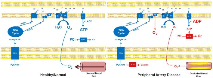
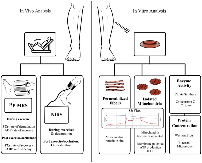
Peripheral artery disease (PAD) is a serious but relatively underdiagnosed and undertreated clinical condition associated with a marked reduction in functional capacity and a heightened risk of morbidity and mortality. The pathophysiology of lower extremity PAD is complex, and extends beyond the atherosclerotic arterial occlusion and subsequent mismatch between oxygen demand and delivery to skeletal muscle mitochondria. In this review, we evaluate and summarize the available evidence implicating mitochondria in the metabolic myopathy that accompanies PAD. Following a short discussion of the available in vivo and in vitro methodologies to quantitate indices of muscle mitochondrial function, we review the current evidence implicating skeletal muscle mitochondrial dysfunction in the pathophysiology of PAD myopathy, while attempting to highlight questions that remain unanswered. Given the rising prevalence of PAD, the detriment in quality of life for patients, and the associated significant healthcare resource utilization, new alternate therapies that ameliorate lower limb symptoms and the functional impairment associated with PAD are needed. A clear understanding of the role of mitochondria in the pathophysiology of PAD may contribute to the development of novel therapeutic interventions.

Keywords: bioenergetics; mitochondria; mitochondrial function; peripheral artery disease; peripheral vascular disease; skeletal muscle.


Figures

Figure 1
Figure 1
Schematic overview of mitochondrial bioenergetics in healthy individuals with normal muscle blood flow, and in patients with peripheral artery disease (PAD) and occluded muscle blood flow. In healthy individuals with normal muscle blood flow, pyruvate can undergo oxidation through pyruvate dehydrogenase (PDH), subsequently participating in the TCA cycle (as acetyl-CoA). Sufficient cellular O2 availability provides a terminal electron acceptor for the electron transport chain (ETC), ultimately allowing electron transfer and the generation of the electro-chemical potential needed for oxidative phosphorylation (ATP production). Further, electron flow to complex IV (IV) of the ETC prevents stagnation of electrons (e) in the chain, thereby preventing superoxide (O2) formation. In this setting, the creatine phosphokinase (CPK) reaction remains at a basal state of flux, where approximately two-thirds of the creatine (Cr) pool is stored as phosphocreatine (PCr). In contrast, reduced muscle blood flow in patients with PAD results in hypoxia and subsequent alterations in muscle bioenergetics. Specifically, in hypoxic tissue, pyruvate is unable to undergo an oxidative fate within the mitochondrion, instead being metabolized by lactate dehydrogenase in the cell cytosol, forming lactate. Similarly, reduced O2 availability in the mitochondria limits electron transfer and respiration resulting in electron accumulation in the ETC, which may lead to O2 production at complex I and complex III, and subsequent oxidative stress. Importantly, reduced O2 availability and the subsequent impairment in oxidative phosphorylation in the muscle of patients with PAD will result in a reduction in ATP levels and a concomitant increase in ADP levels. This change in the cellular ATP to ADP ratio will drive the CPK reaction to breakdown PCr in order to buffer cellular ATP. This phenotype is most pronounced in muscle of patients with PAD when ATP turnover rates are higher, i.e., during muscle contraction associated with physical activity/exercise, and can lead to the localized muscle cramping and pain (claudication) experienced by individuals with PAD.
Figure 2
Figure 2
Schematic overview of available methods for the assessment of skeletal muscle mitochondrial respiratory capacity and function. Abbreviations: ADP, adenosine diphosphate; ATP, adenosine triphosphate; H2O2, hydrogen peroxide; NIRS, near-infrared spectroscopy; PCr, phosphocreatine; 31PMRS, 31Phosphorus magnetic resonance spectroscopy.

Similar articles

Cited by

References

    1. Aboyans V., Criqui M. H., Abraham P., Allison M. A., Creager M. A., Diehm C., et al. . (2012). Measurement and interpretation of the ankle-brachial index: a scientific statement from the American Heart Association. Circulation 126, 2890–2909. 10.1161/CIR.0b013e318276fbcb - DOI - PubMed
    1. Ackerman J. J., Grove T. H., Wong G. G., Gadian D. G., Radda G. K. (1980). Mapping of metabolites in whole animals by 31P NMR using surface coils. Nature 283, 167–170. 10.1038/283167a0 - DOI - PubMed
    1. Allison M. A., Ho E., Denenberg J. O., Langer R. D., Newman A. B., Fabsitz R. R., et al. . (2007). Ethnic-specific prevalence of peripheral arterial disease in the United States. Am. J. Prev. Med. 32, 328–333. 10.1016/j.amepre.2006.12.010 - DOI - PubMed
    1. Amoh-Tonto C. A., Malik A. R., Kondragunta V., Ali Z., Kullo I. J. (2009). Brachial-ankle pulse wave velocity is associated with walking distance in patients referred for peripheral arterial disease evaluation. Atherosclerosis 206, 173–178. 10.1016/j.atherosclerosis.2009.02.003 - DOI - PMC - PubMed
    1. Anderson J. D., Epstein F. H., Meyer C. H., Hagspiel K. D., Wang H., Berr S. S., et al. . (2009). Multifactorial determinants of functional capacity in peripheral arterial disease: uncoupling of calf muscle perfusion and metabolism. J. Am. Coll. Cardiol. 54, 628–635. 10.1016/j.jacc.2009.01.080 - DOI - PMC - PubMed

LinkOut - more resources