

Neurophysiological Correlates of Featural and Spacing Processing for Face and Non-face Stimuli
- PMID: 28348535
- PMCID: PMC5346548
- DOI: 10.3389/fpsyg.2017.00333
Neurophysiological Correlates of Featural and Spacing Processing for Face and Non-face Stimuli
Abstract
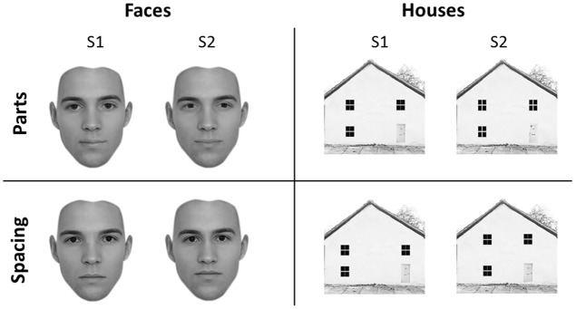
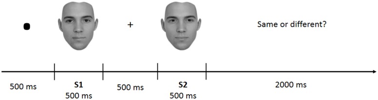
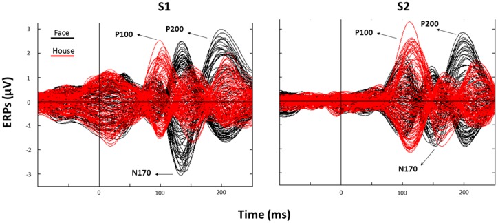
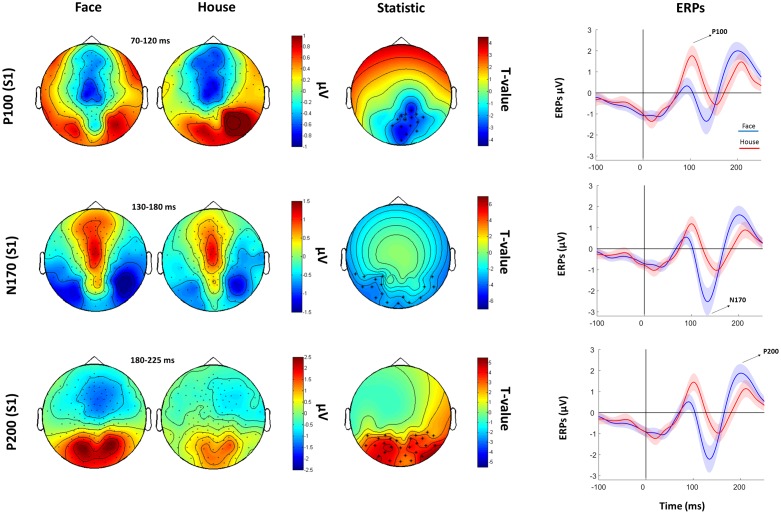
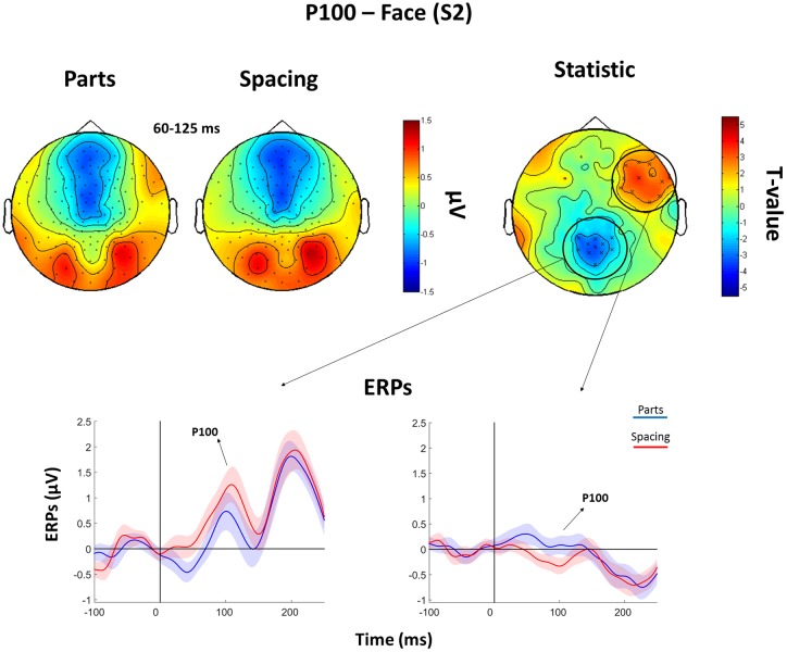
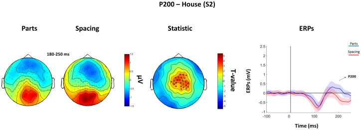
The peculiar ability of humans to recognize hundreds of faces at a glance has been attributed to face-specific perceptual mechanisms known as holistic processing. Holistic processing includes the ability to discriminate individual facial features (i.e., featural processing) and their spatial relationships (i.e., spacing processing). Here, we aimed to characterize the spatio-temporal dynamics of featural- and spacing-processing of faces and objects. Nineteen healthy volunteers completed a newly created perceptual discrimination task for faces and objects (i.e., the "University of East London Face Task") while their brain activity was recorded with a high-density (128 electrodes) electroencephalogram. Our results showed that early event related potentials at around 100 ms post-stimulus onset (i.e., P100) are sensitive to both facial features and spacing between the features. Spacing and features discriminability for objects occurred at circa 200 ms post-stimulus onset (P200). These findings indicate the existence of neurophysiological correlates of spacing vs. features processing in both face and objects, and demonstrate faster brain processing for faces.
Keywords: EEG; N170; P100; configural processing; face perception; holistic processing; object perception.
Figures
Similar articles
-
Neural correlates of processing facial identity based on features versus their spacing.Neuropsychologia. 2007 Apr 8;45(7):1438-51. doi: 10.1016/j.neuropsychologia.2006.11.016. Epub 2007 Jan 3. Neuropsychologia. 2007. PMID: 17204295
-
Neurophysiological correlates of configural face processing in schizotypy.Front Psychiatry. 2014 Aug 12;5:101. doi: 10.3389/fpsyt.2014.00101. eCollection 2014. Front Psychiatry. 2014. PMID: 25161628 Free PMC article.
-
Early holistic face-like processing of Arcimboldo paintings in the right occipito-temporal cortex: evidence from the N170 ERP component.Int J Psychophysiol. 2013 Nov;90(2):157-64. doi: 10.1016/j.ijpsycho.2013.06.024. Epub 2013 Jun 29. Int J Psychophysiol. 2013. PMID: 23816562
-
[Neural mechanisms of face recognition: an event-related potential study].Brain Nerve. 2012 Jul;64(7):717-26. Brain Nerve. 2012. PMID: 22764343 Review. Japanese.
-
Why does picture-plane inversion sometimes dissociate perception of features and spacing in faces, and sometimes not? Toward a new theory of holistic processing.Psychon Bull Rev. 2009 Oct;16(5):778-97. doi: 10.3758/PBR.16.5.778. Psychon Bull Rev. 2009. PMID: 19815781 Review.
Cited by
-
The Time Sequence of Face Spatial Frequency Differs During Working Memory Encoding and Retrieval Stages.Front Psychol. 2022 May 20;13:853992. doi: 10.3389/fpsyg.2022.853992. eCollection 2022. Front Psychol. 2022. PMID: 35668961 Free PMC article.
-
Preliminary Evidence of "Other-Race Effect"-Like Behavior Induced by Cathodal-tDCS over the Right Occipital Cortex, in the Absence of Overall Effects on Face/Object Processing.Front Neurosci. 2017 Nov 30;11:661. doi: 10.3389/fnins.2017.00661. eCollection 2017. Front Neurosci. 2017. PMID: 29249931 Free PMC article.
-
Face memory and facial expression recognition are both affected by wearing disposable surgical face masks.Cogn Process. 2023 Feb;24(1):43-57. doi: 10.1007/s10339-022-01112-2. Epub 2022 Oct 15. Cogn Process. 2023. PMID: 36242672 Free PMC article.
-
Neural Correlates of Single- and Dual-Task Walking in the Real World.Front Hum Neurosci. 2017 Sep 14;11:460. doi: 10.3389/fnhum.2017.00460. eCollection 2017. Front Hum Neurosci. 2017. PMID: 28959199 Free PMC article.
-
Theta- and Gamma-Band Activity Discriminates Face, Body and Object Perception.Front Hum Neurosci. 2020 Mar 12;14:74. doi: 10.3389/fnhum.2020.00074. eCollection 2020. Front Hum Neurosci. 2020. PMID: 32226369 Free PMC article.
References
LinkOut - more resources
Full Text Sources
Other Literature Sources