

Nasal Septal Deviation and Concha Bullosa - Do They Have an Impact on Maxillary Sinus Volumes and Prevalence of Maxillary Sinusitis?
- PMID: 28348652
- PMCID: PMC5347520
- DOI: 10.12659/PJR.900634
Nasal Septal Deviation and Concha Bullosa - Do They Have an Impact on Maxillary Sinus Volumes and Prevalence of Maxillary Sinusitis?
Abstract
Background: The aim of the study was to assess if the presence of nasal septal deviation and concha bullosa is connected with the development of sinuses and the incidence of inflammation within them.
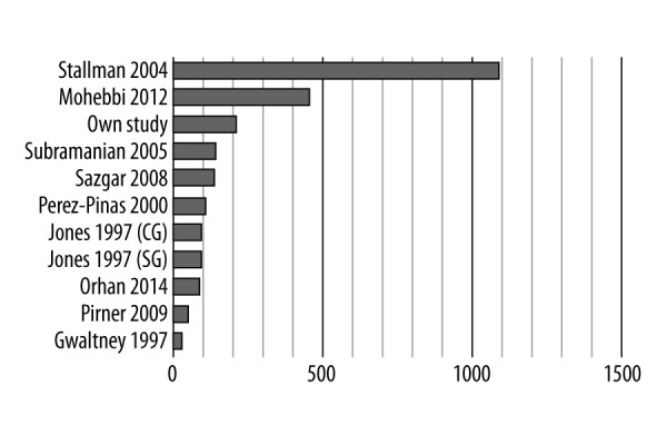
Material/methods: We retrospectively analysed 214 patients who underwent paranasal sinus computed tomography. There were 125 females and 89 males, the mean age being 47.67±16.74 years (range 18-97). Exclusion criteria included: age under 18 years, prior sinonasal surgery and S-shaped septum.
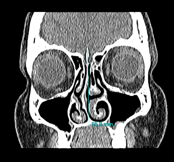
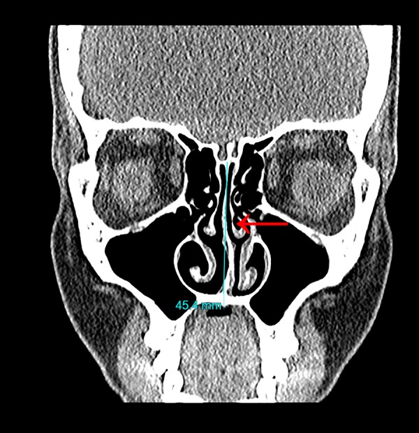
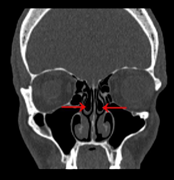
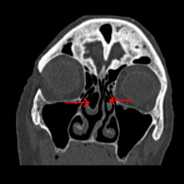
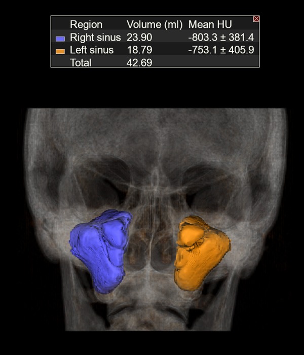
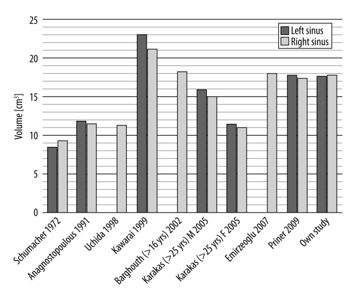
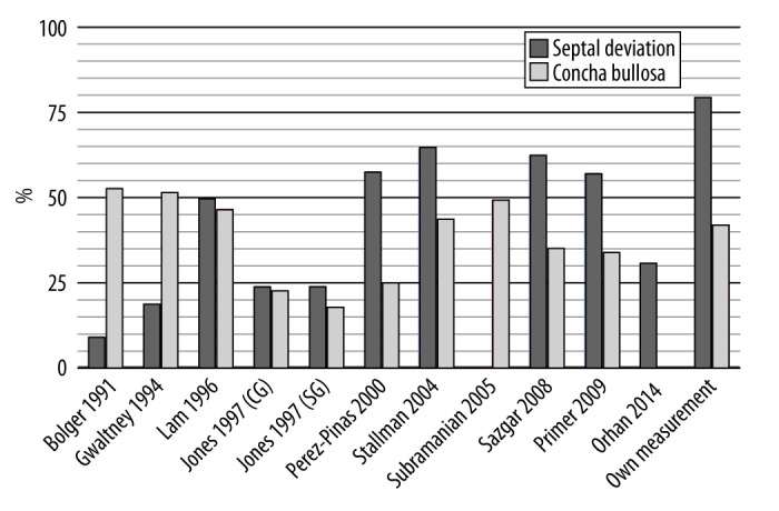
Results: Mean volume of the right maxillary sinus was 17.794 cm3, while for the left one it was 17.713 cm3. Nasal septal deviation was found in 79.9% of computed tomography examinations and concha bullosa was observed in 42.1% of the patients' examinations. There was an association between the presence of unilateral or dominant concha bullosa and contralateral direction of septal deviation [right-sided (p=0.039), left-sided (p=0.003)]. There was higher incidence of bilateral maxillary sinusitis in patients with septal deviation (p=0.007). Bilateral concha bullosa did not influence the incidence of bilateral maxillary sinusitis (p=0.495). Neither septal deviation (right sided: p=0.962; left-sided: p=0.731), nor unilateral/dominant concha bullosa (right: p=0.512; left: p=0,430) affected the asymmetry in volumes of maxillary sinuses. Bilateral concha bullosa was connected with larger volume of maxillary sinuses (right sinus: p=0.005; left sinus: p=0.048).
Conclusions: Nasal septal deviation, contrary to concha bullosa, has influence on the development of maxillary sinusitis. There is a connection between the presence of concha bullosa and direction of septal deviation. Only bilateral concha bullosa affects maxillary sinus volumes.
Keywords: Imaging, Three-Dimensional; Maxillary Sinus; Maxillary Sinusitis; Nasal Septum; Sinusitis; Turbinates.
Figures
References
-
- Onodi A, Thomson SC. The anatomy of the nasal cavity and its accessory sinuses: An atlas for practitioners and students. J Anat Physiol. 1895;29( Pt 3):471.
-
- Vaid S, Vaid N. Normal anatomy and anatomic variants of the paranasal sinuses on computed tomography. Neuroimaging Clin N Am. 2015;25(4):527–48. - PubMed
-
- Wang RG, Jiang SC, Gu R. The cartilaginous nasal capsule and embryonic development of human paranasal sinuses. J Otolaryngol. 1994;23(4):239–43. - PubMed
-
- Marquez S, Lawson W, et al. Anatomy of the nasal accessory sinuses. In: Wackym PA, Rice D, Schaefer SD, editors. Minimally invasive surgery of the head, neck, and cranial base. Lippincott; 2002. pp. 153–93.
-
- Kapusuz Gencer Z, Ozkırış M, Okur A, et al. The effect of nasal septal deviation on maxillary sinus volumes and development of maxillary sinusitis. Eur Arch Otorhinolaryngol. 2013;270(12):3069–73. - PubMed
LinkOut - more resources
Full Text Sources
Other Literature Sources