. 2017 Mar 1;8(3):2107-2114.
doi: 10.1039/C6SC02297A. Epub 2016 Nov 25.

Copper-Catalyzed Click Reaction on/in Live Cells

Affiliations

Copper-Catalyzed Click Reaction on/in Live Cells

Siheng Li et al. Chem Sci. .

Abstract

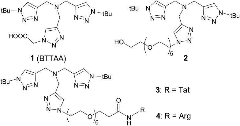
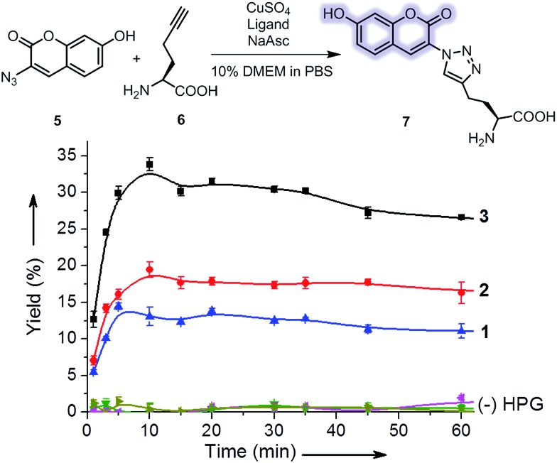
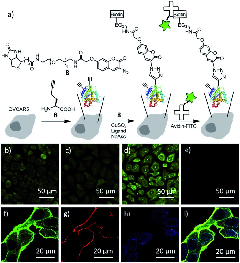
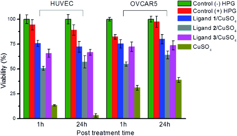
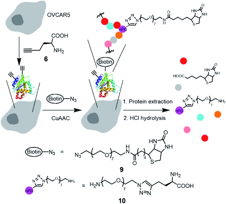
We demonstrated that copper-catalyzed azide-alkyne cycloaddition (CuAAC) reaction could be performed inside live mammalian cells without using a chelating azide. Under optimized conditions, the reaction was performed in human ovary cancer cell line OVCAR5 in which newly synthesized proteins were metabolically modified with homopropargylglycine (HPG). This model system allowed us to estimate the efficiency of the reaction on the cell membranes and in the cytosol using mass spectrometry. We found that the reaction was greatly promoted by a tris(triazolylmethyl)amine CuI ligand tethering a cell-penetrating peptide. Uptake of the ligand, copper, and a biotin-tagged azide in the cells was determined to be 69 ± 2, 163 ± 3 and 1.3 ± 0.1 µM, respectively. After 10 minutes of reaction, the product yields on the membrane and cytosolic proteins were higher than 18% and 0.8%, respectively, while 75% cells remained viable. By reducing the biothiols in the system by scraping or treatment with N-ethylmalemide, the reaction yield on the cytosolic proteins was greatly improved to ~9% and ~14%, respectively, while the yield on the membrane proteins remained unchanged. The results indicate that out of many possibilities, deactivation of the current copper catalysts by biothiols is the major reason for the low yield of CuAAC reaction in the cytosol. Overall, we have improved the efficiency for CuAAC reaction on live cells by 3-fold. Despite the low yielding inside live cells, the products that strongly bind to the intracellular targets can be detected by mass spectrometry. Hence, the in situ CuAAC reaction can be potentially used for screening of cell-specific enzyme inhibitors or biomarkers containing 1,4-substituted 1,2,3-triazoles.


Figures

Fig. 1
Fig. 1. Structural formulas of tris(triazolylmethyl)amine-based ligands used in this study.
Fig. 2
Fig. 2. Evaluation of the CuAAC reactivity of ligands 1–3 using a fluorogenic CuAAC reaction. Reaction conditions: 100 μM 5, 50 μM 6, 100 μM CuSO4, 200 μM ligand, 500 μM sodium ascorbate (NaAsc) in 10/90 (v/v) methionine-free DMEM/PBS, room temperature. Yield was derived from the measured mean fluorescence intensity of 7 (Fig. S4†). Controls were performed without HPG. Error bars represent the standard deviation of data from three samples.
Fig. 3
Fig. 3. Reactivity of ligands 1–3 for CuAAC reaction in live cells. (a) Schematic of metabolic incorporation of HPG (6) to the newly synthesized proteins, CuAAC reaction and fluorescence labeling with avidin–FITC. (b)–(h) Representative wide field (b–e) and confocal (f–h) fluorescence images of OVCAR5 cells after treatment with a mixture of 8 (100 μM), CuSO4 (100 μM), 200 μM of the ligand 1 (b), 2 (c), 3 (d and f–i), and sodium ascorbate (500 μM) in 10/90 (v/v) methionine-free DMEM/PBS for 10 minutes, followed by fixing and labeling with avidin–FITC (b–f), a plasma membrane dye (g), and DAPI (h). (e) Control without ligand and CuSO4. (i) Overlay of (f)–(h). For the control without avidin–FITC, see Fig. S9.
Fig. 4
Fig. 4. Cell viability after treatment with 100 μM 8, 100 μM CuSO4, 200 μM ligand, 500 μM sodium ascorbate in 10/90 (v/v) methionine-free DMEM/PBS for 10 minutes. Cell viability is normalized with the absorbance of the negative control of cells without any treatment. Error bars represent the standard deviation of the data from three samples.
Fig. 5
Fig. 5. Illustration of the incorporation of HPG to proteins, CuAAC reaction in live cells, and subsequent hydrolysis of the biotinylated proteins to the amino acid derivative 10 for LC-ESI-MS/MS quantification. Reaction conditions: 100 μM 9, 100 μM CuSO4, 200 μM 1 or 3, 500 μM sodium ascorbate in 10/90 (v/v) methionine-free DMEM/PBS for 10 minutes. Color dots represent amino acid residues.

Similar articles

Cited by

References

    1. McKay C. S., Finn M. G. Chem. Biol. 2014;21:1075–1101. - PMC - PubMed
    1. Prescher J. A., Bertozzi C. R. Nat. Chem. Biol. 2005;1:13–21. - PubMed
    1. Hao Z., Hong S., Chen X., Chen P. R. Acc. Chem. Res. 2011;44:742–751. - PubMed
    1. Lang K., Chin J. W. Chem. Rev. 2014;114:4764–4806. - PubMed
    1. Sletten E. M., Bertozzi C. R. Angew. Chem., Int. Ed. 2009;48:6974–6998. - PMC - PubMed