. 2015 Aug 3;1(2):e000026.
doi: 10.1099/mgen.0.000026. eCollection 2015 Aug.

Large tandem chromosome expansions facilitate niche adaptation during persistent infection with drug-resistant Staphylococcus aureus

Affiliations

Large tandem chromosome expansions facilitate niche adaptation during persistent infection with drug-resistant Staphylococcus aureus

Wei Gao et al. Microb Genom. .

Abstract

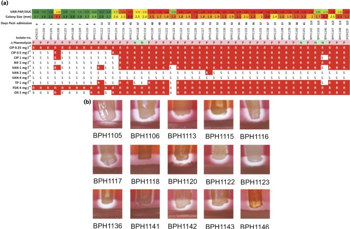
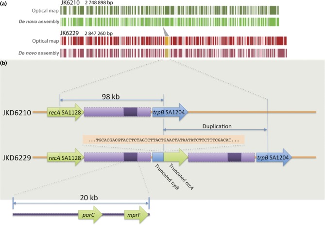
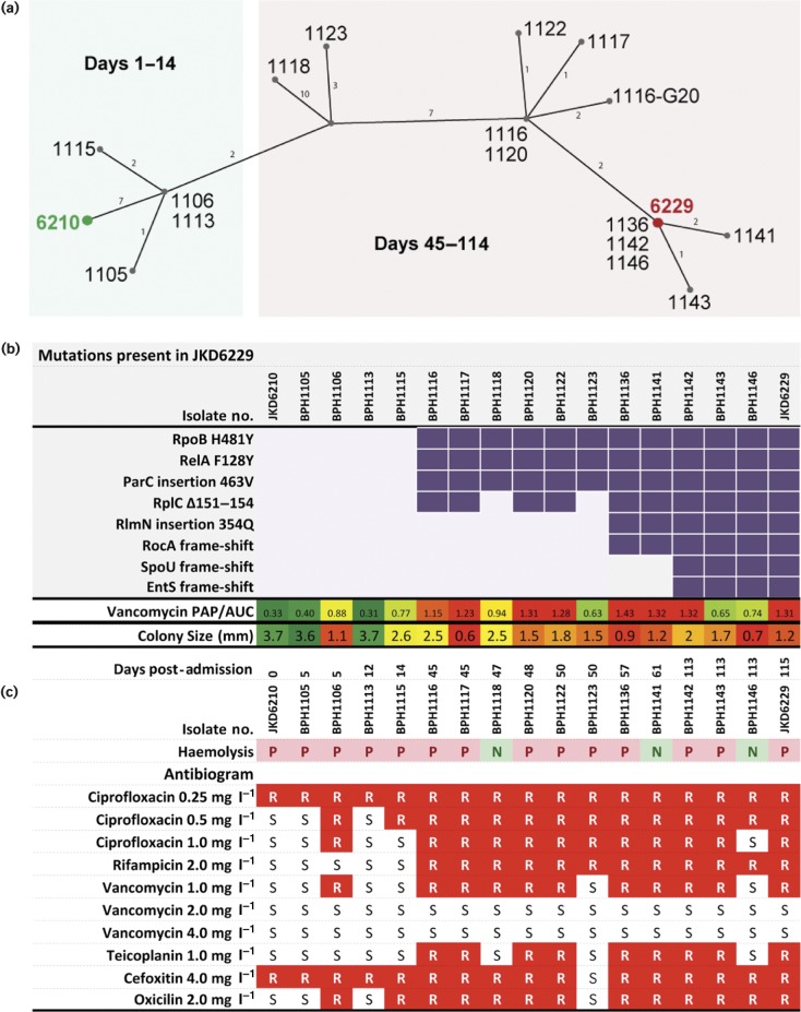
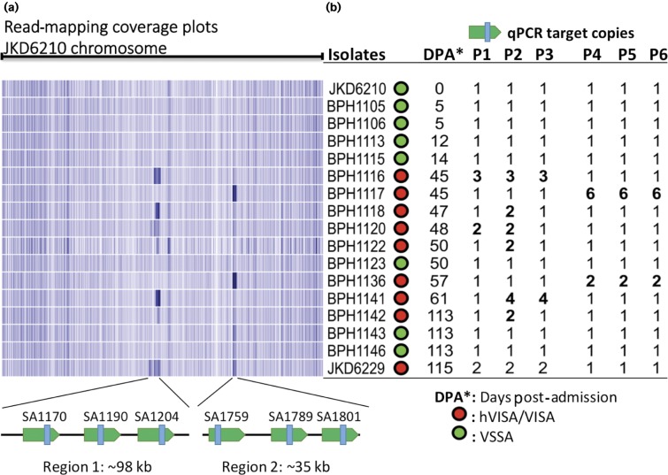
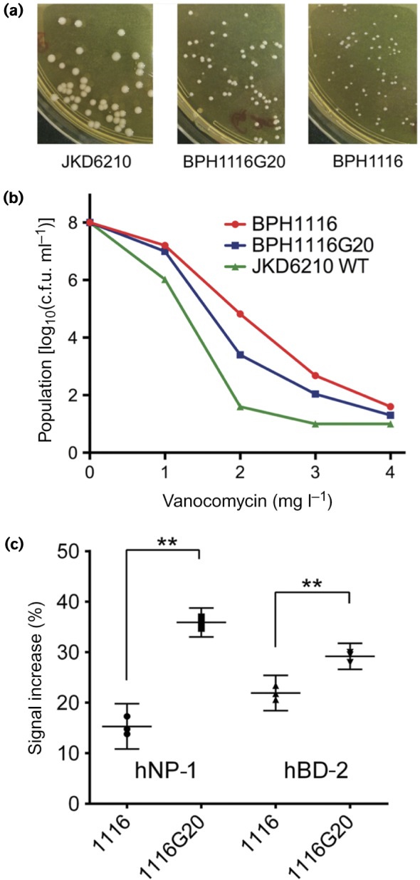
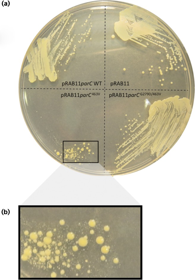
We used genomics to study the evolution of meticillin-resistant Staphylococcus aureus (MRSA) during a complex, protracted clinical infection. Preparing closed MRSA genomes from days 0 and 115 allowed us to precisely reconstruct all genetic changes that occurred. Twenty-three MRSA blood cultures were also obtained during treatment, yielding 44 colony morphotypes that varied in size, haemolysis and antibiotic susceptibility. A subset of 15 isolates was sequenced and shown to harbour a total of 37 sequence polymorphisms. Eighty per cent of all mutations occurred from day 45 onwards, which coincided with the appearance of discrete chromosome expansions, and concluded in the day 115 isolate with a 98 kb tandem DNA duplication. In all heterogeneous vancomycin-intermediate Staphylococcus aureus isolates, the chromosomal amplification spanned at least a 20 kb region that notably included mprF, a gene involved in resistance to antimicrobial peptides, and parC, an essential DNA replication gene with an unusual V463 codon insertion. Restoration of the chromosome after serial passage under non-selective growth was accompanied by increased susceptibility to antimicrobial peptide killing and reduced vancomycin resistance, two signature phenotypes that help explain the clinical persistence of this strain. Elevated expression of the V463 parC was deleterious to the cell and reduced colony size, but did not alter ciprofloxacin susceptibility. In this study, we identified large DNA expansions as a clinically relevant mechanism of S. aureus resistance and persistence, demonstrating the extent to which bacterial chromosomes remodel in the face of antibiotic and host immune pressures.

Keywords: Staphylococcus aureus; antibiotic resistance; chromosome duplication; evolution; genomics; mutation.


Figures

Fig. 1.
Fig. 1.
Phenotypic diversity of isolates during the persistent infection. (a) The colony size, antibiogram, vancomycin population analysis profile area under curve (VAN PAP/AUC) result and δ toxin activity of all 44 isolates are summarized. Increasing vancomycin resistance is indicated by colour (green is susceptible and red is resistant). Likewise, reduced colony size is indicated by red, whilst normal colony size is indicated by green. The sequential increase in antibiotic resistance is highlighted in red, with R indicating no growth and S indicating growth on the relevant antibiotic concentration. CIP, ciprofloxacin; RIF, rifampicin; VAN, vancomycin; TP, teicoplanin; FOX, cefoxitin; OX, oxicillin. (b) Analysis of δ-haemolysin production in 15, sequenced intermediate isolates demonstrated significant heterogeneity. Clinical isolate BPH1118 had no δ-haemolysin activity, whilst BPH1141 and BPH1141 had weak activity.
Fig. 2.
Fig. 2.
Complete and correct assembly of JKD6210 and JKD6229. (a) Comparison of high-resolution NcoI optical maps with in silicoNcoI predictions confirming correct assembly of the chromosome for each isolate. Depicted also is the 98 kb duplicated region. (b) Expanded view of the 98 kb duplication showing the flanking protein-coding DNA sequence impacted by the mutation, and the location of parC and mprF within a 20 kb internal segment. Shown also is 50 bp of sequence either side of the trpB/recA junction.
Fig. 3.
Fig. 3.
Population genotype and phenotype summary of all sequenced isolates. (a) Median-joining network graph showing the genetic relationship between the 17 clinical S. aureus isolates obtained over the duration of treatment, inferred from whole-genome sequence alignments against JKD6210.Numbers on graph edges indicate SNPs. (b) Mutation summary, culminating in SCV JKD6229 as shown by purple blocks. Lower row contains a heatmap summary of changing vancomycin susceptibility. (c) Subset of the phenotype data for each isolate presented in Fig. 1 for ease of comparison with the corresponding genotype, including heatmap of mean colony size (mm) fluctuations, haemolysis and antibiogram results generated by agar dilution. Red blocks denote resistance.
Fig. 4.
Fig. 4.
Detection of chromosomal expansions in intermediate isolates. (a) Heatmap sequence coverage summary of the 15 isolates against the chromosome of JKD6210, showing a region of likely chromosomal amplification of variable length from 10 to 98 kb (region 1) present in six isolates and a ∼35 kb region (region 2), corresponding to phiSA3. (b) Three coding sequences from region 1 and 2 were selected as qPCR targets for verifying the copy number of the amplified regions shown in (a). The copy numbers of these coding sequences were aligned with coverage summary and confirm the expected copy number changes. Isolates that demonstrated reduced vancomycin susceptibility are highlighted in red, whilst green indicates vancomycin susceptibility. All isolates containing chromosomal expansions had reduced vancomycin susceptibility. P1–P3, region 1; P4–P6, region 2 (phiSA3).
Fig. 5.
Fig. 5.
Impact of chromosomal expansion on key phenotypes. (a) Comparisons of colony morphology, showing partial restoration of colony size after loss of the chromosomal amplification spanning parC and mprF in isolate BPH1116G20.(b) Increased susceptibility to vancomycin upon loss of the chromosomal amplification as measured by population analysis profile. (c) Increased sensitivity to antimicrobial peptide killing upon loss of the chromosomal amplification as indicated by enhanced propidium iodide staining, as measured by flow cytometry. The y-axis shows the increase in propidium iodide-mediated fluorescence for antimicrobial peptide-exposed versus non-exposed S. aureus. Results indicate means and 95 % confidence intervals for biological triplicates. **Significantly different, P < 0.001, unpaired t-test.
Fig. 6.
Fig. 6.
Impact of ParC mutations on growth of S. aureus JKD6210.(a) Expression of three different parC alleles in JKD6210. The alleles were cloned into the Tet-inducible expression vector pRAB11 and transformed into JKD6210. Shown is growth of each strain on Mueller–Hinton agar in the presence of 5 ng anhydrotetracycline ml− 1.(b) Impact of overexpression of parC with the V463 mutation on growth and colony size of JKD6210.

References

    1. Adler M., Anjum M., Berg O.G., Andersson D.I., Sandegren L. (2014). High fitness costs and instability of gene duplications reduce rates of evolution of new genes by duplication-divergence mechanisms Mol Biol Evol 311526–153510.1093/molbev/msu111. - DOI - PubMed
    1. Andersson D.I., Hughes D. (2009). Gene amplification and adaptive evolution in bacteria Annu Rev Genet 43167–19510.1146/annurev-genet-102108-134805. - DOI - PubMed
    1. Bae T., Schneewind O. (2006). Allelic replacement in Staphylococcus aureus with inducible counter-selection Plasmid 5558–6310.1016/j.plasmid.2005.05.005. - DOI - PubMed
    1. Bayer A.S., Mishra N.N., Sakoulas G., Nonejuie P., Nast C.C., Pogliano J., Chen K.T., Ellison S.N., Yeaman M.R., Yang S.J. (2014). Heterogeneity of mprF sequences in methicillin-resistant Staphylococcus aureus clinical isolates: role in cross-resistance between daptomycin and host defense antimicrobial peptides Antimicrob Agents Chemother 587462–746710.1128/AAC.03422-14. - DOI - PMC - PubMed
    1. Berglund C., Söderquist B. (2008). The origin of a methicillin-resistant Staphylococcus aureus isolate at a neonatal ward in Sweden-possible horizontal transfer of a staphylococcal cassette chromosome mec between methicillin-resistant Staphylococcus haemolyticusStaphylococcus aureus Clin Microbiol Infect 141048–105610.1111/j.1469-0691.2008.02090.x. - DOI - PubMed

Data Bibliography

    1. Stinear, T. FigShare http://dx.doi.org/10.6084/m9.figshare.1394626 (2015) Table S4. - DOI
    1. Stinear, T. FigShare http://dx.doi.org/10.6084/m9.figshare.1391325 (2015) JKD6229.gb. - DOI
    1. Stinear, T. FigShare http://dx.doi.org/10.6084/m9.figshare.1391322 (2015) JKD6210.gb. - DOI
    1. Stinear, T. FigShare http://dx.doi.org/10.6084/m9.figshare.1391323 (2015) JKD6210p.gb. - DOI
    1. Seeman, T. GitHub https://github.com/tseemann/prokka (2015).