Case Reports
. 2017 Feb 13;9(2):e1026.
doi: 10.7759/cureus.1026.

Tumoral Melanosis Associated with Pembrolizumab-Treated Metastatic Melanoma

Affiliations
Case Reports

Tumoral Melanosis Associated with Pembrolizumab-Treated Metastatic Melanoma

Omar Bari et al. Cureus. .

Abstract

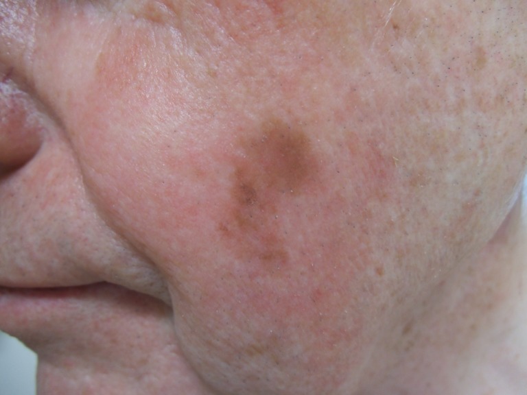
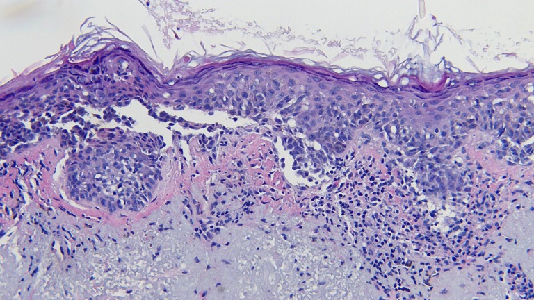
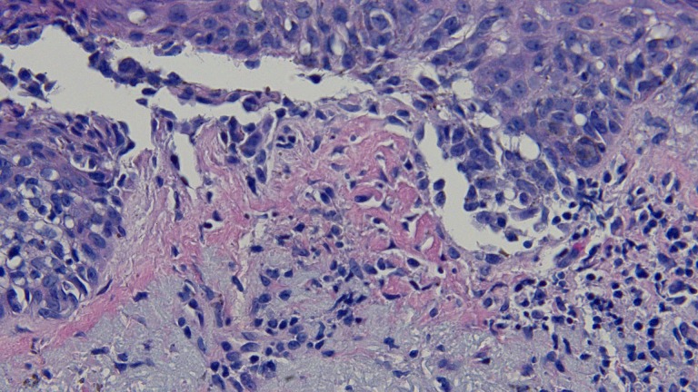
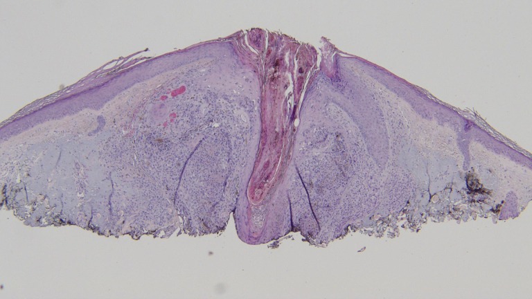
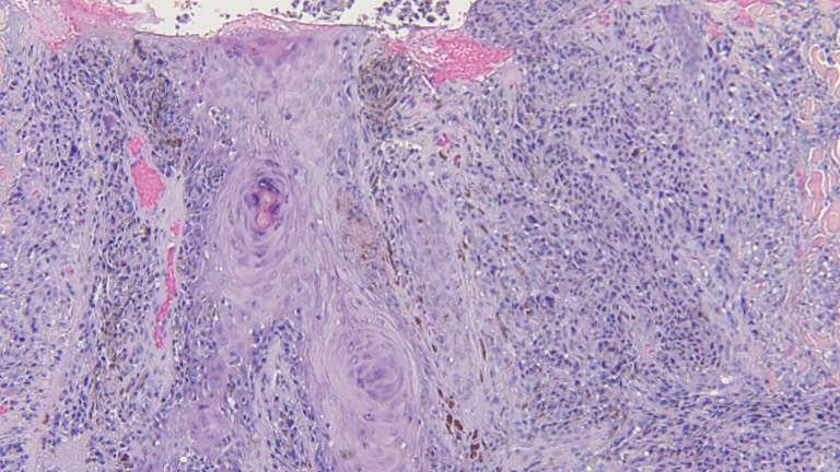
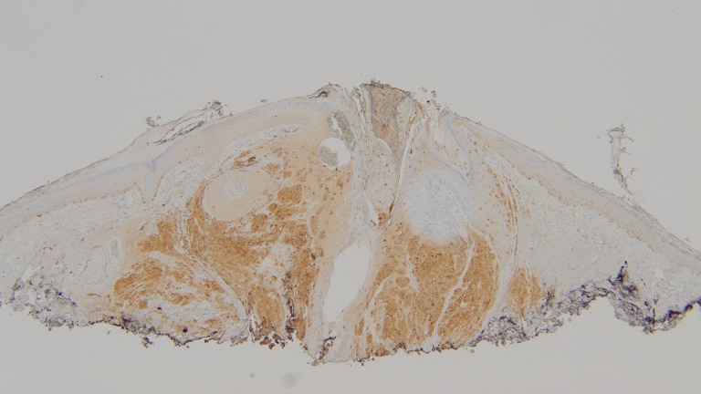
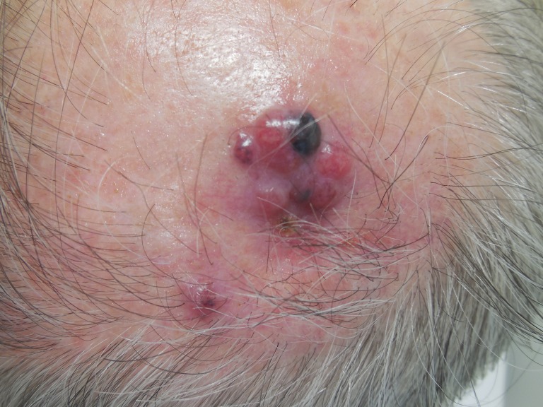
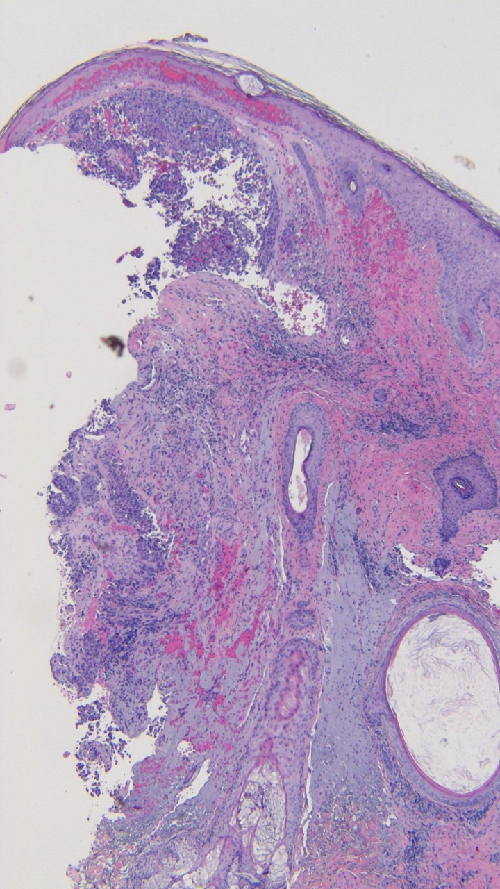
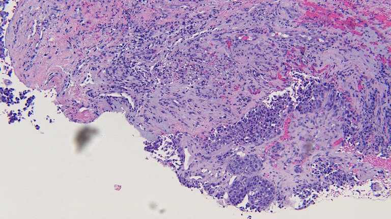
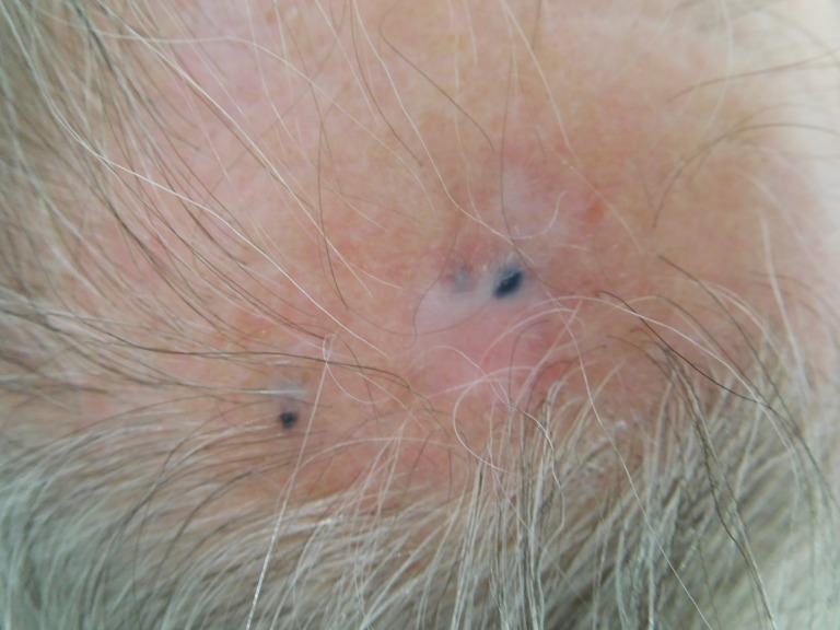
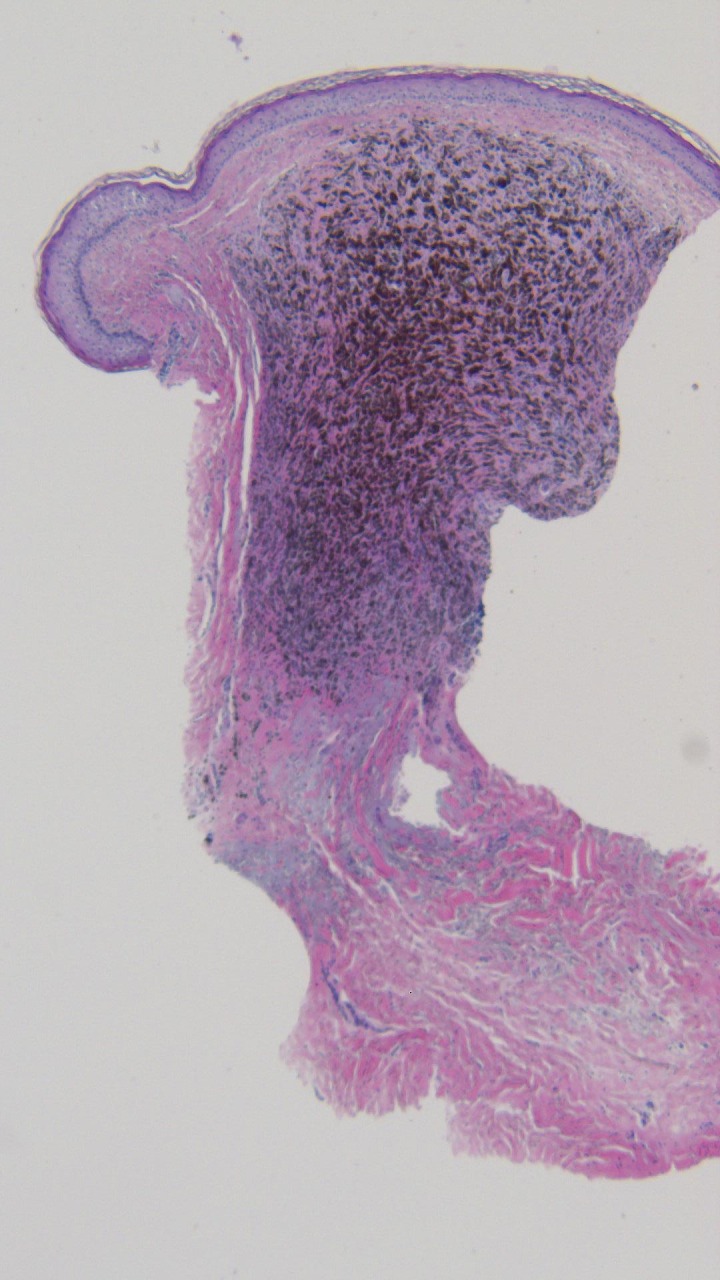
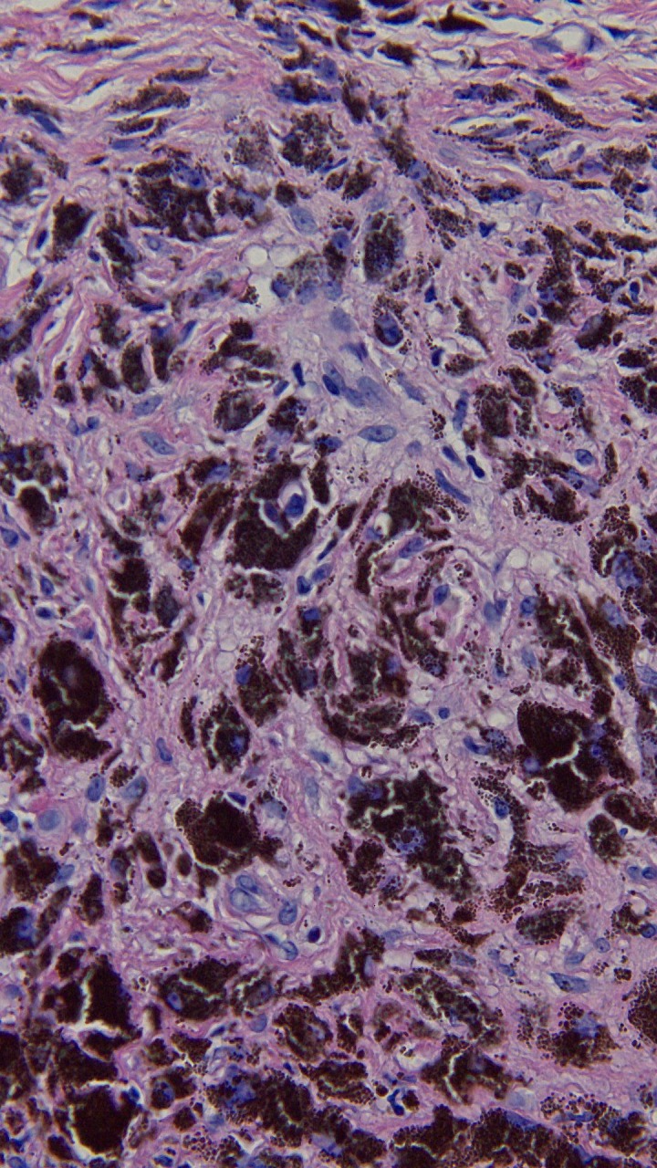
Tumoral melanosis is a form of completely regressed melanoma that usually presents as darkly pigmented lesions suspicious for malignant melanoma. Histology reveals dense dermal and subcutaneous infiltration of melanophages. Pembrolizumab is an antibody directed against programmed death receptor-1 (PD1) and is frontline treatment for advanced melanoma. An 81-year-old man with metastatic melanoma treated with pembrolizumab who developed tumoral melanosis at previous sites of metastases is described. The PubMed database was searched with the key words: antibody, immunotherapy, melanoma, melanosis, metastasis, pembrolizumab, and tumoral. The papers generated by the search and their references were reviewed. The patient was initially diagnosed with lentigo maligna melanoma on the left cheek three years earlier, and he was treated with wide local excision. The patient was subsequently diagnosed with epidermotropic metastatic malignant melanoma on the left parietal scalp 14 months later and was treated with wide local excision. Three months later, the patient was found to have metastatic melanoma in the same area of the scalp and was started on pembrolizumab immunotherapy. The patient was diagnosed with tumoral melanosis in the site of previous metastases nine months later. The patient remained free of disease 13 months after starting pembrolizumab. Tumoral melanosis may mimic malignant melanoma; hence a workup, including skin biopsy, should be undertaken. Extensive tumoral melanosis has been reported with ipilimumab, and we add a case following treatment with pembrolizumab. Additional cases of tumoral melanosis may present since immunotherapy has become frontline therapy for advanced melanoma.

Keywords: antibody; immunotherapy; melanoma; melanosis; metastasis; pembrolizumab; tumoral.


Conflict of interest statement

The authors have declared that no competing interests exist.

Figures

Figure 1
Figure 1. Lentigo maligna melanoma on the left cheek.
A 2 x 1 cm brown patch is observed on the patient’s left cheek.
Figure 2
Figure 2. Microscopic examination of the lentigo maligna melanoma biopsy.
Atypical melanocytes are seen along the dermal-epidermal junction. There is also extension of the tumor cells down adnexal structures (hematoxylin and eosin; x = 20).
Figure 3
Figure 3. Closer magnification of the microscopic examination of the lentigo maligna melanoma biopsy.
Closer view of the atypical melanocytes along the dermal-epidermal junction (hematoxylin and eosin; x = 40).
Figure 4
Figure 4. Epidermotropic metastatic malignant melanoma on the left parietal scalp.
Metastatic malignant melanoma presents here as a 2 mm black papule on the left parietal scalp, mimicking an open comedone.
Figure 5
Figure 5. Microscopic examination of the initial shave biopsy of the epidermotropic metastatic malignant melanoma.
Surrounding a central follicle, a dense infiltrate of melanocytes in the dermis and epidermis is seen (hematoxylin and eosin; x = 4).
Figure 6
Figure 6. Distant view of the subsequent punch biopsy to evaluate the residual pigment observed clinically in the center of the biopsy site after the shave biopsy was performed.
There is a nodular configuration of atypical melanocytes in the papillary and reticular dermis with small areas of junctional involvement and folliculotropism (hematoxylin and eosin; x = 4).
Figure 7
Figure 7. Closer magnification of the microscopic examination of the punch biopsy of the epidermotropic metastatic malignant melanoma.
Atypical tumor melanocytes fill the dermis and extend into the follicular epithelium and overlying epidermis (hematoxylin and eosin; x = 20).
Figure 8
Figure 8. S100 stain of the shave biopsy of the epidermotropic metastatic malignant melanoma.
The tumor cells demonstrate positive S100 staining, confirming the identification of the neoplasm as a melanoma (hematoxylin and eosin; x = 4).
Figure 9
Figure 9. Metastatic melanoma on the left parietal scalp.
Multiple red nodules, ranging from 6 to 8 mm in diameter, with a focal black patch, are observed.
Figure 10
Figure 10. Microscopic examination of the punch biopsy of the metastatic melanoma on the left parietal scalp.
Atypical melanocytes are seen in a nodular configuration in the papillary dermis and extend deeply into the reticular dermis (hematoxylin and eosin; x = 4).
Figure 11
Figure 11. Closer magnification of the microscopic examination of the punch biopsy of the metastatic melanoma on the left parietal scalp.
Atypical melanocytes in the deep dermis are seen in this closer view (hematoxylin and eosin; x = 20).
Figure 12
Figure 12. Tumoral melanosis on the left parietal scalp.
Three blue patches, ranging from 2 to 4 mm in diameter are observed.
Figure 13
Figure 13. Microscopic examination of the punch biopsy of the tumoral melanosis on the left parietal scalp.
Dense collections of melanophages are seen in a nodular aggregate; tumor melanocytes are not observed (hematoxylin and eosin; x = 4).
Figure 14
Figure 14. Closer magnification of the microscopic examination of the punch biopsy of the tumoral melanosis on the left parietal scalp.
The superficial and deep reticular dermis are filled with melanophages, and residual melanoma is absent (hematoxylin and eosin; x = 40).

References

    1. Extensive tumoral melanosis associated with ipilimumab-treated melanoma. Staser K, Chen D, Solus J, et al. Br J Dermatol. 2016;175:391–393. - PubMed
    1. Pembrolizumab for the treatment of advanced melanoma. Burns MC, O'donnell A, Puzanov I, et al. Expert Opin Orphan Drugs. 2016;4:867–873. - PMC - PubMed
    1. Melanophagic dermatitis and panniculitis. A condition revealing an occult metastatic malignant melanoma. Piérard GE. http://journals.lww.com/amjdermatopathology/Abstract/1988/04000/Melanoph.... Am J Dermatopathol. 1988;10:133–136. - PubMed
    1. Lymph node melanosis in a patient with metastatic melanoma of unknown primary. Malafronte P, Sorrells T. http://www.archivesofpathology.org/doi/full/10.1043/1543-2165-133.8.1332. Arch Pathol Lab Med. 2009;133:1332–1334. - PubMed
    1. Tumoral melanosis involving the sentinel lymph nodes: a case report. Satzger I, Völker B, Kapp A, Gutzmer R, et al. J Cutan Pathol. 2007;34:284–286. - PubMed

Publication types

LinkOut - more resources