. 2017 Aug;134(2):221-240.
doi: 10.1007/s00401-017-1703-0. Epub 2017 Mar 27.

Amyloid-β accumulation in the CNS in human growth hormone recipients in the UK

Affiliations

Amyloid-β accumulation in the CNS in human growth hormone recipients in the UK

Diane L Ritchie et al. Acta Neuropathol. 2017 Aug.

Abstract

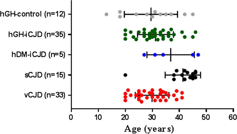
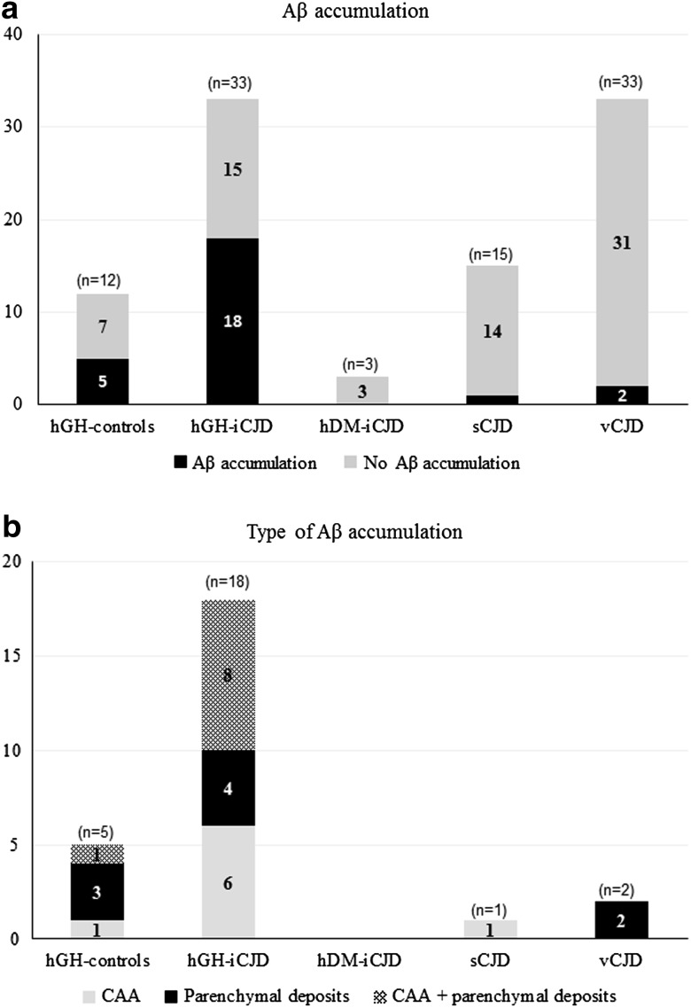
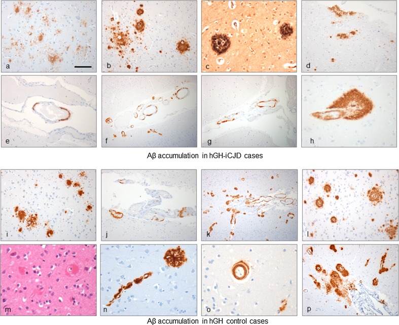
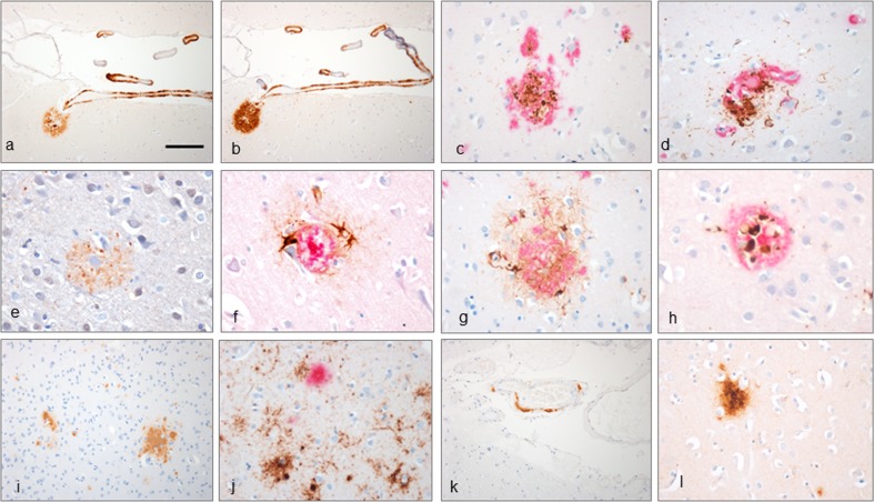
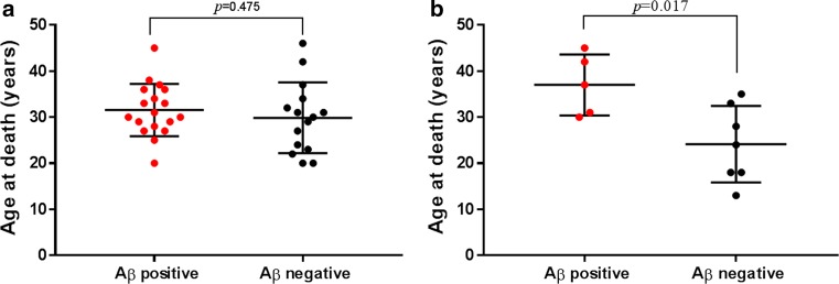
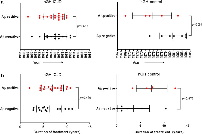
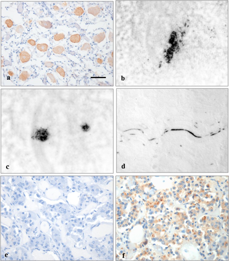
Human-to-human transmission of Creutzfeldt-Jakob disease (CJD) has occurred through medical procedures resulting in iatrogenic CJD (iCJD). One of the commonest causes of iCJD was the use of human pituitary-derived growth hormone (hGH) to treat primary or secondary growth hormone deficiency. As part of a comprehensive tissue-based analysis of the largest cohort yet collected (35 cases) of UK hGH-iCJD cases, we describe the clinicopathological phenotype of hGH-iCJD in the UK. In the 33/35 hGH-iCJD cases with sufficient paraffin-embedded tissue for full pathological examination, we report the accumulation of the amyloid beta (Aβ) protein associated with Alzheimer's disease (AD) in the brains and cerebral blood vessels in 18/33 hGH-iCJD patients and for the first time in 5/12 hGH recipients who died from causes other than CJD. Aβ accumulation was markedly less prevalent in age-matched patients who died from sporadic CJD and variant CJD. These results are consistent with the hypothesis that Aβ, which can accumulate in the pituitary gland, was present in the inoculated hGH preparations and had a seeding effect in the brains of around 50% of all hGH recipients, producing an AD-like neuropathology and cerebral amyloid angiopathy (CAA), regardless of whether CJD neuropathology had occurred. These findings indicate that Aβ seeding can occur independently and in the absence of the abnormal prion protein in the human brain. Our findings provide further evidence for the prion-like seeding properties of Aβ and give insights into the possibility of iatrogenic transmission of AD and CAA.

Keywords: Amyloid β; Cerebral amyloid angiopathy; Human growth hormone; Iatrogenic Creutzfeldt–Jakob disease; Neuropathology; Prion protein.


Conflict of interest statement

The authors declare that they have no conflicts of interest. The human tissue examined in this study was provided by the MRC Edinburgh Brain Bank and its use was covered by ethical approval from the East of Scotland Research Ethics Service REC 1 (reference number 16/ES/0084). Informed consent for the research use of autopsy tissue was obtained from the relatives of the deceased whenever necessary. This article does not contain any studies with animals performed by any of the authors.

Figures

Fig. 1
Fig. 1
Age distributions of hGH-iCJD and control cases. The sCJD and vCJD control patients were chosen to be as close as possible in range to the hGH-iCJD cases. Cases of sCJD under the age of 50 years are rare; the sCJD control cases are clustered in the 40–50 years age range with higher mean age values. In contrast, cases of vCJD over the age of 40 years are rare; the upper age limit for the vCJD cases is 41 years. Vertical bars represent the mean with standard deviation values
Fig. 2
Fig. 2
Relationship of PRNP codon 129 polymorphisms to hGH-iCJD incubation period and disease duration. a Differences in the incubation periods for hGH-iCJD in relation to PRNP codon 129 genotype. b Differences in the hGH-iCJD disease duration in relation to PRNP codon 129 genotype. Statistical analyses were performed using one-way ANOVA followed by Tukey’s multiple comparison test; horizontal bars represent mean with standard deviation values
Fig. 3
Fig. 3
Typical neuropathological phenotypes in UK hGH-iCJD. PRNP codon 129 VV hGH-iCJD cases show microvacuolation in the cerebral cortex (a) with a combination of granular, perineuronal and plaque-like accumulations of PrP in the cerebral cortex (e). The cerebellar cortex in VV hGH-iCJD cases shows a predominantly granular pattern of PrP accumulation (i). PRNP codon 129 MV hGH-iCJD were characterised by the presence of kuru plaques in the cerebellar and occasionally in the cerebral cortex (b, f, j). PRNP codon 129 MM hGH-iCJD cases show widespread microvacuolation in the cerebral cortex (c) with a predominantly granular accumulation of PrP in the cerebral (g) and cerebellar cortex (k). Case hGH-iCJD31 shows atypical neuropathological features in comparison with the codon 129 MM hGH-iCJD cases. Microvacuolation was observed in the lower layers of the cerebral cortex (d) with a combination of granular, perineuronal and plaque-like accumulations of PrP in the cerebral and cerebellar cortex (h, l). No kuru-type amyloid plaques were present in any brain region. These features show some similarities to the VV2 histotype in sCJD. Sections a, c, e, and g, are stained with H&E and sections b, d, f, and h are stained with the KG9 antibody. The bar in a represents 25 µm for a, cl; and 15 µm for b
Fig. 4
Fig. 4
Frequency of Aβ accumulation in hGH-iCJD and control cases. There is a significant difference in the percentage of cases with CNS Aβ deposition in the two groups treated with hGH (51%) compared to the three groups not treated with hGH (6%); p < 0.001 (Fisher’s exact test). Comparison of the hGH-iCJD, hGH control and non hGH-treated groups confirms the association between CNS Aβ accumulation and hGH treatment; p < 0.001 (Chi-squared test)
Fig. 5
Fig. 5
Aβ accumulation in the CNS in hGH-iCJD and hGH control patients. Aβ immunohistochemistry (6F/3D antibody) in hGH-iCJD (ah) and hGH control (ip) cases. Diffuse plaques were a feature of all hGH-iCJD cases in which CNS Aβ deposition was observed (a). Cored plaques were less frequently observed in hGH-iCJD cases (b) with neuritic plaques demonstrated with the Bielschowsky silver stain (c). Patchy diffuse subpial deposits of Aβ were also a feature of hGH-iCJD cases (d). Patchy deposition of Aβ in the wall of a meningeal vessel with a more extensive Aβ deposition in occipital vessels in the meninges and adjacent cortex in hGH-iCJD (e, f). Patchy Aβ deposition was observed in the wall of meningeal vessels overlying the cerebellar cortex in a single hGH-iCJD case (g). Circumferential deposition in a cortical arteriole with extensive perivascular Aβ forming a cored plaque-like structure (h). Diffuse Aβ deposits and plaques were also found in the cerebral cortex in hGH control cases (i). CAA with patchy meningeal deposits and circumferential deposition were observed in the superficial cortical vessels in hGH control cases (j). hGH control11 showed extensive CAA in the meninges and cortex with diffuse and perivascular Aβ deposits and multiple cored plaques and smaller diffuse Aβ deposits (k, l). This case also showed severe capillary CAA with marked thickening of vessel walls, the presence of dyshoric cortical vessel and vasculopathy shown with the splitting of an intracortical arteriole wall (mo). Meningeal and intracortical CAA with diffuse subpial deposits, perivascular deposits and diffuse and cored Aβ plaques in hGH control case11 (p). The bar in a represents 50 µm for a, b, de, g, i, l, p; 20 µm for h, mo; 25 µm for c; and 100 µm for f, j, k
Fig. 6
Fig. 6
Aβ pathology in hGH recipients, sCJD and vCJD. Immunohistochemistry with Aβ 1–40 antibody shows intense labelling of Aβ within cerebral vessels and plaque cores (a). A serial section labelled with the Aβ 1–42 antibody showing intense labelling of a large diffuse Aβ deposit (b). Phospho-tau positivity (brown) in neurites around Aβ (red) in a cored plaque and dyshoric vessel with CAA in hGH control11 (c, d). The ubiquitin antibody labels extensive neurites around a cored Aβ plaque in hGH-iCJD (e). Reactive astrocytes around a cored Aβ plaque revealed on double labelling for Aβ (6F/3D antibody-red) and GFAP (GFAP antibody-brown) in hHG-iCJD (f). Astrocytes (brown) surround a cored Aβ plaque (red) (g) and microglial cells (brown) are present within a cored Aβ plaque (red) in hGH control10 (h). Diffuse Aβ deposits in the parietal cortex in vCJD27 (i). The diffuse cortical Aβ deposits in vCJD 37 (red) are shown not to co-localise with the abundant PrP deposits (brown) (j). Patchy localised meningeal CAA in the occipital region in sCJD14 (k). APOE-4 positivity in diffuse Aβ deposits in hGH control6 (l). The bar in a represents 100 µm for ab; 20 µm for f, h; 25 µm for ce, g, j, l; and 50 µm for i, k
Fig. 7
Fig. 7
CNS accumulation and age at death in hGH-iCJD and hGH control cases. Comparisons of the age at death for a hGH-iCJD and b hGH-control patients in relation to accumulation of Aβ. No significant difference in the age at death was found between the Aβ-positive and Aβ-negative hGH-iCJD cases. However, a significant difference was found between the Aβ-positive and Aβ-negative hGH control cases, with the Aβ-positive cases showing a higher age at death. Statistical analysis was performed using an unpaired t test
Fig. 8
Fig. 8
Treatment times with hGH-iCJD and hGH control patients. a Shows the year of first treatment for hGH-iCJD and hGH-control patients in relation to accumulation of Aβ and b shows duration of treatment for hGH-iCJD and hGH-control patients in relation to CNS Aβ accumulation. Differences were found in both the year of first hGH treatment and duration of treatment in the hGH control cases with and without CNS Aβ accumulation, but these did not reach levels of statistical significance. Statistical analysis was performed using an unpaired t test
Fig. 9
Fig. 9
PrP accumulation in non-CNS tissues in hGH-iCJD cases. a Immunohistochemistry for the prion protein shows labelling of the ganglion cells in a dorsal root ganglion. b PET blot analysis with the 12F10 anti-PrP antibody shows intense labelling (black) of a group of chromaffin cells within the adrenal medulla, c in germinal centres within an abdominal lymph node and d in a small nerve within skeletal muscle. e Immunolabelling for Aβ using the 6F/3D antibody shows no labelling in the anterior pituitary gland, but f the 4G8 anti-Aβ antibody shows some diffuse fine granular and discrete dot-like intracellular positivity in the endocrine cells. The bar in a represents 50 µm for a, b, d; 25 µm for e, f; and 75 µm for c

References

    1. 1000 Genomes Project Consortium. Abecasis GR, Auton A, Brooks LD, DePristo MA, Durbin RM, Handsaker RE, et al. An integrated map of genetic variation from 1,092 human genomes. Nature. 2012;491:56–65. doi: 10.1038/nature11632. - DOI - PMC - PubMed
    1. Benitez BA, Karch CM, Cai Y, Jin SC, Cooper B, Carrell D, et al. The PSEN1, p. E318G variant increases the risk of Alzheimer’s disease in APOE-ɛ4 carriers. PLoS Genet. 2013;9:e1003685. doi: 10.1371/journal.pgen.1003685. - DOI - PMC - PubMed
    1. Blennow K, Hampel H, Weiner M, Zetterberg H. Cerebrospinal fluid and plasma biomarkers in Alzheimer’s disease. Nat Rev Neurol. 2010;6:131–144. doi: 10.1038/nrneurol.2010.4. - DOI - PubMed
    1. Brandel JP, Peckeu L, Haïk S. The French surveillance network of Creutzfeldt–Jakob disease. Epidemiological data in France and worldwide. Transfus Clin Biol. 2013;20:395–397. doi: 10.1016/j.tracli.2013.02.029. - DOI - PubMed
    1. Braak Braak E. Neuropathological staging of Alzheimer-related changes. Acta Neuropathol. 1991;82:239–259. doi: 10.1007/BF00308809. - DOI - PubMed

MeSH terms