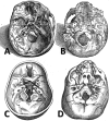
The "polymorphous" history of a polymorphous skull bone: the sphenoid
- PMID: 28349500
- PMCID: PMC5741782
- DOI: 10.1007/s12565-017-0399-5
The "polymorphous" history of a polymorphous skull bone: the sphenoid
Abstract
For a long time, because of its location at the skull base level, the sphenoid bone was rather mysterious as it was too difficult for anatomists to reach and to elucidate its true configuration. The configuration of the sphenoid bone led to confusion regarding its sutures with the other skull bones, its shape, its detailed anatomy, and the vascular and nervous structures that cross it. This article takes the reader on a journey through time and space, charting the evolution of anatomists' comprehension of sphenoid bone morphology from antiquity to its conception as a bone structure in the eighteenth century, and ranging from ancient Greece to modern Italy and France. The journey illustrates that many anatomists have attempted to name and to best describe the structural elements of this polymorphous bone.
Keywords: Anatomy; History; Sella turcica; Skull base; Sphenoid bone.
Conflict of interest statement
The authors declare no conflict of interest.
Figures


References
-
- Ball MJ (1910) Andreas Vesalius, the reformer of anatomy. Medical Science Press, St. Louis
-
- Bell J, Bell C (1827) The anatomy and physiology of the human body. Collins & Co., New York
-
- Cloquet H, Knox R. A system of human anatomy. Edinburgh: Maclachlan & Stewart; 1828.
-
- Columbo R. De re anatomica libri XV. Venetiis: Ex Typographia Nicolai Beuilacquæ; 1559.
Publication types
MeSH terms
LinkOut - more resources
Full Text Sources
Other Literature Sources

