. 2017 Mar 28:7:45263.
doi: 10.1038/srep45263.

Adventitial lymphatic capillary expansion impacts on plaque T cell accumulation in atherosclerosis

Affiliations

Adventitial lymphatic capillary expansion impacts on plaque T cell accumulation in atherosclerosis

Timo Rademakers et al. Sci Rep. .

Abstract

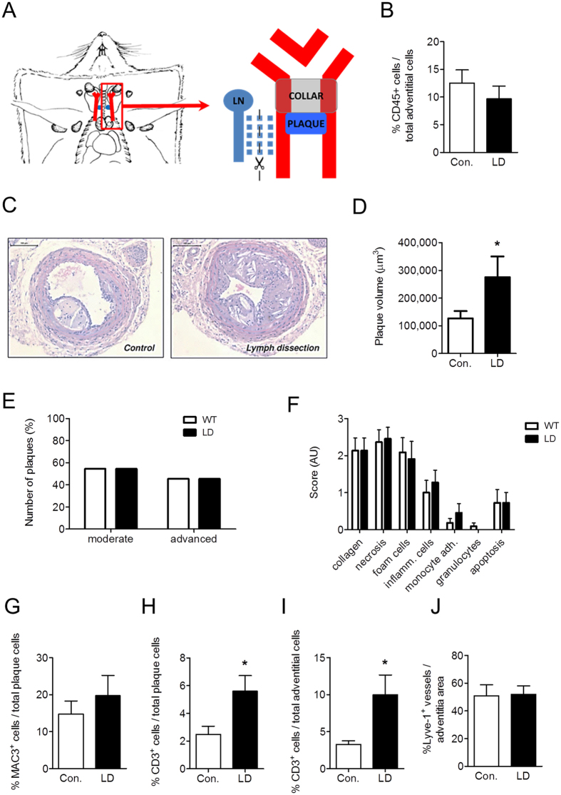
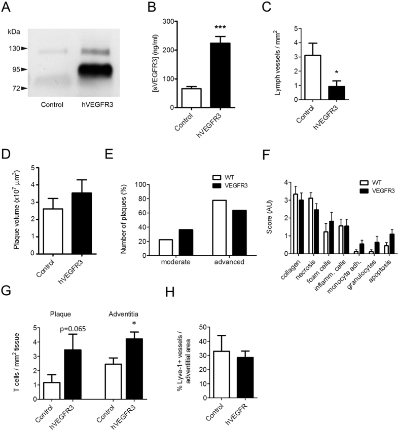
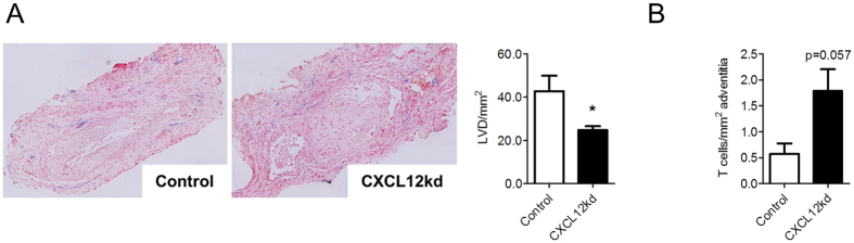
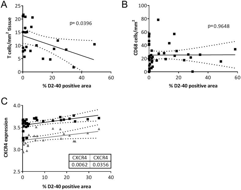
During plaque progression, inflammatory cells progressively accumulate in the adventitia, paralleled by an increased presence of leaky vasa vasorum. We here show that next to vasa vasorum, also the adventitial lymphatic capillary bed is expanding during plaque development in humans and mouse models of atherosclerosis. Furthermore, we investigated the role of lymphatics in atherosclerosis progression. Dissection of plaque draining lymph node and lymphatic vessel in atherosclerotic ApoE-/- mice aggravated plaque formation, which was accompanied by increased intimal and adventitial CD3+ T cell numbers. Likewise, inhibition of VEGF-C/D dependent lymphangiogenesis by AAV aided gene transfer of hVEGFR3-Ig fusion protein resulted in CD3+ T cell enrichment in plaque intima and adventitia. hVEGFR3-Ig gene transfer did not compromise adventitial lymphatic density, pointing to VEGF-C/D independent lymphangiogenesis. We were able to identify the CXCL12/CXCR4 axis, which has previously been shown to indirectly activate VEGFR3, as a likely pathway, in that its focal silencing attenuated lymphangiogenesis and augmented T cell presence. Taken together, our study not only shows profound, partly CXCL12/CXCR4 mediated, expansion of lymph capillaries in the adventitia of atherosclerotic plaque in humans and mice, but also is the first to attribute an important role of lymphatics in plaque T cell accumulation and development.


Conflict of interest statement

The authors declare no competing financial interests.

Figures

Figure 1
Figure 1. Increased presence of adventitial lymphatic capillaries during human and mouse atherosclerosis development.
(A) Immunohistochemical staining of podoplanin+ (D2-40) lymphatic capillaries (Brown) in human aortic atherosclerotic lesions obtained during kidney transplantation (10x magnification, Insert = 20x magnification). *Shows CD31+ adventitial microvessels (blue). (B) Quantification of adventitial lymph vessel density (LVD) in different stages of human atherosclerotic lesion development. (C) Immunohistochemical staining of Lyve-1+ lymphatic capillaries (Purple) and CD31+ vasa vasorum (Blue) in mouse carotid atherosclerotic lesions. Continued line indicates the outer border of the media; the area between the continuous and dotted line indicates the adventitial area used for quantification (10x magnification, Insert = 20x magnification). (D) Quantification of the percentage Lyve-1+ lymphatic capillaries per adventitial area of different stages of mouse lesion development.
Figure 2
Figure 2. Lymph node dissection results in an aggravation of plaque development in mice, characterized by an accumulation of CD3+ T cells within the vessel wall.
(A) Draining lymph node and efferent vessel were excised in the lymph node dissection group. (B) Percentage of CD45+ leukocytes in the adventitia of control and lymph node dissected (LD) mice. (C) Representative pictures of Hematoxylin-Eosin-stained sections of the carotid arteries from control and lymph node dissected mice. Quantification of (D) plaque volume, (E) plaque classification and (F) routine pathological examination of plaque composition. Percentage of Mac-3+ macrophages in the plaques (G). Percentage of CD3+ T cells in (H) plaque and (I) adventitia. (J) Percentage of Lyve-1+ lymphatic capillary area in the adventitia.
Figure 3
Figure 3. Soluble hVEGFR3 gene therapy did not affect plaque burden and plaque-associated lymphangiogenesis, yet induced an increase in plaque T cell content.
(A) Western blot showing cropped image of blot and (B) ELISA analysis of hVEGFR3 expression in plasma after AAV-hVEGFR3-Ig gene transfer. (C) Lymph vessel formation and ingrowth in an in-vivo matrigel plug assay, four weeks after s.c. administration of AAV-hVEGFR3-Ig gene transfer. Quantification of (D) plaque volume, (E) plaque classification and (F) routine pathological examination of plaque composition of lesion formation in carotid arteries. (G) Quantification of adventitial and plaque T cell content. (H) Percentage of Lyve-1+ lymphatic capillary area in the adventitia.
Figure 4
Figure 4. Chemokine and lymph vessel dependent accumulation of T cells.
(A) Representative Lyve-1 staining and quantification of lymph vessel density (LVD) after perivascular treatment with siRNA targeting CXCL12. (B) Representative CD3 staining and quantification of adventitial T cell content after perivascular treatment with siRNA targeting CXCL12.
Figure 5
Figure 5. Correlation analysis in human endarterectomy plaque tissues.
Correlation analysis between T cells (A) and macrophages (B) per plaque area and D2-40 positive, lymphatic area in human atherosclerotic lesions. (C) Correlation analysis of CXCR4 expression and D2-40 positive, lymphatic area in human endarterectomy plaque tissue.

References

    1. Weber C., Zernecke A. & Libby P. The multifaceted contributions of leukocyte subsets to atherosclerosis: lessons from mouse models. Nature reviews. Immunology 8, 802–815, doi: 10.1038/nri2415 (2008). - DOI - PubMed
    1. Bot I. & Biessen E. A. Mast cells in atherosclerosis. Thrombosis and haemostasis 106, 820–826, doi: 10.1160/TH11-05-0291 (2011). - DOI - PubMed
    1. Hansson G. K. & Hermansson A. The immune system in atherosclerosis. Nature immunology 12, 204–212, doi: 10.1038/ni.2001 (2011). - DOI - PubMed
    1. Businaro R. et al.. Cellular and molecular players in the atherosclerotic plaque progression. Annals of the New York Academy of Sciences 1262, 134–141, doi: 10.1111/j.1749-6632.2012.06600.x (2012). - DOI - PubMed
    1. Libby P. Inflammation in atherosclerosis. Arteriosclerosis, thrombosis, and vascular biology 32, 2045–2051, doi: 10.1161/ATVBAHA.108.179705 (2012). - DOI - PMC - PubMed

Publication types

MeSH terms