. 2017 Mar 28:7:45508.
doi: 10.1038/srep45508.

Organization of prefrontal network activity by respiration-related oscillations

Affiliations

Organization of prefrontal network activity by respiration-related oscillations

Jonatan Biskamp et al. Sci Rep. .

Abstract

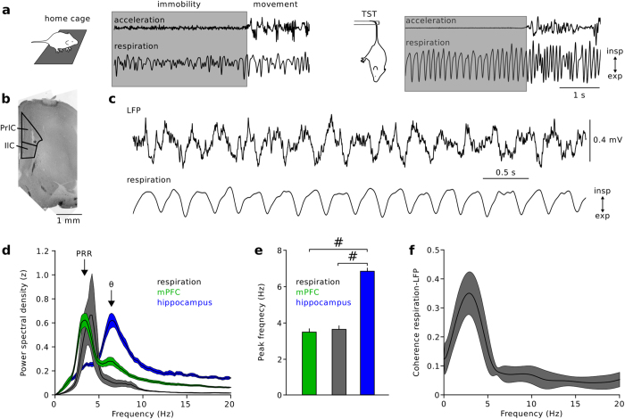
The medial prefrontal cortex (mPFC) integrates information from cortical and sub-cortical areas and contributes to the planning and initiation of behaviour. A potential mechanism for signal integration in the mPFC lies in the synchronization of neuronal discharges by theta (6-12 Hz) activity patterns. Here we show, using in vivo local field potential (LFP) and single-unit recordings from awake mice, that prominent oscillations in the sub-theta frequency band (1-5 Hz) emerge during awake immobility in the mPFC. These oscillation patterns are distinct from but phase-locked to hippocampal theta activity and occur synchronized with nasal respiration (hence termed prefrontal respiration rhythm [PRR]). PRR activity modulates the amplitude of prefrontal gamma rhythms with greater efficacy than theta oscillations. Furthermore, single-unit discharges of putative pyramidal cells and GABAergic interneurons are entrained by prefrontal PRR and nasal respiration. Our data thus suggest that PRR activity contributes to information processing in the prefrontal neuronal network.


Conflict of interest statement

The authors declare no competing financial interests.

Figures

Figure 1
Figure 1. Respiration-related LFP oscillations in the medial prefrontal cortex of awake mice.
(a) Recording respiration with a nasal thermocouple during free home cage behaviour (left) and tail suspension test (TST, right) revealed stable respiration frequency during immobility in the TST. The traces on top display movement extracted from the reading of an accelerometer mounted on the recording amplifier. Grey areas correspond to immobile epochs. (b) LFP recordings were targeted to the prelimbic (PrlC) and infralimbic (IlC) portions of the mPFC. Asterisk denotes the location of the recording electrode. (c) Example LFP (1–200 Hz-filtered, top) and respiration recording (1–10 Hz-filtered, bottom) during TST immobility. Inspiration faces upwards. (d) Power spectral density analysis revealed prefrontal respiration rhythms (PRRs, green) that spectrally overlap with respiration (grey) but not with theta activity in the mPFC (small peak at ~7 Hz) or hippocampus (blue). (e) Summary plot of peak frequency of prefrontal LFP (green, n = 25 mice), respiration (grey, n = 4) and hippocampal LFP (blue, n = 13). Mann-Whitney U-tests with Bonferroni correction. (f) Prefrontal LFPs are highly coherent with nasal respiration (n = 4 mice). Data are mean ± sem. #p < 0.001.
Figure 2
Figure 2. PRR activity requires intact olfactory bulbs.
(a) Example LFP traces recorded after bilateral bulbectomy (red, bottom) and under control conditions (grey, top). Grey superimposed traces are 1–5 Hz filtered to emphasize PRR activity. (b) Power spectral density of prefrontal LFPs after bulbectomy reveals a reduction in PRR power (1–5 Hz). (c) Summary graph of PRR (1–5 Hz), theta (6–12 Hz), and gamma power (30–100 Hz) after bulbectomy (red) and under control conditions (grey). Bulbectomy strongly reduces PRR (unpaired t-test) but leaves theta and gamma power unaffected (Mann-Whitney U-tests). (d) Frontal slice of the mPFC shows the area of red retroBead injection for the retrograde tracing of mPFC inputs. Cg: cingulate cortex, IlC: infralimbic cortex, PrlC: prelimbic cortex, M1, 2: primary and secondary motor cortex. (e) Retrogradely traced cells (red) were detected in olfactory cortices. Blue: DAPI signal. Representative for 3 mice. #p < 0.001. Data represent mean ± sem.
Figure 3
Figure 3. Phase-phase coupling of PRR with hippocampal theta.
(a) Simultaneous recording of prefrontal and hippocampal LFPs. Traces show frequency-filtered signals (PRR: 1–5 Hz, theta: 6–12 Hz) and the respective instantaneous phases obtained by Hilbert transformation. (b) Joint phase histograms of hippocampal theta phase as a function of PRR phase revealed diagonal stripes of increased probability. Left: data from one mouse. Right: Average of 12 mice. (c) Rn:m phase-phase coupling analysis revealed a maximum at m = 2, suggesting that two cycles of hippocampal theta occur within one cycle of PRR. (d) Statistical comparison to surrogate data (time-shifted by a random interval between 5 and 10 s) indicated significant phase-phase coupling (paired t-test). *p < 0.05. Data represent mean ± sem.
Figure 4
Figure 4. PRR activity modulates the amplitude of prefrontal gamma oscillations.
(a) Schematic illustration of the analysis. The phase of the slow modulating PRR or theta oscillation and the amplitude of the fast modulated gamma frequency components were obtained by Hilbert transformation. (b) Modulation of the amplitudes of gamma frequency activity patterns (30–100 Hz) by the phase of ongoing PRR and theta frequency oscillations during TST immobility. The colour code indicates modulation depth, quantified as a modulation index (0: no modulation, 1: maximal modulation, see Methods). Average of 25 mice. (c) Gamma oscillation amplitude is enriched at the peak of RR phase. Zero denotes the peak, −π and π the troughs of the PRR cycle. (d) Restricting the analysis to time epochs of strong theta activity (red areas on the left, see Methods) still revealed prominent PRR-gamma coupling. n = 12 mice. (e) Gamma oscillations were modulated by theta activity in the hippocampus (CA1). n = 13 mice. (f) Summary of gamma amplitude modulation by PRR and prefrontal as well as hippocampal theta. Wilcoxon signed-rank and Mann-Whitney U-tests with Bonferroni correction. #p < 0.001. Data represent mean ± sem.
Figure 5
Figure 5. PRR activity entrains prefrontal single-unit activity.
(a) Single-units were recorded with stereotrodes or tetrodes lowered to the mPFC on a microdrive (n = 224 units). Examples show average waveforms of the four channels of a tetrode recording and the respective autocorrelation function of a putative pyramidal cell (PYR, left) and a putative GABAergic interneuron (IN, right). (b) INs discharged at significantly higher frequency than PYRs. Mann-Whitney U-test. (c) Example of PRR-filtered LFP (1-5 Hz), theta-filtered LFP (6-12 Hz), simultaneously recorded nasal respiration, and spiking activity of a PYR unit (bottom). (d) Spike histograms of the unit shown in (c) as a function of PRR, theta, and respiration phase indicated significant coupling of the discharge activity to the various rhythms (Rayleigh’s test, p < 0.05). (e) Summary graph of the percentage of significantly coupled PYR (black) and IN units (grey). Chisquare and Fisher’s exact tests with Bonferroni correction. (f) Summary histograms of the preferred phase of all significantly phase-coupled PYR (black, top) and IN units (grey, bottom) to the three types of rhythms. Zero denotes the peak, −π and π the troughs of the oscillation cycles. #p < 0.001. Data represent mean ± sem.

References

    1. Jones M. W. & Wilson M. A. Theta rhythms coordinate hippocampal-prefrontal interactions in a spatial memory task. PLoS Biol. 3, e402 (2005). - PMC - PubMed
    1. Sigurdsson T., Stark K. L., Karayiorgou M., Gogos J. A. & Gordon J. A. Impaired hippocampal-prefrontal synchrony in a genetic mouse model of schizophrenia. Nature 464, 763–767 (2010). - PMC - PubMed
    1. Lee M. G., Chrobak J. J., Sik A., Wiley R. G. & Buzsáki G. Hippocampal theta activity following selective lesion of the septal cholinergic system. Neuroscience 62, 1033–1047 (1994). - PubMed
    1. Wang X.-J. Pacemaker neurons for the theta rhythm and their synchronization in the septohippocampal reciprocal loop. J. Neurophysiol. 87, 889–900 (2002). - PubMed
    1. Dégenètais E., Thierry A.-M., Glowinski J. & Gioanni Y. Synaptic influence of hippocampus on pyramidal cells of the rat prefrontal cortex: an in vivo intracellular recording study. Cereb. Cortex N. Y. N 1991 13, 782–792 (2003). - PubMed

Publication types

LinkOut - more resources