

Neural Crest Cells Contribute an Astrocyte-like Glial Population to the Spleen
- PMID: 28349968
- PMCID: PMC5368681
- DOI: 10.1038/srep45645
Neural Crest Cells Contribute an Astrocyte-like Glial Population to the Spleen
Abstract
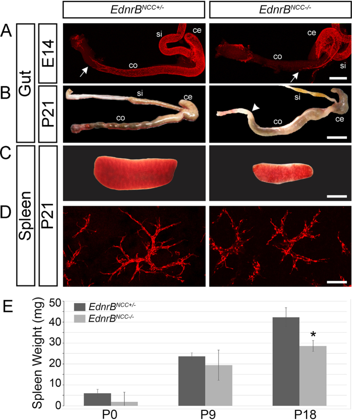
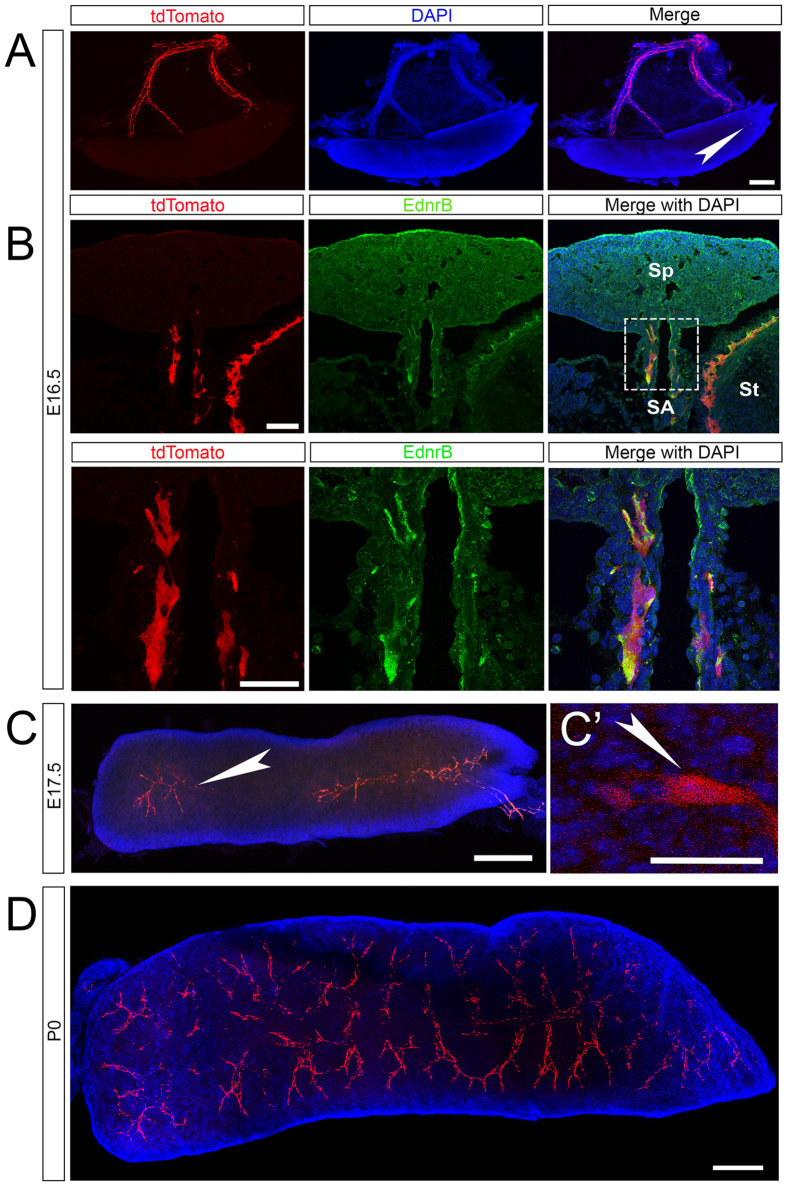
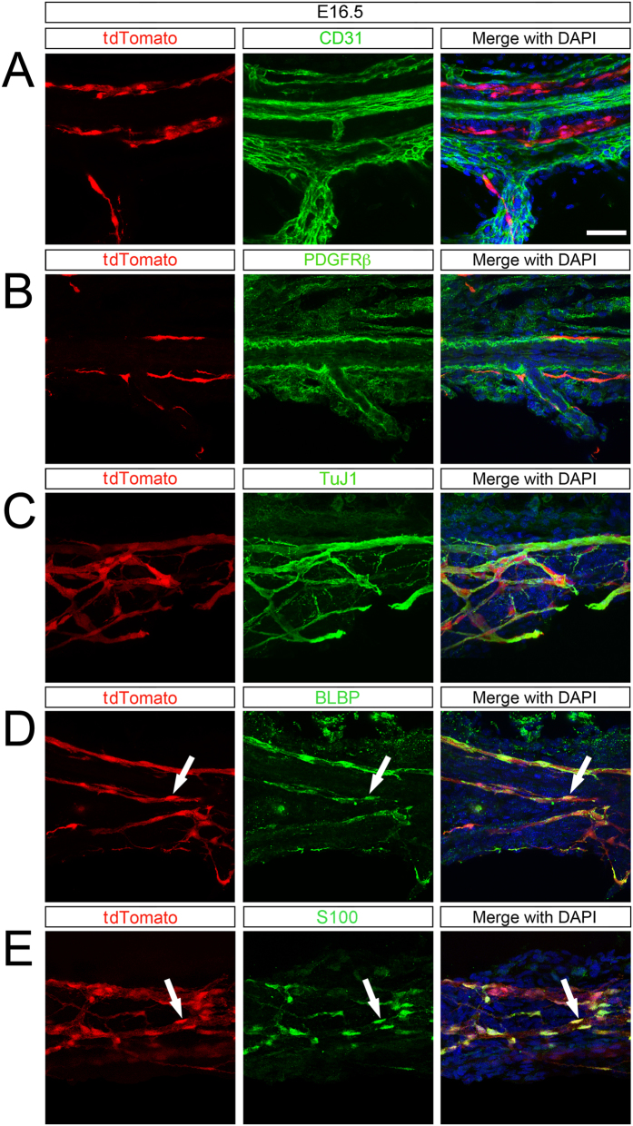
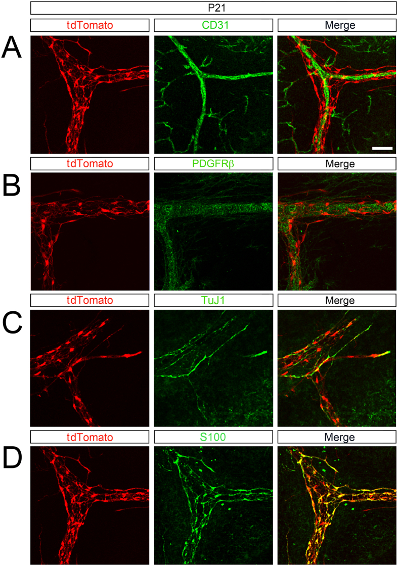
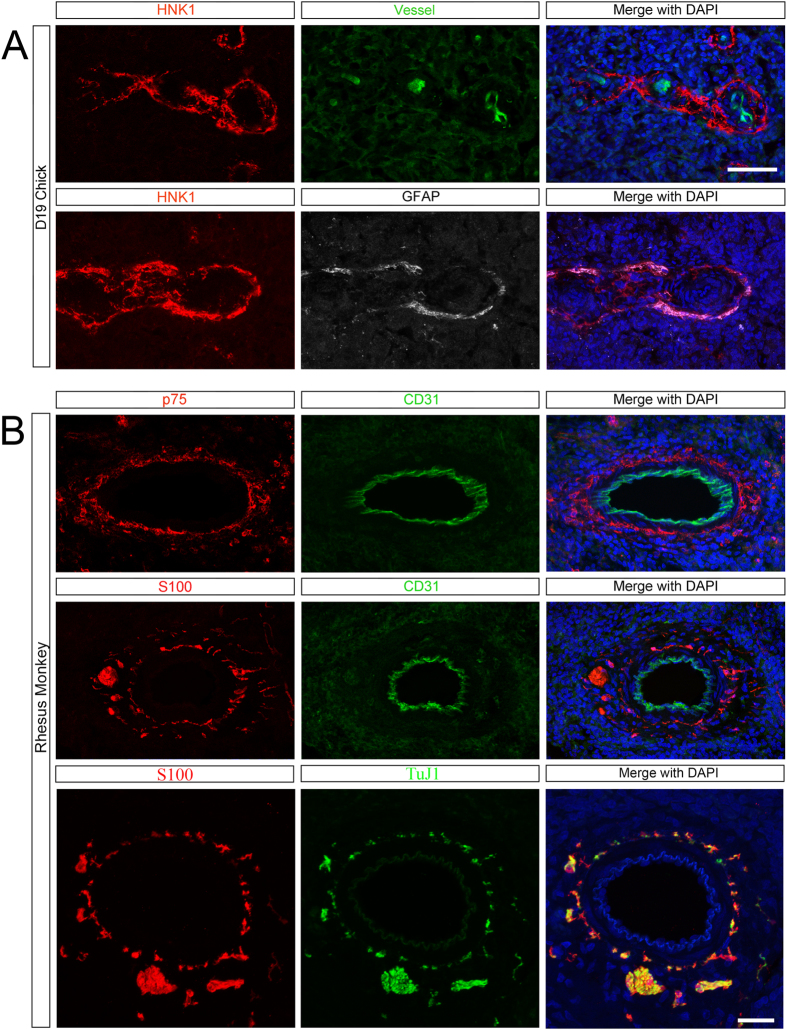
Neural crest cells (NCC) are multi-potent cells of ectodermal origin that colonize diverse organs, including the gastrointestinal tract to form the enteric nervous system (ENS) and hematopoietic organs (bone marrow, thymus) where they participate in lymphocyte trafficking. Recent studies have implicated the spleen as an anatomic site for integration of inflammatory signals from the intestine with efferent neural inputs. We have previously observed alterations in splenic lymphocyte subsets in animals with defective migration of NCC that model Hirschsprung's disease, leading us to hypothesize that there may be a direct cellular contribution of NCC to the spleen. Here, we demonstrate that NCC colonize the spleen during embryogenesis and persist into adulthood. Splenic NCC display markers indicating a glial lineage and are arranged anatomically adjacent to blood vessels, pericytes and nerves, suggesting an astrocyte-like phenotype. Finally, we identify similar neural-crest derived cells in both the avian and non-human primate spleen, showing evolutionary conservation of these cells.
Conflict of interest statement
The authors declare no competing financial interests.
Figures
Similar articles
-
The sacral neural crest contributes neurons and glia to the post-umbilical gut: spatiotemporal analysis of the development of the enteric nervous system.Development. 1998 Nov;125(21):4335-47. doi: 10.1242/dev.125.21.4335. Development. 1998. PMID: 9753687
-
Altered differentiation of enteric neural crest-derived cells from endothelin receptor-B null mouse model of Hirschsprung's disease.Pediatr Surg Int. 2016 Dec;32(12):1095-1101. doi: 10.1007/s00383-016-3964-4. Epub 2016 Sep 23. Pediatr Surg Int. 2016. PMID: 27663687
-
Enteric nervous system development: analysis of the selective developmental potentialities of vagal and sacral neural crest cells using quail-chick chimeras.Anat Rec. 2001 Jan 1;262(1):16-28. doi: 10.1002/1097-0185(20010101)262:1<16::AID-AR1007>3.0.CO;2-O. Anat Rec. 2001. PMID: 11146425
-
Neuron-Glia Interaction in the Developing and Adult Enteric Nervous System.Cells. 2020 Dec 31;10(1):47. doi: 10.3390/cells10010047. Cells. 2020. PMID: 33396231 Free PMC article. Review.
-
Neural crest and the development of the enteric nervous system.Adv Exp Med Biol. 2006;589:181-96. doi: 10.1007/978-0-387-46954-6_11. Adv Exp Med Biol. 2006. PMID: 17076282 Review.
Cited by
-
Spleen glia are a transcriptionally unique glial subtype interposed between immune cells and sympathetic axons.Glia. 2021 Jul;69(7):1799-1815. doi: 10.1002/glia.23993. Epub 2021 Mar 12. Glia. 2021. PMID: 33710690 Free PMC article.
-
Evolutionarily conserved concepts in glial cell biology.Curr Opin Neurobiol. 2023 Feb;78:102669. doi: 10.1016/j.conb.2022.102669. Epub 2022 Dec 27. Curr Opin Neurobiol. 2023. PMID: 36577179 Free PMC article. Review.
-
Cluster expansion of apolipoprotein D (ApoD) genes in teleost fishes.BMC Evol Biol. 2019 Jan 8;19(1):9. doi: 10.1186/s12862-018-1323-x. BMC Evol Biol. 2019. PMID: 30621595 Free PMC article.
-
Development and In Vitro Differentiation of Schwann Cells.Cells. 2022 Nov 24;11(23):3753. doi: 10.3390/cells11233753. Cells. 2022. PMID: 36497014 Free PMC article. Review.
-
A Review on the Vagus Nerve and Autonomic Nervous System During Fetal Development: Searching for Critical Windows.Front Neurosci. 2021 Sep 20;15:721605. doi: 10.3389/fnins.2021.721605. eCollection 2021. Front Neurosci. 2021. PMID: 34616274 Free PMC article.
References
-
- van der Zanden E. P. et al.. Vagus nerve activity augments intestinal macrophage phagocytosis via nicotinic acetylcholine receptor alpha4beta2. Gastroenterology 137, 1029–39– 1039.e1–4 (2009). - PubMed
-
- Takahashi Y., Sipp D. & Enomoto H. Tissue Interactions in Neural Crest Cell Development and Disease. Science 341, 860–863 (2013). - PubMed
Publication types
MeSH terms
Substances
Grants and funding
LinkOut - more resources
Full Text Sources
Other Literature Sources
