

Silybin attenuates LPS-induced lung injury in mice by inhibiting NF-κB signaling and NLRP3 activation
- PMID: 28350048
- PMCID: PMC5403282
- DOI: 10.3892/ijmm.2017.2935
Silybin attenuates LPS-induced lung injury in mice by inhibiting NF-κB signaling and NLRP3 activation
Abstract
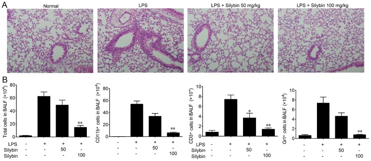
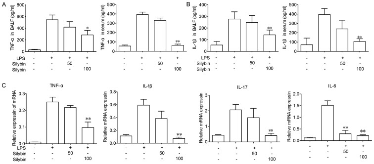
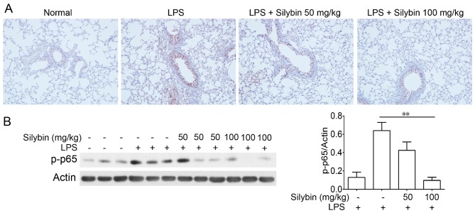
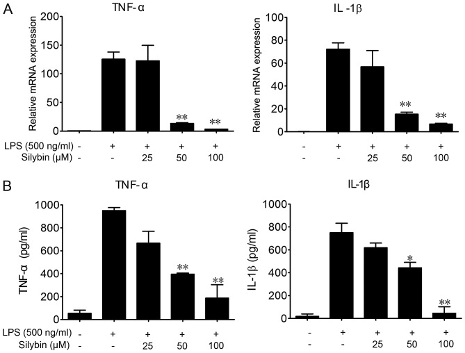
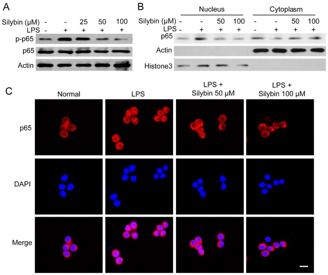
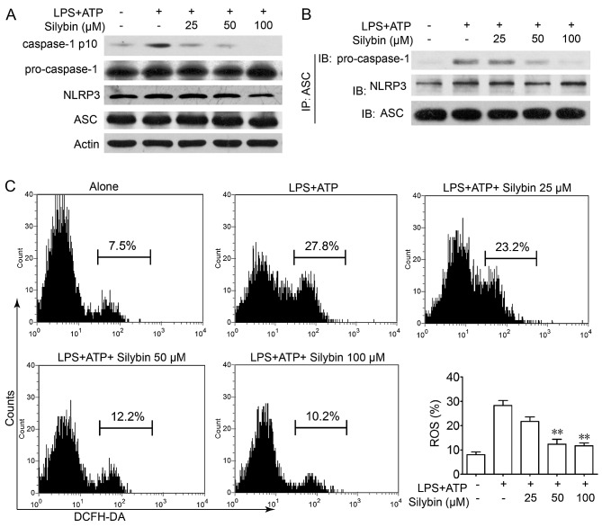
Silybin is one of the main flavonoids produced by milk thistle, which has been used in the treatment of liver diseases. In this study, we examined the protective effects and possible mechanisms of action of silybin in lipopolysaccharide (LPS)‑induced lung injury and inflammation. Pre-treatment of mice with silybin significantly inhibited LPS-induced airway inflammatory cell recruitment, including macrophages, T cells and neutrophils. The production of cytokines, such as interleukin-1β (IL-1β) and tumor necrosis factor-α (TNF-α) in bronchoalveolar fluid and serum was also decreased following treatment with silybin. Elevated cytokine mRNA levels induced by LPS in lung tissue were all suppressed by silybin and lung histological alterations were also improved. In addition, experiments using cells indicated that silybin significantly decreased the mRNA levels and secretion of IL-1β and TNF-α in THP-1 cells. Moreover, the mechanisms responsible for these effects were attributed to the inhibitory effect of silybin on nuclear factor-κB (NF-κB) signaling and NLR family pyrin domain containing 3 (NLRP3) inflammasome activation. The data form our study thus support the utility of silybin as a potential medicine for the treatment of acute lung injury‑associated inflammation and pathological changes. Silybin exerts protective effects against lung injury by regulating NF-κB signaling and the NLRP3 inflammasome activation.
Figures
Similar articles
-
Hydrogen-Rich Saline Attenuated Subarachnoid Hemorrhage-Induced Early Brain Injury in Rats by Suppressing Inflammatory Response: Possible Involvement of NF-κB Pathway and NLRP3 Inflammasome.Mol Neurobiol. 2016 Jul;53(5):3462-3476. doi: 10.1007/s12035-015-9242-y. Epub 2015 Jun 20. Mol Neurobiol. 2016. PMID: 26091790
-
CORM-2 inhibits TXNIP/NLRP3 inflammasome pathway in LPS-induced acute lung injury.Inflamm Res. 2016 Nov;65(11):905-915. doi: 10.1007/s00011-016-0973-7. Epub 2016 Jul 13. Inflamm Res. 2016. PMID: 27412237
-
Apelin-13 Administration Protects Against LPS-Induced Acute Lung Injury by Inhibiting NF-κB Pathway and NLRP3 Inflammasome Activation.Cell Physiol Biochem. 2018;49(5):1918-1932. doi: 10.1159/000493653. Epub 2018 Sep 20. Cell Physiol Biochem. 2018. PMID: 30235451
-
NLRP inflammasome as a key role player in the pathogenesis of environmental toxicants.Life Sci. 2019 Aug 15;231:116585. doi: 10.1016/j.lfs.2019.116585. Epub 2019 Jun 18. Life Sci. 2019. PMID: 31226415 Review.
-
Silybin and silymarin--new and emerging applications in medicine.Curr Med Chem. 2007;14(3):315-38. doi: 10.2174/092986707779941159. Curr Med Chem. 2007. PMID: 17305535 Review.
Cited by
-
Flavonoids as potential phytotherapeutics to combat cytokine storm in SARS-CoV-2.Phytother Res. 2021 Aug;35(8):4258-4283. doi: 10.1002/ptr.7092. Epub 2021 Mar 30. Phytother Res. 2021. PMID: 33786876 Free PMC article. Review.
-
The effect of rutin on pulmonary contusion induced by blunt trauma in rats: Biochemical and histopathological evaluation.Turk Gogus Kalp Damar Cerrahisi Derg. 2022 Jul 29;30(3):431-439. doi: 10.5606/tgkdc.dergisi.2022.21992. eCollection 2022 Jul. Turk Gogus Kalp Damar Cerrahisi Derg. 2022. PMID: 36303692 Free PMC article.
-
Chromolaena odorata flavonoids attenuate experimental nephropathy: Involvement of pro-inflammatory genes downregulation.Toxicol Rep. 2020 Oct 8;7:1421-1427. doi: 10.1016/j.toxrep.2020.10.006. eCollection 2020. Toxicol Rep. 2020. PMID: 33102146 Free PMC article.
-
Silibinin and SARS-CoV-2: Dual Targeting of Host Cytokine Storm and Virus Replication Machinery for Clinical Management of COVID-19 Patients.J Clin Med. 2020 Jun 7;9(6):1770. doi: 10.3390/jcm9061770. J Clin Med. 2020. PMID: 32517353 Free PMC article.
-
MST1 modulates inflammatory responses by targeting the NF-κB/NLRP3 pathway in LPS-induced acute lung injury.Histochem Cell Biol. 2025 May 28;163(1):60. doi: 10.1007/s00418-025-02383-4. Histochem Cell Biol. 2025. PMID: 40434446
References
-
- Global Burden of Disease Study 2013 Collaborators Global, regional, and national incidence, prevalence, and years lived with disability for 301 acute and chronic diseases and injuries in 188 countries, 1990–2013: A systematic analysis for the Global Burden of Disease Study 2013. Lancet. 2015;386:743–800. doi: 10.1016/S0140-6736(15)60692-4. - DOI - PMC - PubMed
MeSH terms
Substances
LinkOut - more resources
Full Text Sources
Other Literature Sources
