. 2017 May;39(5):1242-1254.
doi: 10.3892/ijmm.2017.2938. Epub 2017 Mar 28.

Advanced glycation end products promote the proliferation and migration of primary rat vascular smooth muscle cells via the upregulation of BAG3

Affiliations

Advanced glycation end products promote the proliferation and migration of primary rat vascular smooth muscle cells via the upregulation of BAG3

Cunshu Li et al. Int J Mol Med. 2017 May.

Abstract

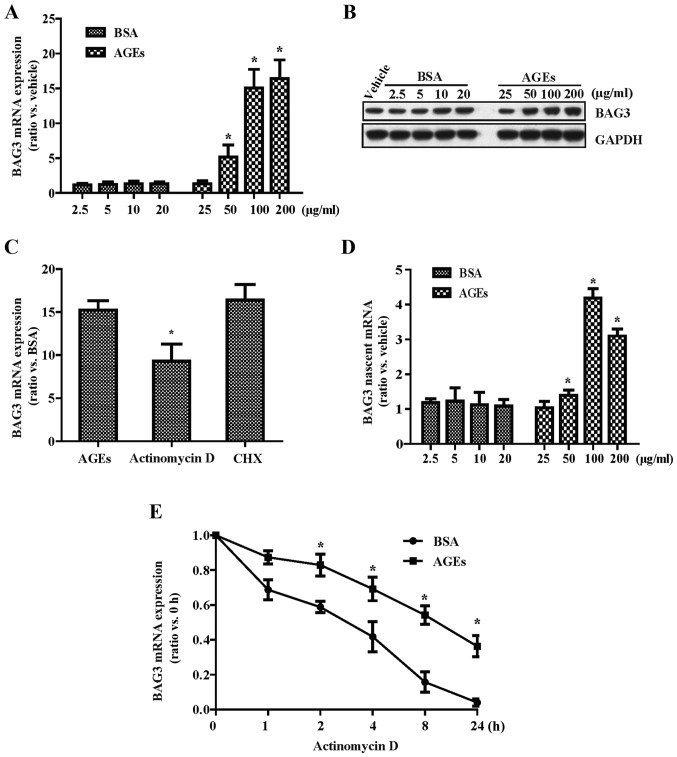
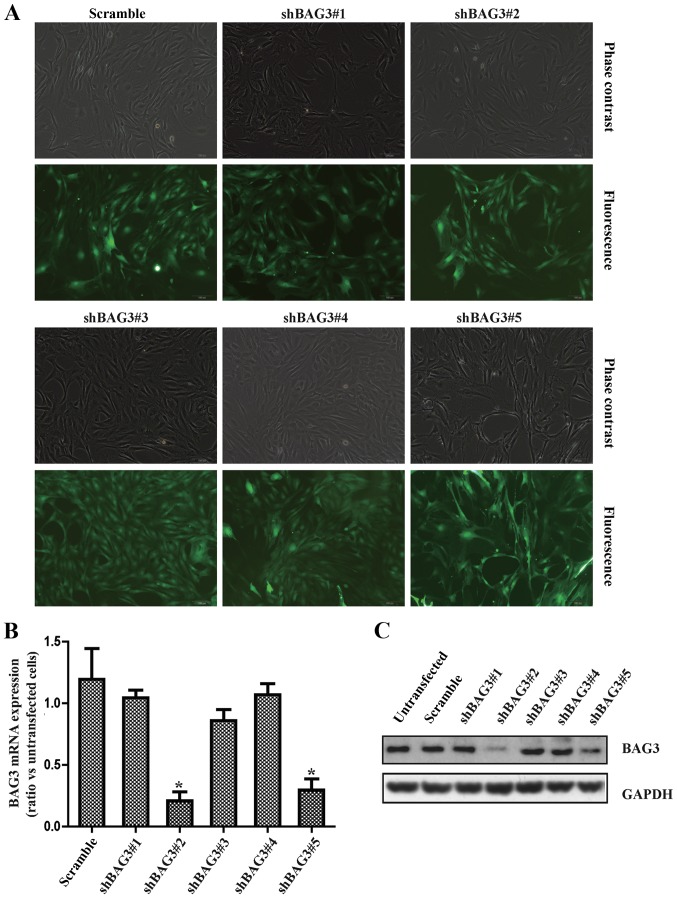
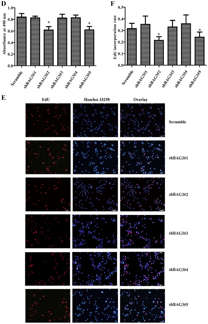
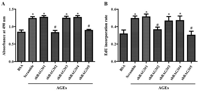
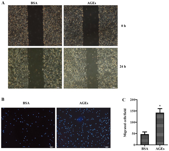
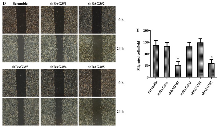
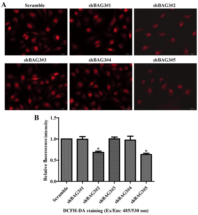
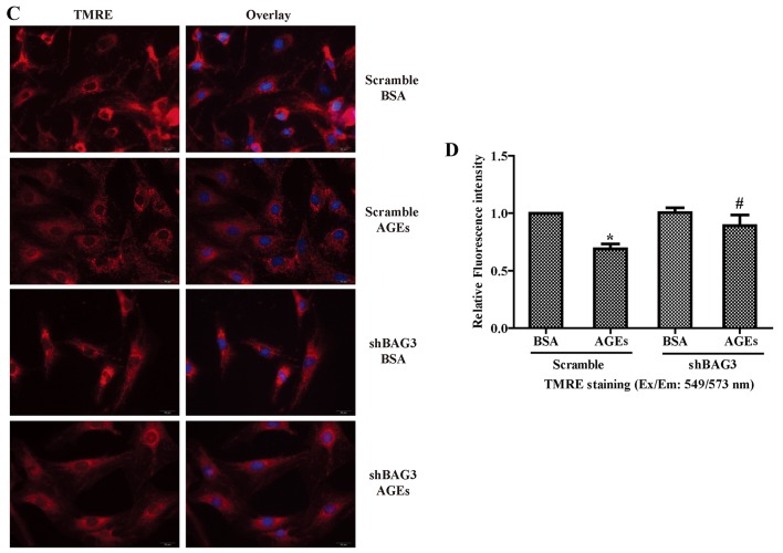
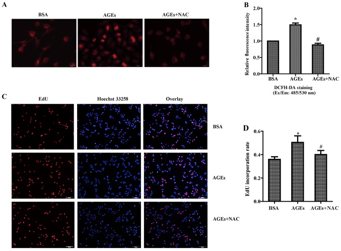
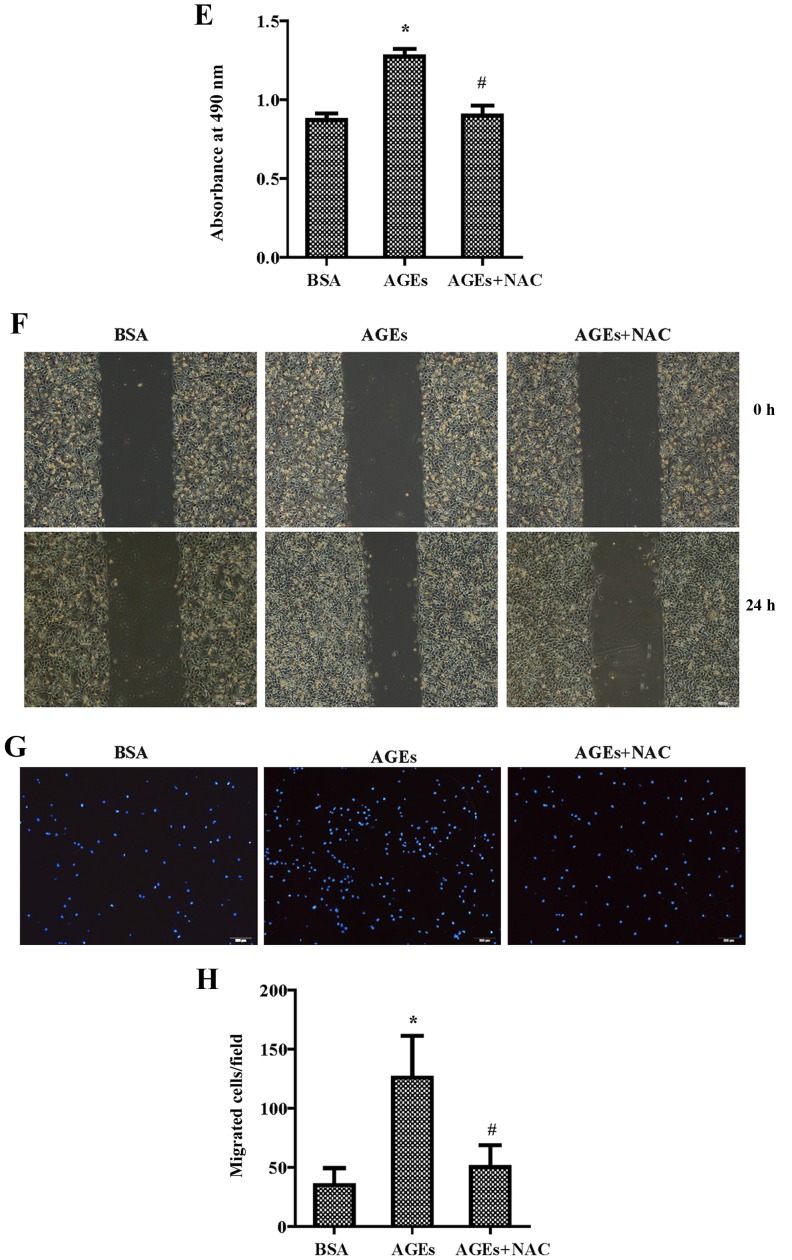
The present study was aimed to investigate the role of reactive oxygen species (ROS) on advanced glycation end product (AGE)-induced proliferation and migration of vascular smooth muscle cells (VSMCs) and whether Bcl-2‑associated athanogene 3 (BAG3) is involved in the process. Primary rat VSMCs were extracted and cultured in vitro. Cell viability was detected by MTT assay and cell proliferation was detected by EdU incorporation assay. Cell migration was detected by wound healing and Transwell assays. BAG3 was detected using qPCR and western blot analysis. Transcriptional and translational inhibitors (actinomycin D and cycloheximide, respectively) were used to study the effect of AGEs on the expression of BAG3 in VSMCs. Lentiviral plasmids containing short hairpin RNA (shRNA) against rat BAG3 or control shRNA were transduced into VSMCs. Cellular ROS were detected by 2',7'-dichlorofluorescein diacetate (DCFH-DA) staining. Mitochondrial membrane potential was detected by tetramethylrhodamine methyl ester (TMRE) staining. AGEs significantly increased the expression of BAG3 in a dose-and time-dependent manner. Furthermore, AGEs mainly increased the expression of BAG3 mRNA by increasing the RNA synthesis rather than inhibiting the RNA translation. BAG3 knockdown reduced the proliferation and migration of VSMCs induced by AGEs. BAG3 knockdown reduced the generation of ROS and sustained the mitochondrial membrane potential of VSMCs. Reduction of ROS production by N-acetylcysteine (NAC), a potent antioxidant, also reduced the proliferation and migration of VSMCs. On the whole, the present study demonstrated for the first time that AGEs could increase ROS production and promote the proliferation and migration of VSMCs by upregulating BAG3 expression. This study indicated that BAG3 should be considered as a potential target for the prevention and/or treatment of vascular complications of diabetes.


Figures

Figure 1
Figure 1
Advanced glycation end products (AGEs) increase the expression of Bcl-2-associated athanogene 3 (BAG3) in cultured primary rat vascular smooth muscle cells (VSMCs). Cells were treated with different concentrations of AGEs (25, 50, 100 and 200 µg/ml) and BSA (2.5, 5, 10 and 20 µg/ml), respectively. The mRNA and protein expression levels of BAG3 were detected by (A) RT-PCR and (B) western blotting, respectively. (C) Actinomycin D (10 µg/ml), a transcriptional inhibitor, and cycloheximide (CHX) (20 µg/ml), a translational inhibitor, were used to study the effect of AGEs on the mRNA expression of BAG3. (D) Click-iT nascent RNA capture kit was used to label and isolate newly synthesized RNA. (E) Cells were incubated with 10 µg/ml actinomycin D and 100 µg/ml AGEs or 10 µg/ml BSA for different times (0, 1, 2, 4, 8 and 24 h), and then the mRNA expression of BAG3 was detected by RT-PCR. The experiments were repeated three times with reproducible results. *p<0.05 compared with the control.
Figure 2
Figure 2
Effects of Bcl-2-associated athanogene 3 (BAG3) on the proliferation of primary rat vascular smooth muscle cells (VSMCs). (A) We generated lentiviral vectors containing shRNAs against BAG3 (shBAG3) to knock down BAG3 expression in VSMCs. The mRNA and protein expression levels of BAG3 were detected by (B) RT-PCR and (C) western blotting, respectively. *p<0.05 compared with the control. (D) Cell viability was determined by 3-(4,5-dimethylthiazol-2-yl)-2,5-diphenyltetrazolium bromide (MTT) assay. Cell proliferation was determined by (E) EdU staining and (F) EdU incorporation was calculated as EdU+ cells/total cells, quantified by ImageJ. The experiments were repeated three times with reproducible results. *p<0.05 compared with the control.
Figure 2
Figure 2
Effects of Bcl-2-associated athanogene 3 (BAG3) on the proliferation of primary rat vascular smooth muscle cells (VSMCs). (A) We generated lentiviral vectors containing shRNAs against BAG3 (shBAG3) to knock down BAG3 expression in VSMCs. The mRNA and protein expression levels of BAG3 were detected by (B) RT-PCR and (C) western blotting, respectively. *p<0.05 compared with the control. (D) Cell viability was determined by 3-(4,5-dimethylthiazol-2-yl)-2,5-diphenyltetrazolium bromide (MTT) assay. Cell proliferation was determined by (E) EdU staining and (F) EdU incorporation was calculated as EdU+ cells/total cells, quantified by ImageJ. The experiments were repeated three times with reproducible results. *p<0.05 compared with the control.
Figure 3
Figure 3
Advanced glycation end products (AGEs) promote the proliferation of primary rat vascular smooth muscle cells (VSMCs) via Bcl-2-associated athanogene 3 (BAG3). VSMCs transfected with shRNAs against BAG3 (shBAG3) were treated with 100 µg/ml AGEs or 10 µg/ml BSA for 24 h. (A) Cell viability was determined by MTT assay. (B) Cell proliferation was determined by EdU staining and EdU incorporation was calculated as EdU+ cells/total cells, quantified by ImageJ. The experiments were repeated three times with reproducible results. *p<0.05 compared with the control; #p<0.05 compared with the scramble + AGEs group.
Figure 4
Figure 4
Advanced glycation end products (AGEs) promote the migration of primary rat vascular smooth muscle cells (VSMCs) via Bcl-2-associated athanogene 3 (BAG3). VSMCs were treated with 100 µg/ml AGEs or 10 µg/ml BSA for 24 h. (A) Cell migration was detected by wound healing assay and (B) Transwell assay. (C) Migrated cells were quantified by ImageJ. Then the migration of VSMCs transfected with shRNAs against BAG3 (shBAG3) was detected by (D) wound healing assay and (E) Transwell assay. The experiments were repeated three times with reproducible results. *p<0.05 compared with the control.
Figure 4
Figure 4
Advanced glycation end products (AGEs) promote the migration of primary rat vascular smooth muscle cells (VSMCs) via Bcl-2-associated athanogene 3 (BAG3). VSMCs were treated with 100 µg/ml AGEs or 10 µg/ml BSA for 24 h. (A) Cell migration was detected by wound healing assay and (B) Transwell assay. (C) Migrated cells were quantified by ImageJ. Then the migration of VSMCs transfected with shRNAs against BAG3 (shBAG3) was detected by (D) wound healing assay and (E) Transwell assay. The experiments were repeated three times with reproducible results. *p<0.05 compared with the control.
Figure 5
Figure 5
Effect of Bcl-2-associated athanogene 3 (BAG3) on the oxidative stress and mitochondrial membrane potential of vascular smooth muscle cells (VSMCs). (A) The cells were labeled with 2′,7′-dichlorofluorescein diacetate (DCFH-DA) (Ex/Em, 485/530 nm) to detect reactive oxygen species (ROS) and analyzed with a fluorescence microscope (scale bar, 20 µm). (B) The fluorescence intensity of DCFH-DA staining was quantified using ImageJ, normalized by the scramble group, *p<0.05 vs. the scramble group. (C) The mitochondria were labeled with tetramethylrhodamine methyl ester (TMRE) (Ex/Em, 549/573 nm) to detect mitochondrial membrane potential and analyzed with a fluorescence micro-scope (scale bar, 20 µm). (D) The fluorescence intensity of TMRE staining was quantified using ImageJ, normalized by the scramble group, *p<0.05 vs. the scramble group; #p<0.05 vs. the scramble + advanced glycation end product (AGE) group.
Figure 5
Figure 5
Effect of Bcl-2-associated athanogene 3 (BAG3) on the oxidative stress and mitochondrial membrane potential of vascular smooth muscle cells (VSMCs). (A) The cells were labeled with 2′,7′-dichlorofluorescein diacetate (DCFH-DA) (Ex/Em, 485/530 nm) to detect reactive oxygen species (ROS) and analyzed with a fluorescence microscope (scale bar, 20 µm). (B) The fluorescence intensity of DCFH-DA staining was quantified using ImageJ, normalized by the scramble group, *p<0.05 vs. the scramble group. (C) The mitochondria were labeled with tetramethylrhodamine methyl ester (TMRE) (Ex/Em, 549/573 nm) to detect mitochondrial membrane potential and analyzed with a fluorescence micro-scope (scale bar, 20 µm). (D) The fluorescence intensity of TMRE staining was quantified using ImageJ, normalized by the scramble group, *p<0.05 vs. the scramble group; #p<0.05 vs. the scramble + advanced glycation end product (AGE) group.
Figure 6
Figure 6
Advanced glycation end products (AGEs) promote the proliferation and migration of vascular smooth muscle cells (VSMCs) via oxidative stress. Cells were incubated with 100 µg/ml N-acetylcysteine (NAC) and 100 µg/ml AGEs or 10 µg/ml BSA for 24 h. (A) Reactive oxygen species (ROS) were detected by DCHF-DA assay. (B) The fluorescence intensity of 2′,7′-dichlorofluorescein diacetate (DCFH-DA) staining was quantified using ImageJ, normalized by the scramble group. (C and D) Cell proliferation was detected by EdU assay. *p<0.05 and #p<0.05 compared with the BSA group. (E) Cell viability was detected by 3-(4,5-dimethylthiazol-2-yl)-2,5-diphenyltetrazolium bromide (MTT) assay. Cell migration was detected by (F) wound healing assay and (G and H) Transwell assay. The experiments were repeated three times with reproducible results. *p<0.05 compared with the BSA control.
Figure 6
Figure 6
Advanced glycation end products (AGEs) promote the proliferation and migration of vascular smooth muscle cells (VSMCs) via oxidative stress. Cells were incubated with 100 µg/ml N-acetylcysteine (NAC) and 100 µg/ml AGEs or 10 µg/ml BSA for 24 h. (A) Reactive oxygen species (ROS) were detected by DCHF-DA assay. (B) The fluorescence intensity of 2′,7′-dichlorofluorescein diacetate (DCFH-DA) staining was quantified using ImageJ, normalized by the scramble group. (C and D) Cell proliferation was detected by EdU assay. *p<0.05 and #p<0.05 compared with the BSA group. (E) Cell viability was detected by 3-(4,5-dimethylthiazol-2-yl)-2,5-diphenyltetrazolium bromide (MTT) assay. Cell migration was detected by (F) wound healing assay and (G and H) Transwell assay. The experiments were repeated three times with reproducible results. *p<0.05 compared with the BSA control.

Similar articles

Cited by

References

    1. Kudryavtseva O, Aalkjaer C, Matchkov VV. Vascular smooth muscle cell phenotype is defined by Ca2+-dependent transcription factors. FEBS J. 2013;280:5488–5499. doi: 10.1111/febs.12414. - DOI - PubMed
    1. Tang DD, Anfinogenova Y. Physiologic properties and regulation of the actin cytoskeleton in vascular smooth muscle. J Cardiovasc Pharmacol Ther. 2008;13:130–140. doi: 10.1177/1074248407313737. - DOI - PMC - PubMed
    1. Owens GK, Kumar MS, Wamhoff BR. Molecular regulation of vascular smooth muscle cell differentiation in development and disease. Physiol Rev. 2004;84:767–801. doi: 10.1152/physrev.00041.2003. - DOI - PubMed
    1. Bochaton-Piallat ML, Gabbiani G. Modulation of smooth muscle cell proliferation and migration: Role of smooth muscle cell heterogeneity. Handb Exp Pharmacol. 2005;170:645–663. doi: 10.1007/3-540-27661-0_24. - DOI - PubMed
    1. Orr AW, Hastings NE, Blackman BR, Wamhoff BR. Complex regulation and function of the inflammatory smooth muscle cell phenotype in atherosclerosis. J Vasc Res. 2010;47:168–180. doi: 10.1159/000250095. - DOI - PMC - PubMed

MeSH terms

Substances