

Comment
doi: 10.7554/eLife.25812.
Transcranial electric stimulation seen from within the brain
Affiliations
- PMID: 28350293
- PMCID: PMC5370182
- DOI: 10.7554/eLife.25812
Item in Clipboard
Comment
Transcranial electric stimulation seen from within the brain
Elife.
.
Abstract
Computer models can make transcranial electric stimulation a better tool for research and therapy.
Keywords: computational current-flow model; human; intracranial recordings; neuroscience; transcranial electric stimulation.
Conflict of interest statement
AVP: An inventor on patents and patent applications and recipient of research and travel support as well as patent royalties from Rogue Research; research and travel support, consulting fees, as well as equipment loan from Tal Medical; patent application support from Magstim; and equipment loans from MagVenture, all related to technology for transcranial magnetic stimulation.
Figures
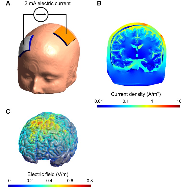
(A) Electrodes (white and orange rectangles) are attached to the scalp and electric current is applied; the model of the head shown here is derived from a structural MRI scan. (B) Simulation showing the electric current per unit area (current density) in a section of the brain during transcranial stimulation: this image shows the scalp (outermost layer), skull, cerebrospinal fluid, gray matter and white matter. The highest current density values in the brain (blue) are 100-fold lower than those in the scalp (red). The high resistance of the skull means that the majority of the current is shunted in the scalp. The cerebrospinal fluid is highly conductive and this takes current away from the brain too. (C) Simulation showing the electric field on the surface of the brain. For this configuration, the electric field is strongest between the two electrodes. The model was created and visualized with the free SimNIBS software package (http://simnibs.de ; Windhoff et al., 2013).
Comment on
-
Measurements and models of electric fields in the in vivo human brain during transcranial electric stimulation.Elife. 2017 Feb 7;6:e18834. doi: 10.7554/eLife.18834. Elife. 2017. PMID: 28169833 Free PMC article.
References
-
- Koessler L, Colnat-Coulbois S, Cecchin T, Hofmanis J, Dmochowski JP, Norcia AM, Maillard LG. In-vivo measurements of human brain tissue conductivity using focal electrical current injection through intracerebral multicontact electrodes. Human Brain Mapping. 2017;38:974–986. doi: 10.1002/hbm.23431. - DOI - PMC - PubMed
Publication types
MeSH terms
Grants and funding
LinkOut - more resources
Full Text Sources
Other Literature Sources
