

A second case of pericardial mesothelioma mimicking systemic lupus erythematosus in the literature in over 30 years: a case report
- PMID: 28351431
- PMCID: PMC5370430
- DOI: 10.1186/s13256-017-1237-z
A second case of pericardial mesothelioma mimicking systemic lupus erythematosus in the literature in over 30 years: a case report
Abstract
Background: Mesothelioma is a rare neoplasm which commonly develops in the pleura of people exposed to asbestos. Pericardial mesothelioma accounts for only 0.7 % of all malignant mesotheliomas and it usually presents with pericardial effusion, mimicking serositis. To date, there are approximately 200 cases of pericardial mesothelioma described in the medical literature, and little knowledge exists about the systemic manifestations of this pathology. The first and only described case of pericardial mesothelioma with autoimmune features dates back to 1984 and, in our case report, we describe the second.
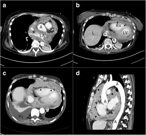
Case presentation: We report a case of a 45-year-old white woman whose pericardial mesothelioma was initially misdiagnosed as pericardial involvement of an autoimmune disease (systemic lupus erythematosus). After several relapses of pericardial effusion, a computed tomography scan and a biopsy with histological analysis were performed revealing neoplastic growth.
Conclusions: We describe a rare case of pericardial mesothelioma in a patient with a clinical presentation compatible with lupus serositis. Clinicians should consider malignant mesothelioma in the differential diagnosis of pericardial effusion, especially when it is recurrent and not clearly explained by other causes. Cytological samples should always be obtained and, if imaging tools are suggestive for solid processes, histological confirmation is mandatory.
Keywords: Asbestos; Case report; Pericardial effusion; Pericardial mesothelioma; Pericarditis; SLE.
Figures
References
-
- Kaul TK, Fields BL, Kahn DR. Primary malignant pericardial mesothelioma: a case report and review. J Cardiovasc Surg (Torino) 1994;35(Suppl 3):261–7. - PubMed
-
- National Cancer Institute. Physician Data Query (PDQ). Malignant Mesothelioma: Treatment. 2014. http://www.cancer.gov/cancertopics/pdq/treatment/malignantmesothelioma/H.... Accessed 4 Mar 2015.
Publication types
MeSH terms
LinkOut - more resources
Full Text Sources
Other Literature Sources
Medical
