. 2017 Apr 1;144(7):1235-1241.
doi: 10.1242/dev.138487.

Cooperative activation of cardiac transcription through myocardin bridging of paired MEF2 sites

Affiliations

Cooperative activation of cardiac transcription through myocardin bridging of paired MEF2 sites

Courtney M Anderson et al. Development. .

Abstract

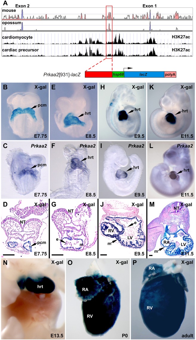
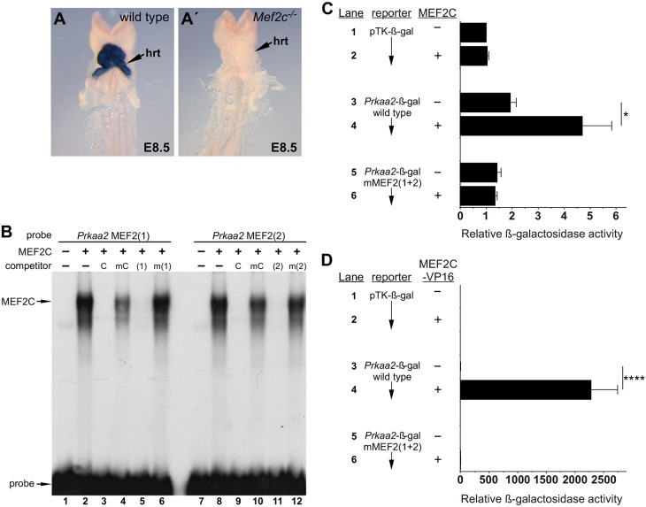
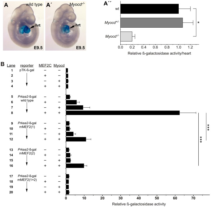
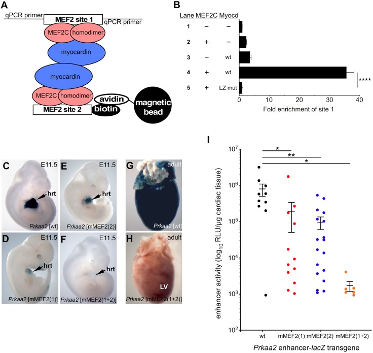
Enhancers frequently contain multiple binding sites for the same transcription factor. These homotypic binding sites often exhibit synergy, whereby the transcriptional output from two or more binding sites is greater than the sum of the contributions of the individual binding sites alone. Although this phenomenon is frequently observed, the mechanistic basis for homotypic binding site synergy is poorly understood. Here, we identify a bona fide cardiac-specific Prkaa2 enhancer that is synergistically activated by homotypic MEF2 binding sites. We show that two MEF2 sites in the enhancer function cooperatively due to bridging of the MEF2C-bound sites by the SAP domain-containing co-activator protein myocardin, and we show that paired sites buffer the enhancer from integration site-dependent effects on transcription in vivo Paired MEF2 sites are prevalent in cardiac enhancers, suggesting that this might be a common mechanism underlying synergy in the control of cardiac gene expression in vivo.

Keywords: AMPK; MEF2; Mouse; Myocardin; Prkaa2; Transcription.


Conflict of interest statement

Competing interests

The authors declare no competing or financial interests.

Figures

Fig. 1.
Fig. 1.
Identification of a cardiac-restricted Prkaa2 enhancer. (A) Human:mouse conservation (top box), human:opossum conservation (second box), H3K27 acetylation (Wamstad et al., 2012) in cardiomyocytes (third box), and H3K27 acetylation (Wamstad et al., 2012) in cardiac precursors (fourth box) in the Prkaa2 locus. The red-boxed peak highlights the 931 bp Prkaa2 enhancer. Red-filled peaks, noncoding sequences conserved between 75% and 100%; blue-filled peaks, coding sequences conserved between 75% and 100%; white peaks, conservation between 50% and 75%. (B-P) Whole-mount (B,E,H,K,N-P) and sections (D,G,J,M) of X-gal-stained Prkaa2[931]-lacZ transgenic embryos and postnatal hearts. Prkaa2[931] enhancer activity recapitulates the expression pattern of endogenous Prkaa2 detected by whole-mount in situ hybridization (C,F,I,L) from E7.75 through E11.5. e, endocardium; hrt, heart; LV, left ventricle; m, myocardium; NT, neural tube; pcm, precardiac mesoderm; RA, right atrium, RV, right ventricle. Scale bars: 100 µm.
Fig. 2.
Fig. 2.
The Prkaa2 cardiac enhancer requires MEF2C for activity. (A,A′) Prkaa2[931]-lacZ transgenic mice were crossed into Mef2c+/+ (wild type, A) and Mef2c−/− (A′) backgrounds, and enhancer activity was examined by X-gal staining at E8.5; hrt, heart. (B) Prkaa2 MEF2 site 1 (lanes 1-6) or Prkaa2 MEF2 site 2 (lanes 7-12) was used in EMSA with reticulocyte lysate (−, lanes 1 and 7) or with recombinant MEF2C (+, lanes 2-6 and 8-12). MEF2C efficiently bound to both of the Prkaa2 MEF2 sites (lanes 2 and 8). Binding to each site was competed by an excess of unlabeled control MEF2 site from the myogenin gene (C) or by unlabeled self probe (1) or (2), respectively, but not by mutant versions of the unlabeled competitors [mC, m(1), m(2)]. (C,D) P19CL6 cells were co-transfected with parental pTK-β-gal reporter (lanes 1, 2), wild-type Prkaa2-β-gal (lanes 3, 4), or a mutant version of the Prkaa2-β-gal reporter with both MEF2 sites disrupted (lanes 5, 6). Co-transfection of an expression plasmid for MEF2C (C) or MEF2C-VP16 (D) is indicated with a plus symbol; a minus symbol indicates that an equivalent amount of the parental expression plasmid was added. Results are reported as mean+s.e.m.; n=14 (C) or n=8 (D) independent biological replicates. Note the difference in the values on the x-axes for transactivation by MEF2C (C) versus MEF2C-VP16 (D). *P<0.05, ****P<0.0001, two-way ANOVA with Bonferroni's post-hoc test.
Fig. 3.
Fig. 3.
Myocardin-935 regulates the Prkaa2 cardiac enhancer. (A-A″) Prkaa2[931]-lacZ transgenic mice were crossed into Myocd+/+ (wild type, A), Myocd+/− and Myocd−/− (A′) genetic backgrounds and enhancer activity was examined qualitatively by X-gal staining (A,A′) or quantitatively by chemiluminescence β-galactosidase assay (A″) at E9.5. hrt, heart. Data in A″ are expressed as the mean β-galactosidase activity (+s.e.m.) with the mean activity on the Myocd+/+ background normalized to a value of 1. n=4 (Myocd+/+), n=5 (Myocd+/−) and n=3 (Myocd−/−) independent biological replicates. *P<0.05, one-way ANOVA with Bonferroni's post-hoc test. (B) P19CL6 cells were co-transfected with the parental pTK-β-gal reporter (lanes 1-4), wild-type Prkaa2-β-gal (lanes 5-8), or mutant versions of the Prkaa2-β-gal reporter containing disruptions in MEF2 site 1 (lanes 9-12), site 2 (lanes 13-16) or both MEF2 sites (lanes 17-20). Co-transfections with expression plasmids for MEF2C and myocardin-935 are indicated with a plus symbol; a minus symbol indicates that an equivalent amount of the parental expression plasmid was added. Results shown are the mean fold activation over the pTK-β-gal reporter in the presence of parental expression vectors+ s.e.m.; n=9 independent biological replicates. ***P<0.001, two-way ANOVA with Bonferroni's post-hoc test.
Fig. 4.
Fig. 4.
Bridging of MEF2C-bound MEF2 sites by myocardin-935. (A) Schematic of the in vitro pull-down assay. (B) qPCR detection of co-precipitated MEF2 site 1 after incubation with biotinylated MEF2 site 2 in the presence of reticulocyte lysate control (lane 1), MEF2C alone (lane 2), myocardin-935 (wt) alone (lane 3), MEF2C plus myocardin-935 (lane 4), or MEF2C plus a leucine zipper mutant form of myocardin-935 (LZ mut) (lane 5). Results are presented as mean fold enrichment over the reticulocyte lysate control+s.d. ****P<0.0001, two-way ANOVA with Bonferroni's post-hoc test. (C-H) The MEF2 sites in the Prkaa2 cardiac enhancer act synergistically in vivo. The wild-type Prkaa2[931]-lacZ transgenic reporter (C) and versions containing mutations in MEF2 site 1 (D), site 2 (E) or both MEF2 sites (F) were used to generate multiple independent transgenic lines or F0 embryos, and representative E11.5 embryos are shown. hrt, heart; LV, left ventricle. (G,H) The presence of both MEF2 sites is required for Prkaa2 enhancer activity in the adult heart. (I) Quantitation of β-galactosidase activity in E11.5 hearts from each of the Prkaa2[931]-lacZ transgenic reporter constructs shown in C-F. Each point on the graph represents a single embryonic heart from an independently generated transgenic embryo. Data are expressed as mean RLU/µg of excised heart tissue+s.e.m. Note the log scale on the y-axis. *P<0.05, **P<0.01, two-way ANOVA with Bonferroni's post-hoc test.

References

    1. Anderson J. P., Dodou E., Heidt A. B., De Val S. J., Jaehnig E. J., Greene S. B., Olson E. N. and Black B. L. (2004). HRC is a direct transcriptional target of MEF2 during cardiac, skeletal, and arterial smooth muscle development in vivo. Mol. Cell. Biol. 24, 3757-3768. 10.1128/MCB.24.9.3757-3768.2004 - DOI - PMC - PubMed
    1. Black B. L. and Cripps R. M. (2010). Myocyte enhancer factor 2 transcription factors in heart development and disease. In Heart Development and Regeneration (ed. Rosenthal N. and Harvey R. P.), pp. 673-699. Oxford: Academic Press.
    1. Black B. L. and Olson E. N. (1998). Transcriptional control of muscle development by myocyte enhancer factor-2 (MEF2) proteins. Annu. Rev. Cell Dev. Biol. 14, 167-196. 10.1146/annurev.cellbio.14.1.167 - DOI - PubMed
    1. Black B. L., Martin J. F. and Olson E. N. (1995). The mouse MRF4 promoter is trans-activated directly and indirectly by muscle-specific transcription factors. J. Biol. Chem. 270, 2889-2892. 10.1074/jbc.270.13.7055 - DOI - PubMed
    1. Black B. L., Ligon K. L., Zhang Y. and Olson E. N. (1996). Cooperative transcriptional activation by the neurogenic basic helix-loop-helix protein MASH1 and members of the myocyte enhancer factor-2 (MEF2) family. J. Biol. Chem. 271, 26659-26663. 10.1074/jbc.271.37.22499 - DOI - PubMed

Publication types

MeSH terms