. 2017 Apr 3;214(4):1111-1128.
doi: 10.1084/jem.20161382. Epub 2017 Mar 28.

Eros is a novel transmembrane protein that controls the phagocyte respiratory burst and is essential for innate immunity

Affiliations

Eros is a novel transmembrane protein that controls the phagocyte respiratory burst and is essential for innate immunity

David C Thomas et al. J Exp Med. .

Abstract

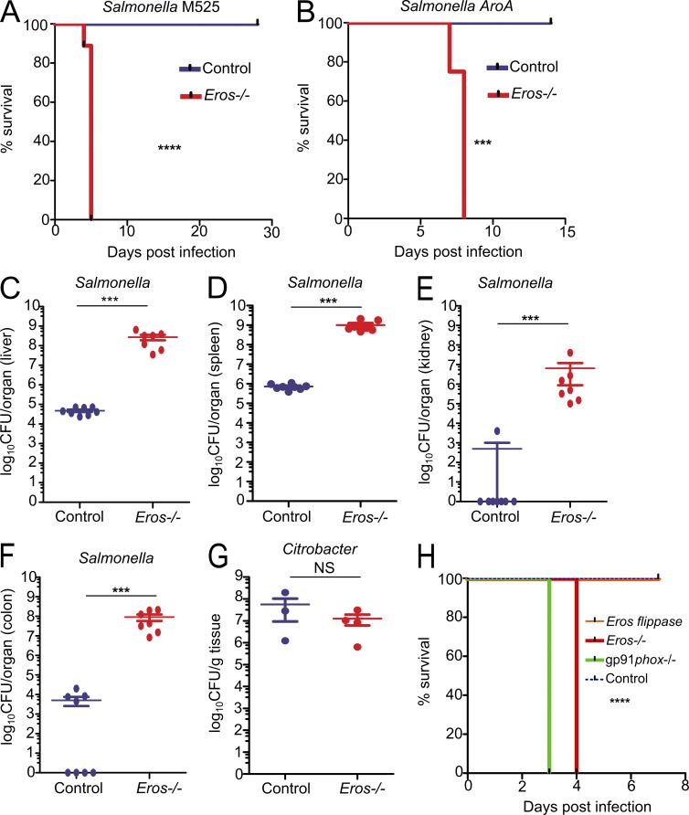
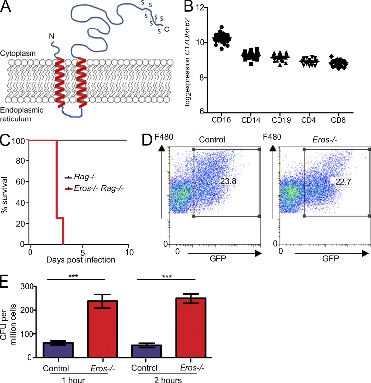
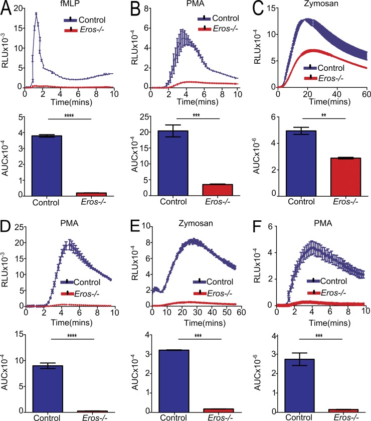
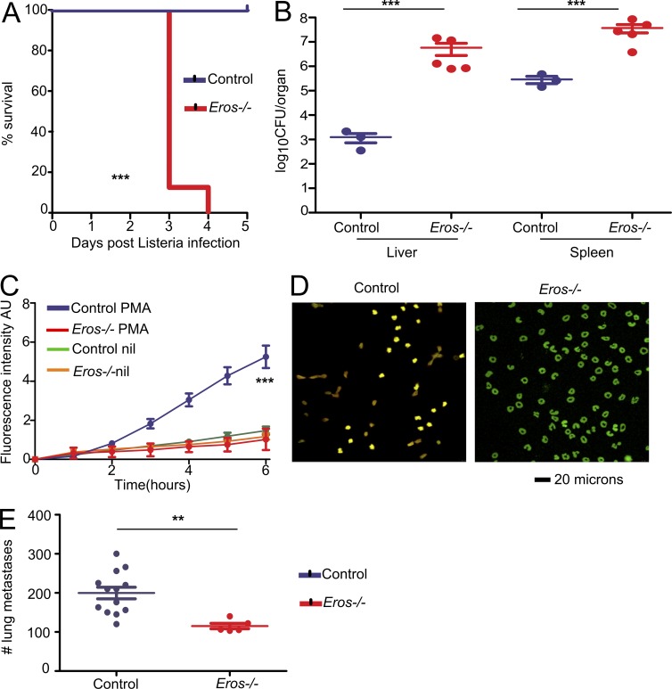
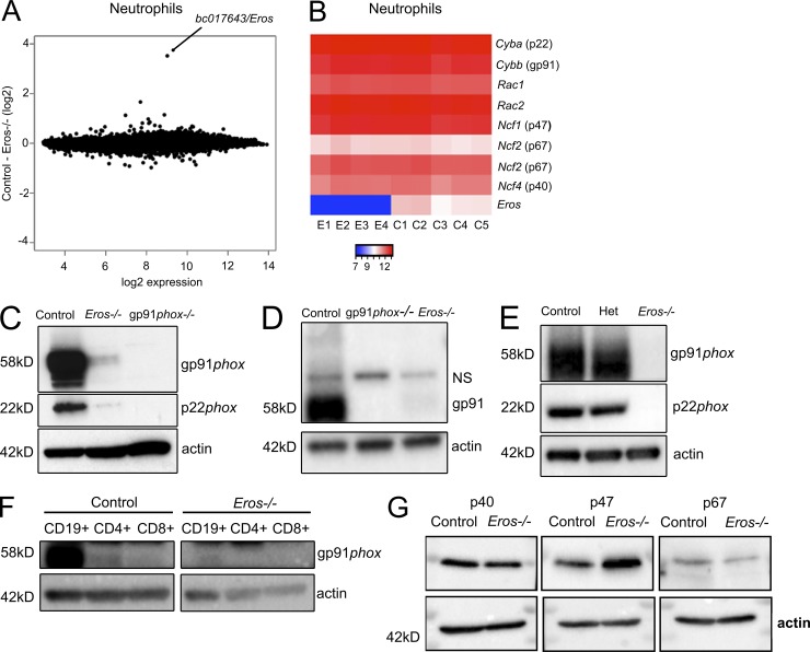
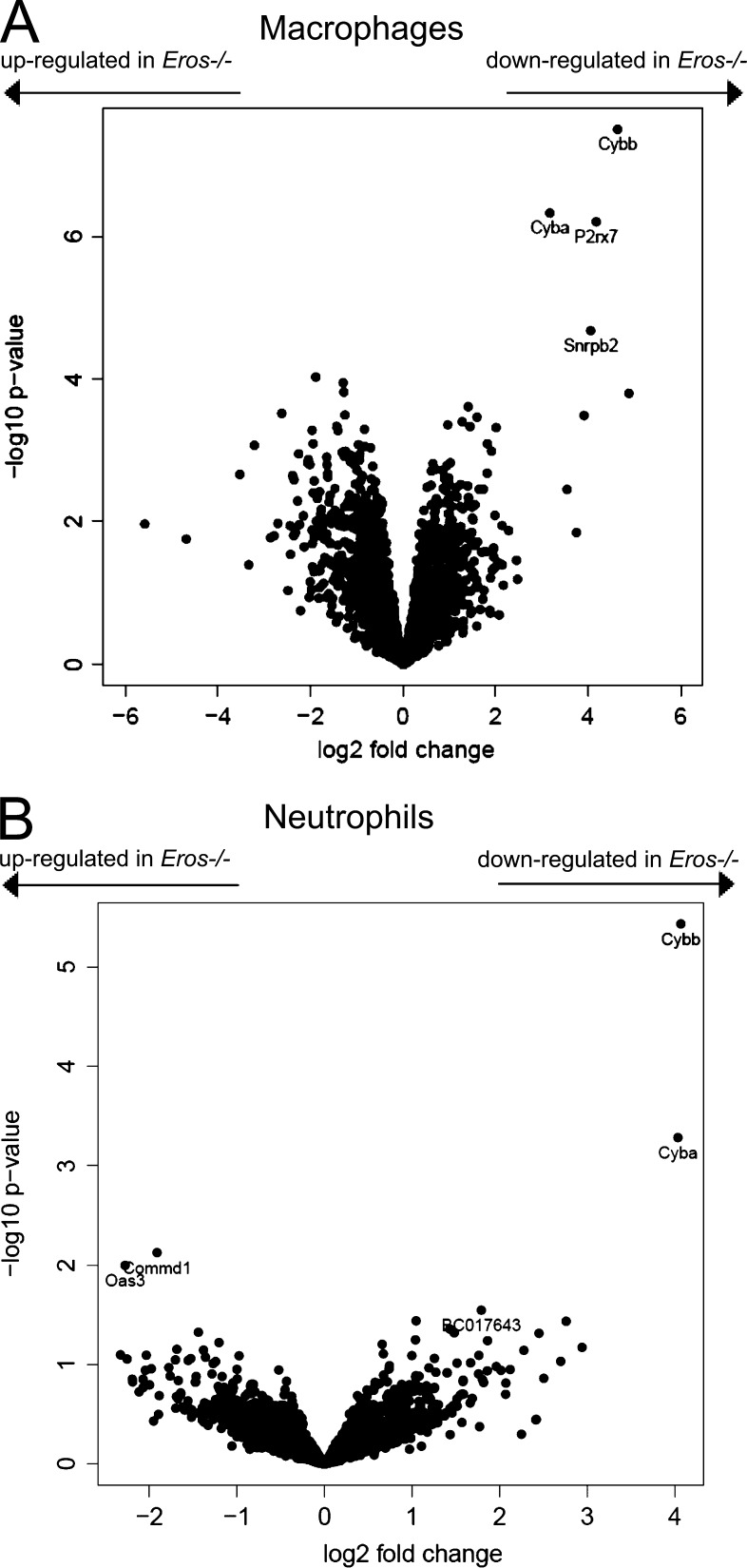
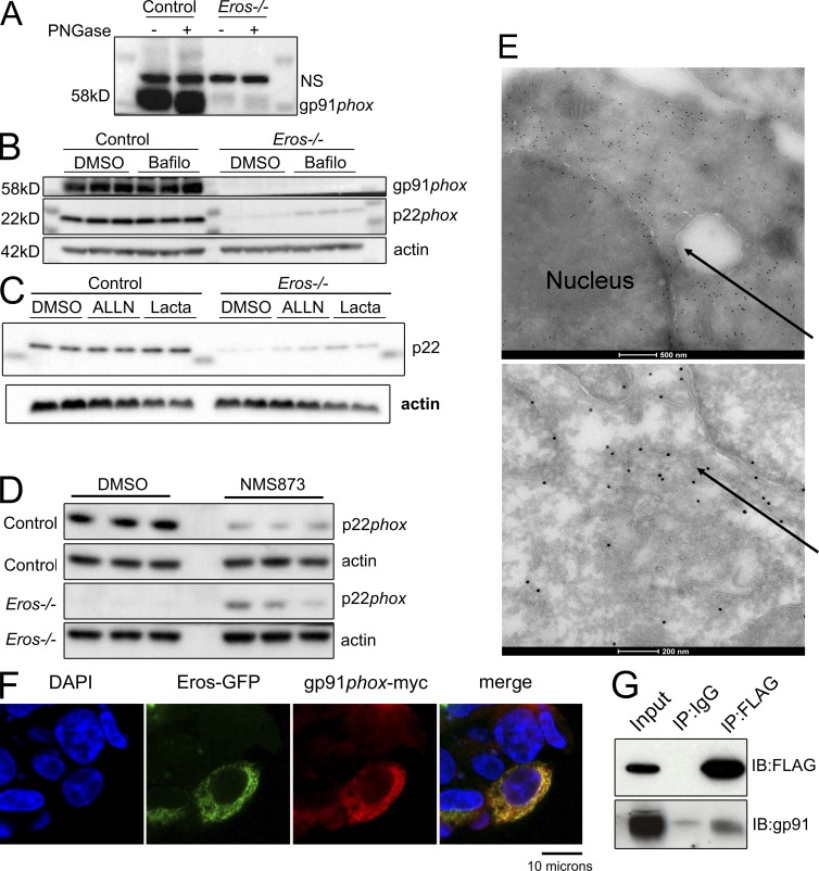
The phagocyte respiratory burst is crucial for innate immunity. The transfer of electrons to oxygen is mediated by a membrane-bound heterodimer, comprising gp91phox and p22phox subunits. Deficiency of either subunit leads to severe immunodeficiency. We describe Eros (essential for reactive oxygen species), a protein encoded by the previously undefined mouse gene bc017643, and show that it is essential for host defense via the phagocyte NAPDH oxidase. Eros is required for expression of the NADPH oxidase components, gp91phox and p22phox Consequently, Eros-deficient mice quickly succumb to infection. Eros also contributes to the formation of neutrophil extracellular traps (NETS) and impacts on the immune response to melanoma metastases. Eros is an ortholog of the plant protein Ycf4, which is necessary for expression of proteins of the photosynthetic photosystem 1 complex, itself also an NADPH oxio-reductase. We thus describe the key role of the previously uncharacterized protein Eros in host defense.


Figures

Figure 1.
Figure 1.
Eros is essential for host defense against S. Typhimurium. (A and B) Survival of Eros−/− mice after i.v. challenge with S. Typhimurium M525 (A) or after oral challenge with S. Typhimurium ΔaroA (B). Data in A show eight mice per group and are representative of greater than five independent experiments. Data in B show eight mice per group and are representative of two independent experiments. (C–F) Bacterial burden expressed as CFU per organ in liver (C), spleen (D), kidney (E), and colon (F) at day 4 after infection with S. Typhimurium M525. Eight mice were used in each group. Results are representative of greater than five independent experiments (G) C. rodentium burden in colon after oral challenge. Eight mice were used per group and data are representative of two independent experiments. (H) Survival of control (eight mice), Eros-deficient (five mice), gp91phox-deficient (four mice), and Eros-sufficient flippase mice (repaired Eros allele; eight mice) after infection with 5 × 10⁵ CFU of S. Typhimurium M525. Data are representative of two independent experiments. The P-value shown in H denotes the significant difference in survival between Eros-flippase and Eros-deficient mice. Error bars represent the SEM. ***, P < 0.001; ****, P < 0.0001. Data in A, B, and H were analyzed by Log-Rank test and data in C–G were analyzed by Mann-Whitney test.
Figure 2.
Figure 2.
Eros is a transmembrane protein, and Eros-deficient macrophages fail to kill S. Typhimurium. (A) Schematic diagram of Eros structure based on prediction algorithms described in text. Serines at the C terminus are denoted by S. (B) Expression of Eros ortholog C17ORF62 in peripheral blood cells of healthy volunteers by probe level expression on microarray. Each dot represents one individual. (C) Survival of Eros−/− Rag−/− double-deficient mice and Rag−/− mice (eight mice per group) after i.v. infection with 106 CFU of S. Typhimurium M525. Results are representative of three independent experiments. (D) Flow cytometric analysis of uptake of opsonized spi2-inducible GFP expressing S. Typhimurium by F480+ peritoneal macrophages. Results are representative of three independent experiments. (E) Killing of M525 S. Typhimurium by control or Eros−/− peritoneal macrophages in a gentamicin protection assay. Macrophages from three independent mice were used in each group, and data are representative of three independent experiments. Error bars represent the SEM. ***, P < 0.001. Data in A were analyzed by log-rank test and data in E were analyzed by unpaired Student’s t test.
Figure 3.
Figure 3.
Eros−/− neutrophils and macrophages have a severely impaired phagocyte respiratory burst in vitro. (A–C) ROS production (three technical replicates, each control and Eros−/− sample is pooled from two mice) measured in relative light units (RLU) by purified BM neutrophils from control or Eros−/− mice in response to (A) fMLP, (B) PMA, or (C) zymosan. Data are representative of at least three independent experiments. (D and E) ROS production (replicates from BM derived from three individual mice, measured in RLU) by purified BM-derived macrophages from control or Eros−/− mice in response to PMA (D) and zymosan (E). (F) ROS production by peritoneal cells from Rag−/− (three mice) or Eros−/− Rag−/− mice (three mice) in response to PMA. Data are representative of at least three independent experiments. Area under the curve (AUC) was calculated for each sample. Error bars represent the SEM. **, P < 0.01; ***, P < 0.001; ****, P < 0.0001, analyzed by unpaired Student’s t test.
Figure 4.
Figure 4.
Eros−/− mice are susceptible to L. monocytogenes infection and fail to form NETS but are resistant to melanoma metastasis. (A) Survival of control (three mice) and Eros−/− (five mice) mice after i.v. injection with L. monocytogenes. (B) Bacterial burden in liver and spleen of control and Eros−/− mice 3 d after infection with L. monocytogenes. Data in A and B are representative of three independent experiments. (C) Neutrophil extracellular trap formation in response to PMA by control and Eros−/− mice. NETosis is measured by absorbance of Sytox Green. (D) Representative staining of control and Eros−/− neutrophils with sytox green and anticitrullinated histone 3 antibody. (E) Number of lung metastases in control or Eros−/− mice 10 d after i.v. injection of B16-F10 melanoma cells. Error bars represent the SEM. **, P < 0.01; ***, P < 0.001. Data in A were analyzed by Log-rank test. Data in B and E were analyzed by Mann Whitney test. Data in C was analyzed by Tukey’s test. Data are representative of at least three independent experiments.
Figure 5.
Figure 5.
Eros−/− mice have severely reduced expression of cytochrome b558 (gp91phox/p22phox heterodimer). (A) mRNA microarray analysis of purified BM neutrophils from control (five individuals) and Eros−/− (four individuals) mice showing bc017643 (Eros) as the only statistically significant differentially expressed gene. 1 (B) Heat map of selected genes from A. Samples from individual Eros–/– mice are denoted E1-E4 and those from control mice are denoted C1-C5. (C) gp91phox and p22phox expression in purified BM neutrophils from control, Eros−/−, and gp91phox−/− neutrophils. Data are representative of at least three independent experiments. (D) gp91phox expression in peritoneal macrophages from control, Eros−/−, and gp91phox−/− mice. NS, nonspecific band. Data are representative of at least three independent experiments. (E) gp91phoxand p22phox expression in BM-derived macrophages from control, Eros−/−, and Eros+/− (het) mice. Data are representative of three independent experiments. (F) gp91phox expression in lymphocytes from control and Eros−/− mice. Data are representative of two independent experiments. (G) Expression of the indicated cytoplasmic subunits of the phagocyte NADPH oxidase in neutrophils from control and Eros−/− mice. Data are representative of three independent experiments.
Figure 6.
Figure 6.
Eros deficiency has very specific effects on gp91phox and p22phox expression. Volcano plots of label-free mass spectrometry from control and Eros−/− innate immune cells. Data are shown for macrophages (A; four biological replicates for control and Eros−/− mice) and neutrophils (B; three biological replicates for control and Eros−/− mice). The volcano plots display statistical significance (Log10 P-value) versus the log2 fold change. Cybb and Cyba are the gene symbols for gp91phox and p22phox respectively.
Figure 7.
Figure 7.
Eros immunoprecipitates with gp91phox and also localizes to the endoplasmic reticulum. (A) Western blot for gp91phox expression in BM-derived macrophages with or without treatment with PNGaseF. NS, nonspecific band. Data are representative of two independent experiments. (B and C) p22phox expression in control and Eros-deficient macrophages after incubation with bafilomycin (B) and ALLN and lactacystin (C) NMS873 (D). Data are representative of three independent experiments. (E) Electron micrographs of RAW264.7 cells transduced with Eros-FLAG, stained with anti-FLAG immunogold. Arrows indicate FLAG localization to the endoplasmic reticulum. Data are representative of three independent experiments. (F) Confocal microscopy of HEK 293T cells transfected with Eros-GFP and gp91phox-myc constructs. Data are representative of three independent experiments. (G) Immunoprecipitation of Eros-FLAG with a control antibody or anti-FLAG and blotting for FLAG or gp91phox, as indicated. Data are representative of three independent experiments.

References

    1. Ayadi A., Birling M.C., Bottomley J., Bussell J., Fuchs H., Fray M., Gailus-Durner V., Greenaway S., Houghton R., Karp N., et al. . 2012. Mouse large-scale phenotyping initiatives: overview of the European Mouse Disease Clinic (EUMODIC) and of the Wellcome Trust Sanger Institute Mouse Genetics Project. Mamm. Genome. 23:600–610. 10.1007/s00335-012-9418-y - DOI - PMC - PubMed
    1. Babior B.M. 1984. The respiratory burst of phagocytes. J. Clin. Invest. 73:599–601. 10.1172/JCI111249 - DOI - PMC - PubMed
    1. Berendes H., Bridges R.A., and Good R.A.. 1957. A fatal granulomatosus of childhood: the clinical study of a new syndrome. Minn. Med. 40:309–312. - PubMed
    1. Boudreau E., Takahashi Y., Lemieux C., Turmel M., and Rochaix J.D.. 1997. The chloroplast ycf3 and ycf4 open reading frames of Chlamydomonas reinhardtii are required for the accumulation of the photosystem I complex. EMBO J. 16:6095–6104. 10.1093/emboj/16.20.6095 - DOI - PMC - PubMed
    1. Broz P., Ruby T., Belhocine K., Bouley D.M., Kayagaki N., Dixit V.M., and Monack D.M.. 2012. Caspase-11 increases susceptibility to Salmonella infection in the absence of caspase-1. Nature. 490:288–291. 10.1038/nature11419 - DOI - PMC - PubMed

MeSH terms