. 2018 Jan;11(1):144-157.
doi: 10.1038/mi.2017.20. Epub 2017 Mar 29.

Helminth-induced alterations of the gut microbiota exacerbate bacterial colitis

Affiliations

Helminth-induced alterations of the gut microbiota exacerbate bacterial colitis

C Su et al. Mucosal Immunol. 2018 Jan.

Abstract

Infection with the intestinal helminth parasite Heligmosomoides polygyrus exacerbates the colitis caused by the bacterial enteropathogen Citrobacter rodentium. To clarify the underlying mechanism, we analyzed fecal microbiota composition of control and helminth-infected mice and evaluated the functional role of compositional differences by microbiota transplantation experiments. Our results showed that infection of Balb/c mice with H. polygyrus resulted in significant changes in the composition of the gut microbiota, characterized by a marked increase in the abundance of Bacteroidetes and decreases in Firmicutes and Lactobacillales. Recipients of the gut microbiota from helminth-infected wide-type, but not STAT6-deficient, Balb/c donors had increased fecal pathogen shedding and significant worsening of Citrobacter-induced colitis compared to recipients of microbiota from control donors. Recipients of helminth-altered microbiota also displayed increased regulatory T cells and IL-10 expression. Depletion of CD4+CD25+ T cells and neutralization of IL-10 in recipients of helminth-altered microbiota led to reduced stool C. rodentium numbers and attenuated colitis. These results indicate that alteration of the gut microbiota is a significant contributor to the H. polygyrus-induced exacerbation of C. rodentium colitis. The helminth-induced alteration of the microbiota is Th2-dependent and acts by promoting regulatory T cells that suppress protective responses to bacterial enteropathogens.


Conflict of interest statement

DISCLOSURE

The authors have no conflict of interest to declare.

Figures
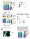
Figure 1
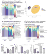
Figure 1. H. polygyrus-infection induces alterations of intestinal microbiota in Balb/c mice
The results are 16S rRNA gene sequence data from fecal pellets of control and H. polygyrus-infected Balb/c mice (2 weeks post-infection). a. Taxonomic classifications at the genus level showing the relative abundance of the top 10 genera in each sample. b. Relative abundance of the top 15 species in each sample. c. Biplot of the Principle Coordinates Analysis (PCoA), based on Bray-Curtis distances. Ellipses represent the 95% confidence interval around group centroids, and the test for group differences shows P=0.005. Arrows indicate the contribution of individual taxa to the PCoA axes, and only those taxa with the largest contributions are shown. d. OTUs that exhibited a significant change between infected and non-infected samples. Each dot represents an individual OTU. Positive values indicate an increase relative to non-infected, and negative values indicate a decrease. OTUs are grouped by the genus (x-axis) in which they belong, and are color coded by phylum. e. Heat map summarizing the relative abundance of the 10 most dominant genera. Samples are sorted based on hierarchical clustering of Bray-Curtis distances, and infection is highlighted via the colors on the left. The order of taxa is determined by a hierarchical clustering of Euclidean distances among taxa. f. Relative abundance of the top 10 genera in each sample showed high similarity in the gut microbiota of helminth-infected mice with and without deworming treatment.
Figure 2
Figure 2. Colonization of recipient mice with microbiota from H. polygyrus-infected or control mice reproduces the microbial composition of the donor
The results are 16S rRNA gene sequence data from fecal pellets of recipient mice that are colonized with microbiota from control (Cont-F) and H. polygyrus-infected (Hp-F) donor mice. a. The relative abundance of the top 10 genera in each sample of the different recipient groups, in which mice were colonized with Hp-F or Cont-F. b. Biplot of the Principle Coordinates Analysis (PCoA), based on Bray-Curtis distances. c. Relative abundance of the top 15 species in each sample from the recipient mice that were colonized with Hp-F and Cont-F. d. The relative abundance of the top 10 genera in each sample from the Hp-F-colonized recipient mice and from helminth-infected donor (Hp). e, f and g. qPCR analysis of fecal Bacteroides, SFB and Lactobacillus level in donor mice: normal and H. polygyrus-infected mice, and in the recipient mice that are colonized with Cont-F and Hp-F.
Figure 3
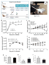
Figure 3. Colonization of mice with microbiota from H. polygyrus-infected mice results in exacerbated C .rodentium infection
a. Microbiota colonization protocol. b. Colonization of mice with Hp-F results in increased fecal C. rodentium output in mice. c. Body weight changes of recipients of Cont-F and Hp-F with and without C. rodentium-infection. d. Development of anal prolapse in mice colonized with Hp-F after C. rodentium-infection. e. Fecal C. rodentium output in mice colonized with microbiota from uninfected (control) or H. polygyrus-infected STAT 6 KO mice (STAT6KO Hp-F). f. Body weight changes of mice colonized with STAT6KO-F or STAT6KO Hp-F with and without C. rodentium infection. Data shown are pooled from three independent experiments and are expressed as a percentage of the initial body weight ± SE (n = 10–15) at each time point. g, h and i: qPCR analysis of fecal Bacteroides, SFB and Lactobacillus level in STAT 6 KO mice with and without H. polygyrus-infection.
Figure 4
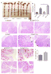
Figure 4. Colonization of mice with microbiota from H. polygyrus-infected mice exacerbates C. rodentium-induced colitis
a. Colon tissues were removed from mice colonized with Cont-F or Hp-F with and without C. rodentium-infection (two weeks post-bacterial-infection). b–f. Histopathological score and pathology analysis of colon. The data shown are pooled from three independent experiments with total (n =9 to 12 per group). *p<0.005. Magnification, x200. c–f: H&E staining of colon section: Mice colonized with Cont-F (c) or Hp-F (e) without C. rodentium-infection. d and f: duplicate samples are presented from C. rodentium-infected mice colonized with Cont-F+Cr (d) or Hp-F+Cr (f). gi: Paraffin-embedded colon tissues from mice colonized with Cont-F (g) or Hp-F (h) with C. rodentium-infection (two weeks post-bacterial-infection) were processed with periodic acid-Schiff (PAS) stain (×200 magnification). i: Quantitation of goblet cells in C. rodentium-infected colons from mice colonized with Cont-F (g) or Hp-F (h). The bars indicate the mean numbers of goblet cells per high power field counted in PAS-stained colon sections. The data shown are the mean ± the SEM (n = 3–5 mice/group) from one of two experiments performed showing similar results. **p < 0.01.
Figure 5
Figure 5. Transfer of intestinal microbes from helminth-infected donors enhances bacterial colitis
a: Bacterial culture of unfractionated fecal material, and the centrifuged pellet and filtrate from the fecal material. b–e. Body weight changes, colon pathology and colon IL-6 expression of recipients of fecal material from normal control donor (Cont-F) or H. polygyrus-infected donor derived unfractionated fecal material (Hp-F), centrifuged pellet (Hp-F-pellet) or fecal material filtrate (Hp-F-filtrate) at 2 weeks post C. rodentium infection. Colonic IL-6 expression was determined by qRT-PCR. Values are the fold increase relative to C. rodentium-infected mice colonized with normal microbiota. The data shown are the mean ± the SEM (n = 3–5 mice/group). *p < 0.05.
Figure 6
Figure 6. Colonization of mice with microbiota from helminth infected donor mice results in down-regulation of C. rodentium-induced colonic anti-microbial responses
Colon tissues were collected from mice colonized with Cont-F and Hp-F with and without C. rodentium-infection for total RNA isolation. Reg3γ (a), CRAMP (b), IL-22 (c), and iNOS (d) expression was determined using qRT-PCR. Values are the fold increase compared with baseline obtained from uninfected mice colonized with normal microbiota. The data shown are the mean ± the SEM (n = 3–5 mice/group) from one of three experiments performed showing similar results. *p < 0.05, **p < 0.01, *** p < 0.005.
Figure 7
Figure 7. Effects of helminth-altered microbiota on Citrobacter infection are mediated by increased Tregs
a: MLN cells collected from mice colonized with Cont-F and Hp-F with and without C. rodentium-infection and were cultured with surface-bound anti-CD3 mAb. Culture supernatants were collected 48 h later for cytokine determination (ELISA). The data shows that colonization of mice with Hp-F did not alter IFN-γ, IL-4 or IL-17 response, and it drives IL-10 response during C. rodentium infection. b: FACS data shows that colonization of mice with Hp-F induces a significant expansion of CD4+CD25+ T cells in MLN during C. rodentium infection. c: FACS data shows that colonization of mice with Hp-F induces a significant expansion of CD4+Foxp3+, CD4+IL-10+, Foxp3+IL-10+ T cells in MLN during C. rodentium infection. The data shown are the mean ± the SEM (n = 3–5 mice/group) from one of three experiments performed showing similar results. *p < 0.05, **p < 0.01, *** p < 0.005.
Figure 8
Figure 8. Treg depletion results in reduced fecal bacterial output and attenuated C. rodentium-mediated intestinal injury in mice colonized with microbiota from helminth-infected donor
a and b: The numbers of Citrobacter recovered from fecal samples were determined at different time points during infection in C. rodentium-infected mice that were colonized with Cont-F or Hp-F and treated with isotype control Ab or Anti-CD25 and IL-10 Ab. c. Histopathological analysis and score of colonic inflammation from mice infected with Citrobacter after treatment with isotype control Ab or Anti-CD25 and IL-10 Ab. d. Colonic IL-6 expression was determined by qRT-PCR. The data shown are the mean ± the SEM (n = 3–5 mice/group) from one of two experiments performed showing similar results. *p < 0.05.

Similar articles

Cited by

References

    1. Bäckhed F, Ley R, Sonnenburg J, Peterson D, Gordon J. Host-bacterial mutualism in the human intestine. Science (New York, NY) 2005;307(5717):1915–1920. - PubMed
    1. Round J, Mazmanian S. Inducible Foxp3+ regulatory T-cell development by a commensal bacterium of the intestinal microbiota. Proceedings of the National Academy of Sciences of the United States of America. 2010;107(27):12204–12209. - PMC - PubMed
    1. Ivanov I, Frutos R, Manel N, Yoshinaga K, Rifkin D, Sartor R, et al. Specific microbiota direct the differentiation of IL-17-producing T-helper cells in the mucosa of the small intestine. Cell host & microbe. 2008;4(4):337–349. - PMC - PubMed
    1. Atarashi K, Tanoue T, Shima T, Imaoka A, Kuwahara T, Momose Y, et al. Induction of colonic regulatory T cells by indigenous Clostridium species. Science (New York, NY) 2011;331(6015):337–341. - PMC - PubMed
    1. O’Hara A, Shanahan F. The gut flora as a forgotten organ. EMBO reports. 2006;7(7):688–693. - PMC - PubMed

Publication types

MeSH terms