Long-term electrical stimulation at ear and electro-acupuncture at ST36-ST37 attenuated COX-2 in the CA1 of hippocampus in kainic acid-induced epileptic seizure rats
- PMID: 28352122
- PMCID: PMC5428861
- DOI: 10.1038/s41598-017-00601-1
Long-term electrical stimulation at ear and electro-acupuncture at ST36-ST37 attenuated COX-2 in the CA1 of hippocampus in kainic acid-induced epileptic seizure rats
Abstract
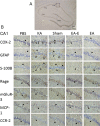
Seizures produce brain inflammation, which in turn enhances neuronal excitability. Therefore, anti-inflammation has become a therapeutic strategy for antiepileptic treatment. Cycloxygenase-2 (COX-2) plays a critical role in postseizure brain inflammation and neuronal hyperexcitability. Our previous studies have shown that both electrical stimulation (ES) at the ear and electro-acupuncture (EA) at the Zusanli and Shangjuxu acupoints (ST36-ST37) for 6 weeks can reduce mossy fiber sprouting, spike population, and high-frequency hippocampal oscillations in kainic acid (KA)-induced epileptic seizure rats. This study further investigated the effect of long-term ear ES and EA at ST36-ST37 on the inflammatory response in KA-induced epileptic seizure rats. Both the COX-2 levels in the hippocampus and the number of COX-2 immunoreactive cells in the hippocampal CA1 region were increased after KA-induced epileptic seizures, and these were reduced through the 6-week application of ear ES or EA at ST36-ST37. Thus, long-term ear ES or long-term EA at ST36-ST37 have an anti-inflammatory effect, suggesting that they are beneficial for the treatment of epileptic seizures.
Conflict of interest statement
The authors declare that they have no competing interests.
Figures


Similar articles
-
Electroacupuncture at ST36-ST37 and at ear ameliorates hippocampal mossy fiber sprouting in kainic acid-induced epileptic seizure rats.Biomed Res Int. 2014;2014:756019. doi: 10.1155/2014/756019. Epub 2014 Jun 22. Biomed Res Int. 2014. PMID: 25045697 Free PMC article.
-
Auricular electroacupuncture reduced inflammation-related epilepsy accompanied by altered TRPA1, pPKCα, pPKCε, and pERk1/2 signaling pathways in kainic acid-treated rats.Mediators Inflamm. 2014;2014:493480. doi: 10.1155/2014/493480. Epub 2014 Jul 24. Mediators Inflamm. 2014. PMID: 25147437 Free PMC article.
-
Electric Stimulation of Ear Reduces the Effect of Toll-Like Receptor 4 Signaling Pathway on Kainic Acid-Induced Epileptic Seizures in Rats.Biomed Res Int. 2018 Feb 26;2018:5407256. doi: 10.1155/2018/5407256. eCollection 2018. Biomed Res Int. 2018. PMID: 29682548 Free PMC article.
-
Electro-acupuncture improves epileptic seizures induced by kainic acid in taurine-depletion rats.Acupunct Electrother Res. 2005;30(3-4):207-17. doi: 10.3727/036012905815901280. Acupunct Electrother Res. 2005. PMID: 16617689
-
Anti-Inflammatory Effects of Acupuncture at ST36 Point: A Literature Review in Animal Studies.Front Immunol. 2022 Jan 12;12:813748. doi: 10.3389/fimmu.2021.813748. eCollection 2021. Front Immunol. 2022. PMID: 35095910 Free PMC article. Review.
Cited by
-
Multi-level exploration of auricular acupuncture: from traditional Chinese medicine theory to modern medical application.Front Neurosci. 2024 Sep 23;18:1426618. doi: 10.3389/fnins.2024.1426618. eCollection 2024. Front Neurosci. 2024. PMID: 39376538 Free PMC article. Review.
-
Efficacy of acupuncture combined with traditional Chinese herbal for primary epilepsy patients with cognitive impairment: A protocol for systematic review and meta-analysis.PLoS One. 2024 Jul 1;19(7):e0297410. doi: 10.1371/journal.pone.0297410. eCollection 2024. PLoS One. 2024. PMID: 38950015 Free PMC article.
-
Current Tracking on Effectiveness and Mechanisms of Acupuncture Therapy: A Literature Review of High-Quality Studies.Chin J Integr Med. 2020 Apr;26(4):310-320. doi: 10.1007/s11655-019-3150-3. Epub 2019 Feb 1. Chin J Integr Med. 2020. PMID: 30707414 Review.
-
New advances in Traditional Chinese Medicine interventions for epilepsy: where are we and what do we know?Chin Med. 2025 Mar 18;20(1):37. doi: 10.1186/s13020-025-01088-z. Chin Med. 2025. PMID: 40098198 Free PMC article. Review.
-
Excitatory neurons in paraventricular hypothalamus contributed to the mechanism underlying acupuncture regulating the swallowing function.Sci Rep. 2022 Apr 6;12(1):5797. doi: 10.1038/s41598-022-09470-9. Sci Rep. 2022. PMID: 35388042 Free PMC article.
References
Publication types
MeSH terms
Substances
LinkOut - more resources
Full Text Sources
Other Literature Sources
Medical
Research Materials
Miscellaneous

