. 2017 Mar 28;7(1):493.
doi: 10.1038/s41598-017-00538-5.

Recruitment of Lyn from endomembranes to the plasma membrane through calcium-dependent cell-cell interactions upon polarization of inducible Lyn-expressing MDCK cells

Affiliations

Recruitment of Lyn from endomembranes to the plasma membrane through calcium-dependent cell-cell interactions upon polarization of inducible Lyn-expressing MDCK cells

Takao Morinaga et al. Sci Rep. .

Abstract

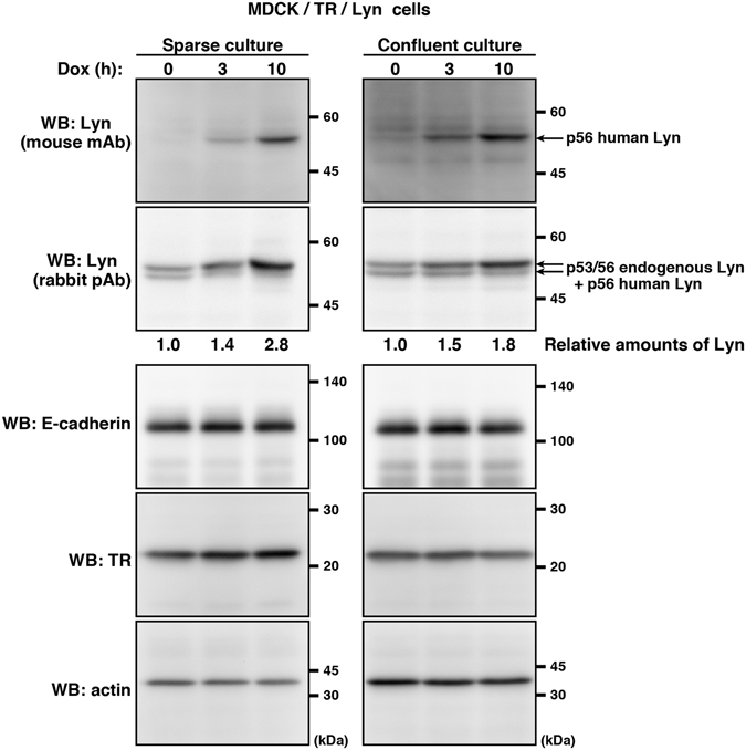
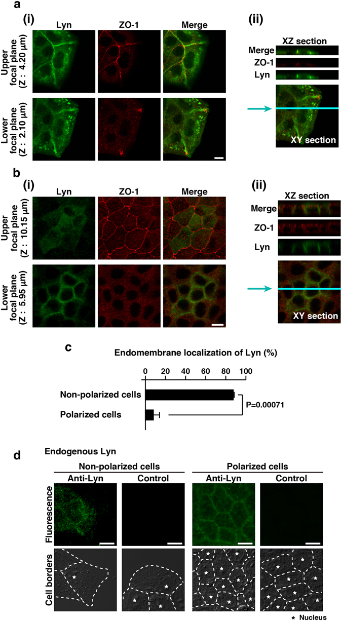
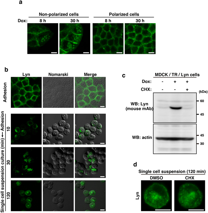
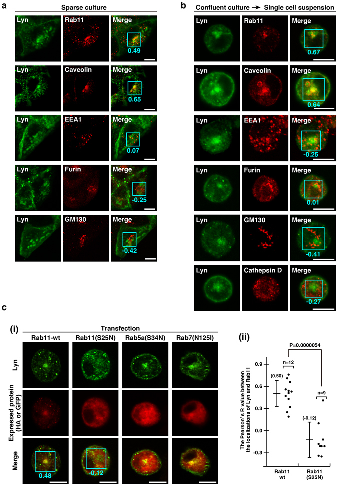
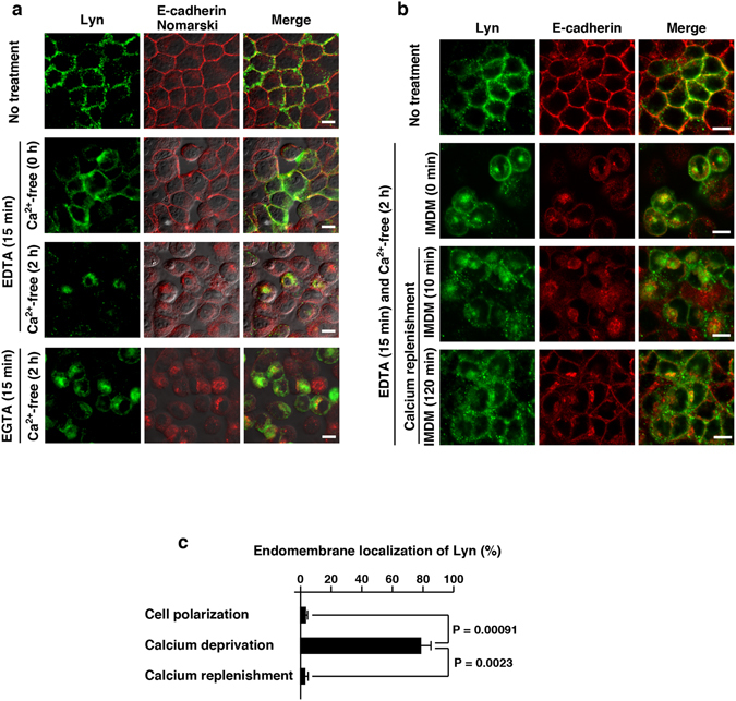
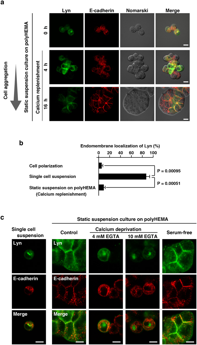
Src-family kinases, expressed in a wide variety of cell types, are anchored to cellular membranes through posttranslational lipid modifications and involved in diverse cellular signalling. In epithelial cells, Src-family kinases are localized at the plasma membrane and participate in epithelial functions. Epithelial cell polarity is achieved through dynamic reorganization of protein trafficking. To examine the trafficking of Src-family kinases between polarized and non-polarized epithelial cells, we generated an MDCK cell line that can inducibly express a protein of interest in a polarized state at any time. We show here that Lyn, a member of Src-family kinases, mainly localizes to the plasma membrane in polarized MDCK cells and to endomembranes in non-polarized MDCK cells. Cell-cell interactions between adjacent MDCK cells recruit Lyn from endomembranes to the plasma membrane even without cell attachment to extracellular matrix scaffolds, and loss of cell-cell interactions by calcium deprivation relocates Lyn from the plasma membrane to endomembranes through Rab11-mediated recycling. Therefore, using our MDCK cells expressing inducible Lyn, we reveal that calcium-dependent cell-cell interactions play a critical role in plasma membrane localization of Lyn in polarized MDCK cells.


Conflict of interest statement

The authors declare that they have no competing interests.

Figures

Figure 1
Figure 1
Establishment of an MDCK cell line expressing inducible Lyn. MDCK/TR/Lyn cells were grown in sparse and confluent cultures, which generates non-polarized cells and polarized cells, respectively. The cells were subsequently treated with 1 μg/ml doxycycline (Dox) for the indicated times. Whole cell lysates were subjected to Western blot analysis using the indicated antibodies. TR, tetracycline repressor. Molecular size markers are shown in kDa. The amount of p53/p56 Lyn in Dox-treated cells is expressed as the value relative to that in cells without Dox treatment after normalization with actin levels. For better clarity and conciseness of the presentation, cropped blots are shown. The full-length blots are presented in Supplementary Fig. S6.
Figure 2
Figure 2
Localizations of Lyn in MDCK cells under the different culture conditions. (ac) MDCK/TR/Lyn cells grown in sparse (non-polarized) (a) or confluent (polarized) (b) culture were treated with 1 μg/ml Dox for 10 h. Cells were fixed and then stained with rabbit polyclonal anti-Lyn antibody (green) and anti-ZO-1 antibody (red). The positions of the focal plane (Upper and Lower) are indicated along the z-axis. Representative XY sections (i) and XZ sections on the blue line (ii) are shown. Scale bar, 10 μm. (c) The number of cells in which Lyn was appreciably accumulated to endomembranes was counted. Cells grown at the cell periphery of each colony were regarded as non-polarized cells. The data represent the mean ratio ± S.D. from three independent experiments in which at least 50 cells for each category were scored. The significant difference is calculated by one-tailed Student’s t test. (d) Parental MDCK cells were grown in sparse (non-polarized) or confluent (polarized) culture. Cells were fixed and stained with rabbit polyclonal anti-Lyn antibody. Control samples were stained with secondary antibody alone. For eye guide, cell borders and nuclei are marked by dotted lines and stars, respectively. Scale bar, 10 μm.
Figure 3
Figure 3
Differential localizations of Lyn in non-polarized and polarized MDCK cells. (a,b,d) Immunofluorescence staining was performed using mouse monoclonal anti-Lyn antibody (green). Scale bars, 10 μm. (a) MDCK/TR/Lyn cells were cultured in sparse or confluent conditions and then treated with 1 μg/ml Dox for 8 h and 30 h. (b) MDCK/TR/Lyn cells cultured to confluence (polarized cells) were treated with 1 μg/ml Dox for 8 h (Adhesion), detached with trypsin treatment and subsequently cultured for the indicated time (min) in a spinner flask with calcium- and serum-free IMDM containing 1 μg/ml Dox (Adhesion → Single cell suspension culture). (c) MDCK/TR/Lyn cells cultured to confluence (polarized cells) were treated with 1 μg/ml Dox in the presence or absence of 200 μg/ml cycloheximide (CHX) for 120 min. Whole cell lysates were subjected to Western blot analysis using the indicated antibodies. Molecular size markers are shown in kDa. The full-length blots are presented in Supplementary Fig. S7. (d) MDCK/TR/Lyn cells cultured to confluence (polarized cells) were treated with 1 μg/ml Dox for 10 h, detached with trypsin treatment and subsequently cultured for 120 min in a spinner flask with calcium- and serum-free IMDM (single cell suspension cultures) in the presence or absence of 200 μg/ml CHX for 120 min.
Figure 4
Figure 4
Endomembrane localization of Lyn through Rab11-positive endosomes. (a,b) Double immunofluorescence staining was performed using mouse monoclonal or rabbit polyclonal anti-Lyn antibody (green) together with the indicated antibody (red). The Pearson’s R value determined from the green and red signals in the region of interest (ROI; blue square) is shown in each image. Scale bars, 10 μm. (a) MDCK/TR/Lyn cells sparsely cultured (non-polarized cells) were treated with 1 μg/ml Dox for 10 h. (b) MDCK/TR/Lyn cells cultured to confluence (polarized cells) were treated with 1 μg/ml Dox for 8 h, detached with trypsin and subsequently cultured in a spinner flask with calcium- and serum-free IMDM containing 1 μg/ml Dox for 2 h. (c) MDCK/TR/Lyn cells transiently transfected with the indicated plasmid were cultured to confluence and then treated with 1 μg/ml Dox for 8 h, detached with trypsin and subsequently cultured in a spinner flask with calcium- and serum-free IMDM for 2 h. (i) HA-tagged Rab11-wt (wild-type Rab11) and HA-tagged Rab11(S25N) (dominant-negative Rab11) were stained with rabbit anti-HA antibody (red). The images of GFP-conjugated Rab5a(S34N) (dominant-negative Rab5) and GFP-conjugated Rab7(N125I) (dominant-negative Rab7) were pseudocoloured in red. Lyn was stained with mouse monoclonal anti-Lyn antibody (green). The Pearson’s R value determined from the green and red signals in the ROI (blue square) is shown in each representative image of Rab11- or Rab11(S25N)-expressing cell. Scale bars, 10 μm. (ii) The plots represent the Pearson’s R values determined from Lyn and Rab11-wt or Lyn and Rab11(S25N) in the ROI in each cell. n, cell number.
Figure 5
Figure 5
Role for palmitoylation of Lyn in its endomembrane localization in MDCK cells. (a,d) MDCK/TR/Lyn, MDCK/TR/c-Src, and MDCK/TR/Lyn(C3S) cells cultured as confluent monolayers (polarized cells) were treated with 1 μg/ml Dox for 10 h. (b,e) MDCK/TR/Lyn, MDCK/TR/c-Src, and MDCK/TR/Lyn(C3S) cells cultured to confluence (polarized cells) were treated with 1 μg/ml Dox for 8 h, detached with trypsin and subsequently cultured in spinner flasks for 2 h. (a,b) Cells were stained with mouse monoclonal anti-Lyn or anti-c-Src antibody. Scale bars, 10 μm. (c) Parental MDCK cells were cultured to confluence (i, polarized cells), cultured sparsely (ii, non-polarized cells), or cultured to confluence and subsequently cultured in suspension for 2 h (iii, single cell suspension cultures). Cells were stained with anti-c-Yes antibody. For eye guide, cell borders and nuclei are marked by dotted lines and stars, respectively (ii). Scale bars, 10 μm. (d,e) Post-nuclear supernatants obtained from polarized (d) or single cell suspension-cultured, non-polarized (e) MDCK cells were subjected to sucrose density gradient fractionation, and each fraction was analysed by Western blotting with the indicated antibodies. Molecular size markers are shown in kDa. The full-length blots are presented in Supplementary Fig. S8.
Figure 6
Figure 6
Translocation of Lyn to endomembranes by calcium deprivation. (a) MDCK/TR/Lyn cells cultured to confluence (polarized cells) were treated with 1 μg/ml Dox for 8 h, then treated with 4 mM EDTA or 4 mM EGTA for 15 min and subsequently cultured in calcium- and serum-free IMDM for 2 h (calcium deprivation). Cells were fixed and stained with rabbit anti-Lyn antibody (green) and anti-E cadherin antibody (red). Scale bars, 10 μm. (b) MDCK/TR/Lyn cells cultured to confluence (polarized cells) were treated with 1 μg/ml Dox for 8 h, then treated with 4 mM EDTA for 15 min and further incubated in calcium- and serum-free IMDM for 2 h (calcium deprivation), and subsequently incubated with IMDM containing 5% bovine serum for 10 min and 120 min (calcium replenishment). Cells were stained with anti-Lyn and anti-E-cadherin antibodies. Scale bars, 10 μm. (c) MDCK/TR/Lyn cells cultured to confluence (polarized cells) were treated with 1 μg/ml Dox for 8 h, then treated with 4 mM EDTA for 15 min and further incubated in calcium- and serum-free IMDM for 2 h (calcium deprivation), and subsequently incubated with IMDM containing 5% bovine serum for 20 h (calcium replenishment). Cells were stained with anti-Lyn antibody (green) and propidium iodide. The number of cells in which Lyn was predominantly accumulated to endomembranes was counted. The data represent the mean ratio ± S.D. from three independent experiments in which at least 50 cells for each category were scored. The significant differences are calculated by one-tailed Student’s t test.
Figure 7
Figure 7
Relocation of Lyn to the plasma membrane by cell-cell interactions. (a,c) Cells cultured as described below were stained with rabbit anti-Lyn and anti-E-cadherin antibodies. Scale bars, 10 μm. (a) MDCK/TR/Lyn cells cultured as confluent monolayers (polarized cells) were treated with 1 μg/ml Dox for 8 h, detached by trypsinization and subsequently cultured in a spinner flask with calcium- and serum-free IMDM (single cell suspension culture). Single suspended cells were transferred into polyHEMA-coated culture dishes and cultured in IMDM-5% bovine serum (calcium replenishment) for the indicated time (static suspension culture). (b) Single suspended cells cultured as described in (a) (single cell suspension culture) were transferred into polyHEMA-coated culture dishes and cultured in IMDM-5% bovine serum (calcium replenishment and static suspension culture) for 20 h. Cells were stained with anti-Lyn antibody and propidium iodide. The number of cells exhibiting predominant accumulation of Lyn to endomembranes was counted. The data represent the mean ratio ± S.D. from three independent experiments in which at least 50 cells for each category were scored. The significant differences are calculated by one-tailed Student’s t test. (c) Cells cultured as described in (a) (single cell suspension culture) were transferred into polyHEMA-coated culture dishes and cultured for 16 h in IMDM containing 5% bovine serum (static suspension culture, Control), IMDM containing 5% bovine serum in the presence of 4 mM or 10 mM EGTA (static suspension culture, Calcium deprivation), or serum-free IMDM (static suspension culture, Serum-free). Scale bars, 10 μm.

Similar articles

Cited by

References

    1. Brown MT, Cooper JA. Regulation, substrates and functions of src. Biochim. Biophys. Acta. 1996;1287:121–149. - PubMed
    1. Resh MD. Myristylation and palmitylation of Src family members: the fats of the matter. Cell. 1994;76:411–413. doi: 10.1016/0092-8674(94)90104-X. - DOI - PubMed
    1. Tsukita S, et al. Specific proto-oncogenic tyrosine kinases of Src family are enriched in cell-to-cell adherens junctions where the level of tyrosine phosphorylation is elevated. J. Cell Biol. 1991;113:867–879. doi: 10.1083/jcb.113.4.867. - DOI - PMC - PubMed
    1. Hayashi T, Umemori H, Mishina M, Yamamoto T. The AMPA receptor interacts with and signals through the protein tyrosine kinase Lyn. Nature. 1999;397:72–76. doi: 10.1038/16269. - DOI - PubMed
    1. Chudakova DA, et al. Integrin-associated Lyn kinase promotes cell survival by suppressing acid sphingomyelinase activity. J. Biol. Chem. 2008;283:28806–28816. doi: 10.1074/jbc.M803301200. - DOI - PMC - PubMed

Publication types

LinkOut - more resources