. 2017 Mar 15:11:783-795.
doi: 10.2147/DDDT.S129913. eCollection 2017.

HMGB1 siRNA can reduce damage to retinal cells induced by high glucose in vitro and in vivo

Affiliations

HMGB1 siRNA can reduce damage to retinal cells induced by high glucose in vitro and in vivo

Shuang Jiang et al. Drug Des Devel Ther. .

Abstract

Background: Diabetic retinopathy (DR), one of the most common complications of late-phase diabetes, is associated with many risk factors, among which continuous low-grade inflammation is one of the principal ones. As such, lowering inflammation levels and maintain the viability of human retinal endothelial cells (HRECs) are critical for DR therapy. HMGB1 is a well-known proinflammatory cytokine. However, whether HMGB1 small interfering RNA (siRNA) can protect retina cells under a high-glucose environment from morphological changes and functional abnormalities remain undetermined. We aimed to investigate the effect of HMGB1 siRNA on retinal cells in DR.

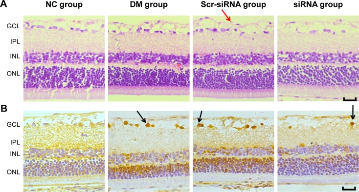
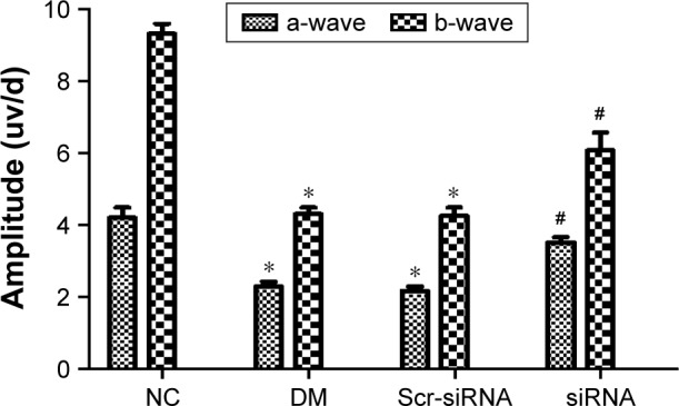
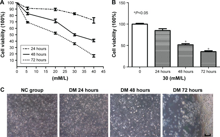
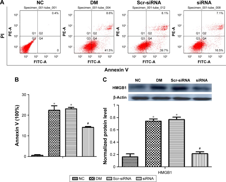
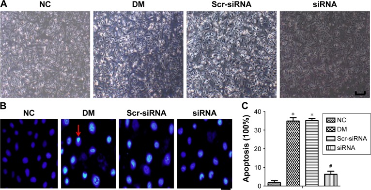
Materials and methods: A total of 80 adult Wistar rats were randomly divided into four groups (n=20 each): normal control, diabetes mellitus (DM), scrambled (Scr) siRNA, and HMGB1 siRNA. Rats in the DM, Scr siRNA, and siRNA groups were established by intraperitoneal injection of streptozotocin. At 16 weeks after injection, rats in the siRNA and Scr-siRNA groups were intravitreally injected with 2 μL HMGB1 siRNA and 2 μL Scr-siRNA, while rats in the control and DM groups were intravitreally injected with the same dose of sterile saline. At 1 week after injections, we performed the following experiments. Immunohistochemical staining and real-time quantitative polymerase chain reaction were performed to test HMGB1 protein and messenger RNA expression in retinas. We performed TUNEL assays to detect retinal cell apoptosis and electroretinography to detect retinal function. In HRECs treated with high glucose, proliferation, morphology, apoptosis, super-oxide dismutase (SOD), and reactive oxygen species production were detected. Western blot was applied to determine the expressions of HMGB1 and its related protein and apoptosis protein.

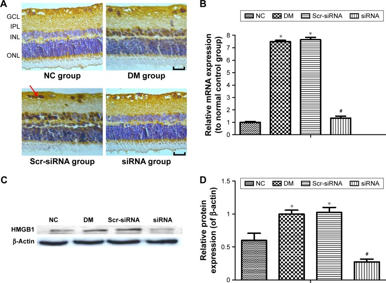
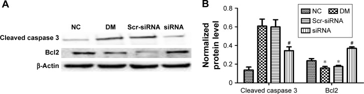
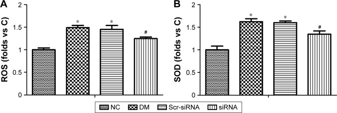
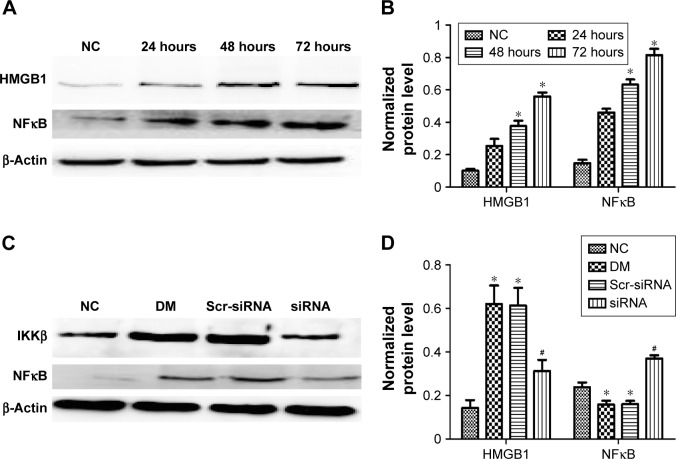
Results: Intravitreal injection of HMGB1 siRNA reduced protein and messenger RNA expression of HMGB1 (both P<0.05). Intravitreal injection of HMGB1 siRNA reduced apoptosis of retinal cells (P<0.05), protected morphological changes in the retina, and improved the function of the retina (P<0.05). In HRECs treated with high glucose, HMGB1 siRNA pretreatment increased cell viability, reduced cell apoptosis, and reduced oxidative damage to cells (all P<0.05). Western blot detection found that HMGB1 siRNA pretreatment can inhibit the expression of cleaved caspase 3 and improve the expression of BCL2 (P<0.05). HMGB1 and NFκB expression increased in a time-dependent manner in the high-glucose environment and IKKβ and NFκB protein expression decreased significantly after HMGB1 silencing.

Conclusion: As a therapeutic target, HMGB1 siRNA can reduce retinal cell damage induced by high glucose in vitro and in vivo and delay DR progress through the HMGB1-IKKβ-NFκB signaling pathway.

Keywords: diabetic retinopathy; high-mobility group box 1; human retinal endothelial cells; inhibitor of nuclear factor κB; nuclear factor κB; small interfering RNA.


Conflict of interest statement

Disclosure The authors report no conflicts of interest in this work.

Figures

Figure 1
Figure 1
Intravitreal injection of HMGB1 siRNA significantly inhibited the expression of HMGB1 in the retina. Notes: *P<0.05 versus NC group, #P<0.05 versus Scr-siRNA group. (A) Immunohistochemical staining for HMGB1 expression (scale bar 50 μm). Arrows show positive expression. There was little expression of HMGB1 in the NC group, and HMGB1 expression in the DM and Scr-siRNA groups was significantly increased, mainly located in the retinal ganglion-cell layer, inner nuclear layer, and outer nuclear layer, while HMGB1 expression in the siRNA group was significantly decreased. (B) HMGB1 mRNA by real-time quantitative polymerase chain reaction. HMGB1 mRNA levels in the siRNA group were reduced compared to the Scr-siRNA group (P<0.05). HMGB1 mRNA levels in the DM and Scr-siRNA groups were upregulated significantly compared to the NC group (all P<0.05), whereas there was no significant difference between the DM and Scr-siRNA groups (P>0.05). (C) Western blot detection. Protein expression of HMGB1 in the siRNA group was reduced compared with the Scr-siRNA group (P<0.05). HMGB1 protein levels in the DM and Scr-siRNA groups were upregulated significantly compared to the NC group (all P<0.05), whereas there was no significant difference between the DM and Scr-siRNA groups (P>0.05). (D) HMGB1 protein expression. Protein expression was normalized to β-actin. Relative protein expression presented as mean ± standard deviation of three independent experiments. HMGB1 expression in the siRNA group was significantly lower than in the DM group. Abbreviations: siRNA, small interfering RNA; NC, normal control; Scr, scrambled; DM, diabetes mellitus; mRNA, messenger RNA; GCL, ganglion cell layer; IPL, inner plexiform layer; INL, inner nuclear layer; ONL, outer nuclear layer.
Figure 2
Figure 2
siRNA HMGB1 significantly inhibited retinal damage and ameliorated retina-function degeneration in vivo. Notes: (A) Histopathological examination of retina (bar 50 μm). Red arrows show vascular endothelial cell nuclei protruding through the inner limiting membrane. (B) TUNEL staining of retinal slices (bar 50 μm). Positive apoptotic cells nuclei stained brown yellow. Black arrows show positive apoptotic cells. Abbreviations: siRNA, small interfering RNA; NC, normal control; Scr, scrambled; DM, diabetes mellitus; GCL, ganglion cell layer; IPL, inner plexiform layer; INL, inner nuclear layer; ONL, outer nuclear layer.
Figure 3
Figure 3
Amplitude by flash electroretinography. The experiment was carried out three times, and average amplitudes of retinas were taken. Notes: *P<0.05 versus NC group; #P<0.05 versus Scr-siRNA group. Amplitudes of the a- and b-waves in the siRNA group increased by 77.3% and 66.9%, respectively, compared with the Scr-siRNA group. Amplitudes of the a- and b-waves in the DM and Scr-siRNA groups were downregulated significantly compared to the NC group (all P<0.05), whereas there was no significant difference between the DM and Scr-siRNA groups (P>0.05). Abbreviations: siRNA, small interfering RNA; NC, normal control; Scr, scrambled; DM, diabetes mellitus.
Figure 4
Figure 4
High-glucose-induced HREC damage in vitro. Notes: (A) MTT assay. High glucose depressed HREC viability in a dose-dependent manner. The IC50 of high glucose was about 30 mM/L at 48 hours and 22 mM/L at 72 hours. (B) MTT assay. High-glucose-induced HREC viability in a time-dependent manner. With time, HREC viability reduced gradually with 30 mM glucose. (C) Morphological changes in HRECs treated with high glucose at different time points (bar 25 μm). The morphology of normal cells was uniform and spindle-shaped, and the cells became smaller and density decreased after treatment with high glucose. With the extension of treatment time, changes in morphology and density were more obvious. *P<0.05 vs 0 hour. Abbreviations: HREC, human retinal endothelial cell; IC50, half-maximal inhibitory concentration; NC, normal control; DM, diabetes mellitus.
Figure 5
Figure 5
Cell apoptosis was determined by flow cytometry using annexin V–PI staining. Notes: (A) Flow-cytometry detection. Annexin V was set as the horizontal axis and PI as the vertical axis. Upper-right (Q2) quadrant, late-apoptotic or necrotic cells; lower-left (Q3) quadrant, dual-negative/normal cells; lower-right (Q4) quadrant, early-apoptotic cells; upper left (Q1) quadrant, mechanically damaged cells. Apoptotic cells were the sum of Q2 and Q4. (B) Apoptosis rate by flow-cytometry detection. HMGB1 siRNA inhibited HREC apoptosis. Total percentages of apoptotic cells are presented as mean ± standard deviation of three independent experiments. HMGB1 siRNA pretreatment significantly inhibited HREC-apoptosis rate for those treated with high glucose. *P<0.05 versus NC group; #P<0.05 versus the Scr-siRNA group. (C) Western blot detection. HRECs were transfected with HMGB1 siRNA and Scr-siRNA for 12 hours, and then cultured under high glucose (30 mM/L) for 48 hours. Protein expression of HMGB1 in the siRNA group was reduced compared with the Scr-siRNA group (P<0.05). Protein levels of HMGB1 in the DM and Scr-siRNA groups were upregulated significantly compared to the NC group (all P<0.05), whereas there was no significant difference between the DM and Scr-siRNA groups (P>0.05). Abbreviations: siRNA, small interfering RNA; HREC, human retinal endothelial cell; Scr, scrambled; DM, diabetes mellitus; NC, normal control; FITC, fluorescein isothiocyanate.
Figure 6
Figure 6
HMGB1 siRNA inhibits HREC apoptosis induced by high glucose in vitro. Notes: *P<0.05 versus NC group; #P<0.05 versus Scr-siRNA group. (A) Cell morphology in different groups (bar 25 μm). HRECs were treated with HMGB1 siRNA and Scr-siRNA for 12 hours, and then cultured under high glucose (30 mM/L) for 48 hours. The morphology of normal cells was uniform and spindle-shaped, and cells treated with high glucose became smaller and fewer. The cell state of the siRNA group was improved compared with the DM and Scr-siRNA groups. (B) Cell nuclear morphology after Hoechst 33342 staining (bar 25 μm). Apoptotic nuclei were stained bright blue. Apoptotic nuclei were densely stained and with crescent condensation. Red arrows show the nuclei of apoptotic cells. (C) Cell viability in different groups. HMGB1 siRNA pretreatment significantly inhibited cell apoptosis under a high-glucose environment. Abbreviations: siRNA, small interfering RNA; HREC, human retinal endothelial cell; NC, normal control; Scr, scrambled; DM, diabetes mellitus.
Figure 7
Figure 7
Mechanism of HMGB1 siRNA against cell apoptosis induced by high glucose. Notes: *P<0.05 versus NC group, #P<0.05 versus Scr-siRNA group. (A) Western blot detection. HMGB1 siRNA inhibited high-glucose-induced cleaved caspase 3 and promoted Bcl2 expression in HRECs. (B) Quantification of A. Protein expression was normalized to β-actin. Abbreviations: siRNA, small interfering RNA; NC, normal control; Scr, scrambled; HRECs, human retinal endothelial cells; DM, diabetes mellitus.
Figure 8
Figure 8
Effect of HMGB1 siRNA against oxidative stress. Notes: *P<0.05 versus NC group, #P<0.05 versus Scr-siRNA group. (A) ROS assay in different groups, showing the protective effect of siRNA HMGB1 in HREC damage caused by high glucose. (B) SOD assay in different groups, showing the protective effect of siRNA HMGB1 in HREC damage caused by high glucose. Abbreviations: siRNA, small interfering RNA; ROS, reactive oxygen species; HREC, human retinal endothelial cell; NC, normal control; Scr, scrambled; DM, diabetes mellitus.
Figure 9
Figure 9
HMGB1 siRNA inhibited diabetic retinopathy through the IKKβ–NFκB signaling pathway. Notes: *P<0.05 versus NC group, #P<0.05 versus Scr-siRNA group. (A) Western blot detection. Under a high-glucose environment, HMGB1 and NFκB protein expression increased gradually in a time-dependent manner. (B) Quantification of A. (C) Western blot detection. After HMGB1 siRNA transfection, IKKβ and NFκb protein expression was inhibited significantly. (D) Quantification of C. Abbreviations: siRNA, small interfering RNA; NC, normal control; Scr, scrambled; DM, diabetes mellitus.

Similar articles

Cited by

References

    1. Agarwal A, Soliman MK, Sepah YJ, Do DV, Nguyen QD. Diabetic retinopathy: variations in patient therapeutic outcomes and pharmacogenomics. Pharmgenomics Pers Med. 2014;7:399–409. - PMC - PubMed
    1. Wang SY, Andrews CA, Herman WH, Gardner TW, Stein JD. Incidence and risk factors for developing diabetic retinopathy among youths with type 1 or type 2 diabetes throughout the United States. Ophthalmology. 2016 Nov 30; Epub. - PMC - PubMed
    1. Schorr SG, Hammes HP, Müller UA, Abholz HH, Landgraf R, Bertram B. The prevention and treatment of retinal complications in diabetes. Dtsch Arztebl Int. 2016;113(48):816–823. - PMC - PubMed
    1. Cheung N, Mitchell P, Wong TY. Diabetic retinopathy. Lancet. 2010;376(9735):124–136. - PubMed
    1. Lohani N, Rajeswari MR. Dichotomous life of DNA binding high mobility group box1 protein in human health and disease. Curr Protein Pept Sci. 2016;17(8):762–775. - PubMed

MeSH terms