Knockdown of IQGAP1 inhibits proliferation and epithelial-mesenchymal transition by Wnt/β-catenin pathway in thyroid cancer
- PMID: 28352188
- PMCID: PMC5359122
- DOI: 10.2147/OTT.S128564
Knockdown of IQGAP1 inhibits proliferation and epithelial-mesenchymal transition by Wnt/β-catenin pathway in thyroid cancer
Abstract
Background: Thyroid cancer is the most common endocrine malignant disease with a high incidence rate. The expression of IQGAP1 is upregulated in various cancers, including thyroid cancer. However, the role and underlying mechanism of IQGAP1 in thyroid cancer are still not clear.
Materials and methods: The expression of IQGAP1 in thyroid cancer tissues and cells was determined by reverse transcription polymerase chain reaction and Western blot analysis. Cells were transfected with different siRNAs using Lipofectamine 2000 or were treated with various concentrations of XAV939. The effects of IQGAP1 knockdown on proliferation and epithelial-mesenchymal transition (EMT) of thyroid cancer cells were determined by MTT assay and Western blot analysis. Animal experiments were performed to investigate the effects of IQGAP1 knockdown on the growth of tumors in vivo.
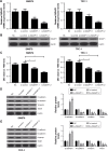
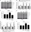
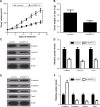
Results: High IQGAP1 expression is found in thyroid cancer tissues and cells. Knockdown of IQGAP1 had inhibitory effects on cell proliferation and EMT, as well as on the Wnt/β-catenin pathway. Additionally, inactivation of the Wnt/β-catenin pathway by XAV939 or si-β-catenin suppressed cell proliferation and EMT. Furthermore, suppression of the Wnt/β-catenin pathway reversed the positive effects of pcDNA-IQGAP1 on cell proliferation and EMT in vitro. Moreover, downregulation of IQGAP1 suppressed tumor growth and EMT in SW579 tumor xenografts through the Wnt/β-catenin pathway in vivo.
Conclusion: Our study demonstrated that knockdown of IQGAP1 inhibited cell proliferation and EMT through blocking the Wnt/β-catenin pathway in thyroid cancer.
Keywords: IQGAP1; Wnt/β-catenin; epithelial–mesenchymal transition; proliferation; thyroid cancer.
Conflict of interest statement
Disclosure The authors report no conflicts of interest in this work.
Figures


Similar articles
-
ING5 inhibits lung cancer invasion and epithelial-mesenchymal transition by inhibiting the WNT/β-catenin pathway.Thorac Cancer. 2019 Apr;10(4):848-855. doi: 10.1111/1759-7714.13013. Epub 2019 Feb 27. Thorac Cancer. 2019. PMID: 30810286 Free PMC article.
-
Role of Wnt/beta-catenin signaling pathway in epithelial-mesenchymal transition of human prostate cancer induced by hypoxia-inducible factor-1alpha.Int J Urol. 2007 Nov;14(11):1034-9. doi: 10.1111/j.1442-2042.2007.01866.x. Int J Urol. 2007. PMID: 17956532
-
Downregulation of IQGAP1 inhibits epithelial-mesenchymal transition via the HIF1α/VEGF-A signaling pathway in gastric cancer.J Cell Biochem. 2019 Sep;120(9):15790-15799. doi: 10.1002/jcb.28849. Epub 2019 May 15. J Cell Biochem. 2019. PMID: 31090961
-
Baicalein suppresses metastasis of breast cancer cells by inhibiting EMT via downregulation of SATB1 and Wnt/β-catenin pathway.Drug Des Devel Ther. 2016 Apr 18;10:1419-41. doi: 10.2147/DDDT.S102541. eCollection 2016. Drug Des Devel Ther. 2016. PMID: 27143851 Free PMC article.
-
Dihydroartemisinin suppresses proliferation, migration, the Wnt/β-catenin pathway and EMT via TNKS in gastric cancer.Oncol Lett. 2021 Oct;22(4):688. doi: 10.3892/ol.2021.12949. Epub 2021 Jul 29. Oncol Lett. 2021. PMID: 34457043 Free PMC article.
Cited by
-
IQGAP1 promotes pancreatic cancer progression and epithelial-mesenchymal transition (EMT) through Wnt/β-catenin signaling.Sci Rep. 2019 May 17;9(1):7539. doi: 10.1038/s41598-019-44048-y. Sci Rep. 2019. PMID: 31101875 Free PMC article.
-
Upregulation of OIP5-AS1 Predicts Poor Prognosis and Contributes to Thyroid Cancer Cell Proliferation and Migration.Mol Ther Nucleic Acids. 2020 Jun 5;20:279-291. doi: 10.1016/j.omtn.2019.11.036. Epub 2020 Jan 11. Mol Ther Nucleic Acids. 2020. Retraction in: Mol Ther Nucleic Acids. 2022 Feb 03;27:969. doi: 10.1016/j.omtn.2022.01.023. PMID: 32193154 Free PMC article. Retracted.
-
Engineering of HN3 increases the tumor targeting specificity of exosomes and upgrade the anti-tumor effect of sorafenib on HuH-7 cells.PeerJ. 2020 Jul 20;8:e9524. doi: 10.7717/peerj.9524. eCollection 2020. PeerJ. 2020. PMID: 33062407 Free PMC article.
-
SOX5 promotes cell invasion and metastasis via activation of Twist-mediated epithelial-mesenchymal transition in gastric cancer.Onco Targets Ther. 2019 Apr 3;12:2465-2476. doi: 10.2147/OTT.S197087. eCollection 2019. Onco Targets Ther. 2019. PMID: 31040690 Free PMC article.
-
SNHG29 regulates miR-223-3p/CTNND1 axis to promote glioblastoma progression via Wnt/β-catenin signaling pathway.Cancer Cell Int. 2019 Dec 19;19:345. doi: 10.1186/s12935-019-1057-x. eCollection 2019. Cancer Cell Int. 2019. PMID: 31889897 Free PMC article.
References
LinkOut - more resources
Full Text Sources
Other Literature Sources
Research Materials
Miscellaneous

