

Adjuvant Role of CT in the Diagnosis of Post-Infarction Left Ventricular Free-Wall Rupture
- PMID: 28352419
- PMCID: PMC5358304
- DOI: 10.4021/cr239w
Adjuvant Role of CT in the Diagnosis of Post-Infarction Left Ventricular Free-Wall Rupture
Abstract
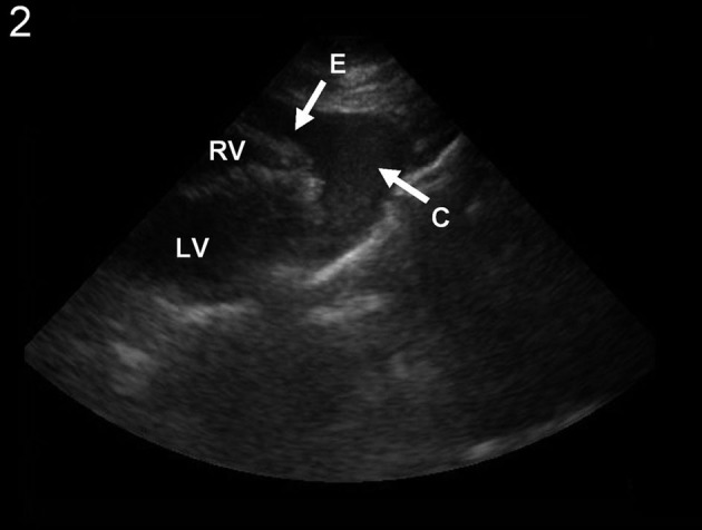
Left ventricular free wall rupture is usually a catastrophic mechanical complication of myocardial infarction. Risk factors include advanced age, female gender and absence of prior infarction. The vast majority of patients succumb rapidly due to cardiac tamponade and electromechanical dissociation. Expedited and accurate diagnosis can improve the chances of survival. Echocardiography has been advocated as the gold standard for diagnosis, but other imaging modalities can provide valuable information in these patients. We present the case of a patient who presented with cardiogenic shock, in which the definitive diagnosis of a left ventricular free wall rupture was accomplished by CT scan with intravenous contrast.
Keywords: Computed tomography; Myocardial infarction; Shock; Ventricular rupture.
Figures


References
-
- Raposo L, Andrade MJ, Ferreira J, Aguiar C, Couto R, Abecasis M, Canada M. et al. Subacute left ventricle free wall rupture after acute myocardial infarction: awareness of the clinical signs and early use of echocardiography may be life-saving. Cardiovasc Ultrasound. 2006;4:46. doi: 10.1186/1476-7120-4-46. - DOI - PMC - PubMed
-
- Slater J, Brown RJ, Antonelli TA, Menon V, Boland J, Col J, Dzavik V. et al. Cardiogenic shock due to cardiac free-wall rupture or tamponade after acute myocardial infarction: a report from the SHOCK Trial Registry. Should we emergently revascularize occluded coronaries for cardiogenic shock? J Am Coll Cardiol. 2000;36(3 Suppl A):1117–1122. doi: 10.1016/S0735-1097(00)00845-7. - DOI - PubMed
Publication types
LinkOut - more resources
Full Text Sources