

Giant Perineal Solitary Fibrous Tumor: A Rare Case Report
- PMID: 28352487
- PMCID: PMC5352885
- DOI: 10.1155/2017/4876494
Giant Perineal Solitary Fibrous Tumor: A Rare Case Report
Abstract
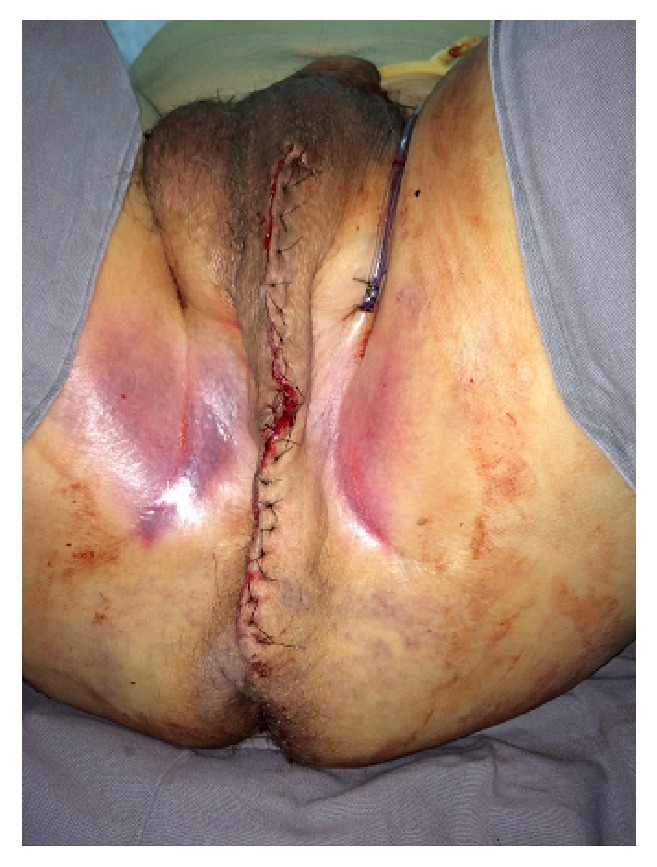
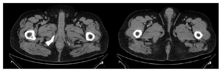
Background. Solitary fibrous tumor (SFT) is a fibroblastic mesenchymal tumor that was initially described from the pleura but currently arises at almost every anatomic site. It is usually benign, and surgical resection is curative. SFT involving the perineum is extremely rare. This is the third case report of a perineal SFT in the literature. Case Presentation. We reported an uncommon case of a 64-year-old man presenting with a huge perineal mass that started growing 3 years before his arrival in our service. He was asymptomatic. A contrast-enhanced CT scan revealed a heterogeneous well-circumscribed perineal mass with soft-tissue density. Invasion of the surrounding organs, distal metastasis, and lymph node swelling were absent. The complete resection of mass was done successfully. The specimen was a 23.0 × 14.0 × 8.0 cm encapsulated tumor. Mass weight was 1,170 g. After pathological analysis, we confirmed that the mass was a solitary fibrous tumor. The diagnosis was based on clinical findings and histological morphology and immunohistochemistry study. Conclusion. SFTs are usually indolent tumors with a favorable prognosis. The perineal location is extremely rare. Complete resection of the mass is the treatment of choice.
Conflict of interest statement
The authors declare that there is no conflict of interests regarding the publication of this paper.
Figures


Similar articles
-
Perianal Solitary Fibrous Tumor in a Rare Anatomical Presentation: A Case Report and Literature Review.Am J Case Rep. 2021 May 19;22:e929742. doi: 10.12659/AJCR.929742. Am J Case Rep. 2021. PMID: 34010267 Free PMC article. Review.
-
Retroperitoneal solitary fibrous tumor of the pelvis with pollakiuria: a case report.BMC Res Notes. 2012 Oct 29;5:593. doi: 10.1186/1756-0500-5-593. BMC Res Notes. 2012. PMID: 23107063 Free PMC article.
-
Extrapleural solitary fibrous tumor of the thyroid gland: A case report and review of literature.World J Clin Cases. 2020 Feb 26;8(4):782-789. doi: 10.12998/wjcc.v8.i4.782. World J Clin Cases. 2020. PMID: 32149061 Free PMC article.
-
Intracranial malignant solitary fibrous tumor metastasized to the chest wall: A case report and review of literature.World J Clin Cases. 2020 Oct 26;8(20):4844-4852. doi: 10.12998/wjcc.v8.i20.4844. World J Clin Cases. 2020. PMID: 33195652 Free PMC article.
-
Solitary fibrous tumor of the scrotum: a case report and review of the literature.BMC Urol. 2019 Dec 30;19(1):138. doi: 10.1186/s12894-019-0573-2. BMC Urol. 2019. PMID: 31888599 Free PMC article. Review.
Cited by
-
A rare solitary fibrous tumor in the ischiorectal fossa: a case report.Surg Case Rep. 2018 Oct 3;4(1):126. doi: 10.1186/s40792-018-0533-1. Surg Case Rep. 2018. PMID: 30284069 Free PMC article.
-
Perianal Solitary Fibrous Tumor in a Rare Anatomical Presentation: A Case Report and Literature Review.Am J Case Rep. 2021 May 19;22:e929742. doi: 10.12659/AJCR.929742. Am J Case Rep. 2021. PMID: 34010267 Free PMC article. Review.
References
-
- Meittinen M. Solitary fibrous tumor, hemaogiopericytoma, and related tumors. In: Miettinen M., editor. Modern Soft Tissue Pathology: Tumors and Non-Neoplastic Conditions. New York, NY, USA: Cambridge University Press; 2010. p. p. 335.
-
- Klemperer P., Rabin C. B. Primary neoplasms of the pleura. A report of five cases. Archives of Pathology. 1931;11:385–412.
Publication types
LinkOut - more resources
Full Text Sources
Other Literature Sources
