. 2017:2017:6590609.
doi: 10.1155/2017/6590609. Epub 2017 Mar 2.

Cardiac Function Remains Impaired Despite Reversible Cardiac Remodeling after Acute Experimental Viral Myocarditis

Affiliations

Cardiac Function Remains Impaired Despite Reversible Cardiac Remodeling after Acute Experimental Viral Myocarditis

Peter Moritz Becher et al. J Immunol Res. 2017.

Abstract

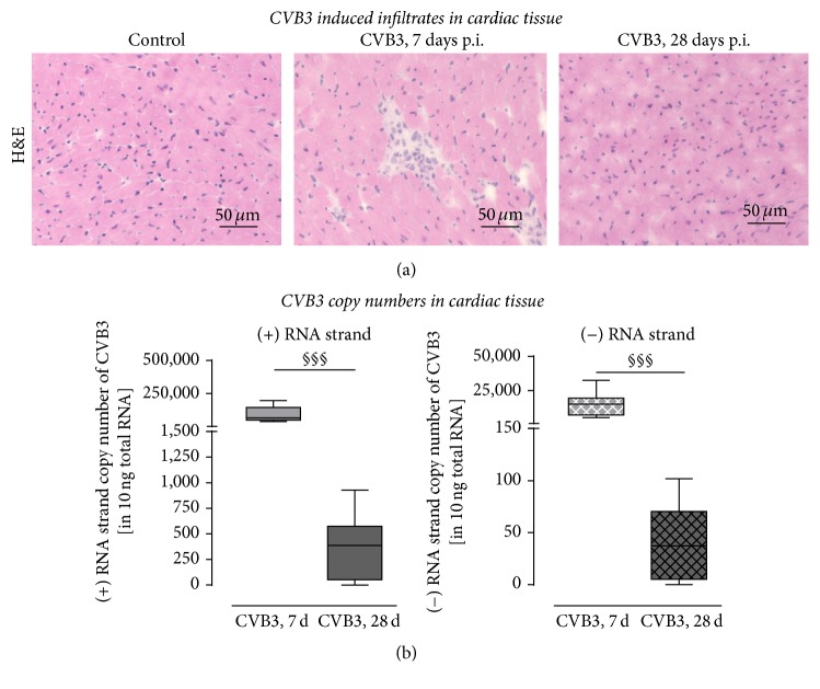
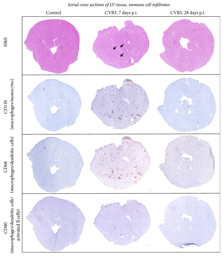
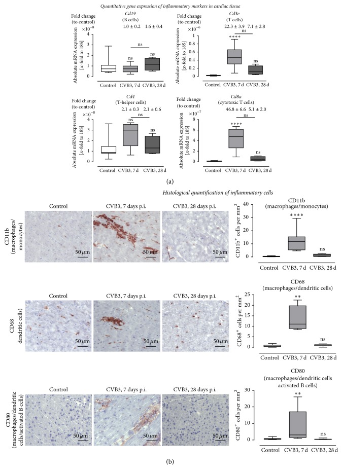
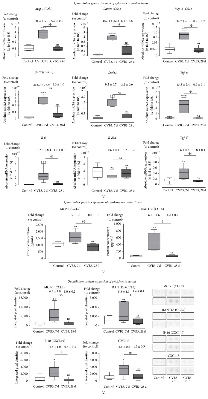
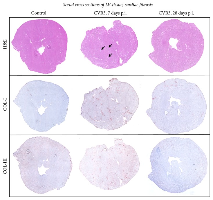
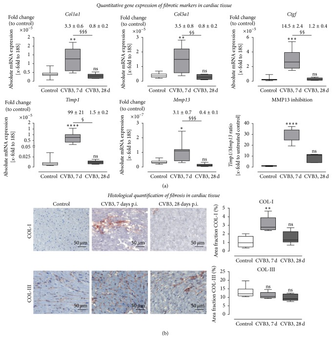
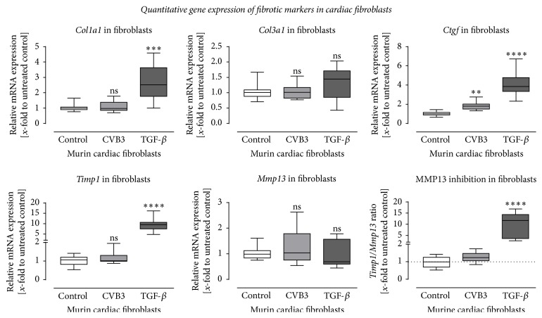
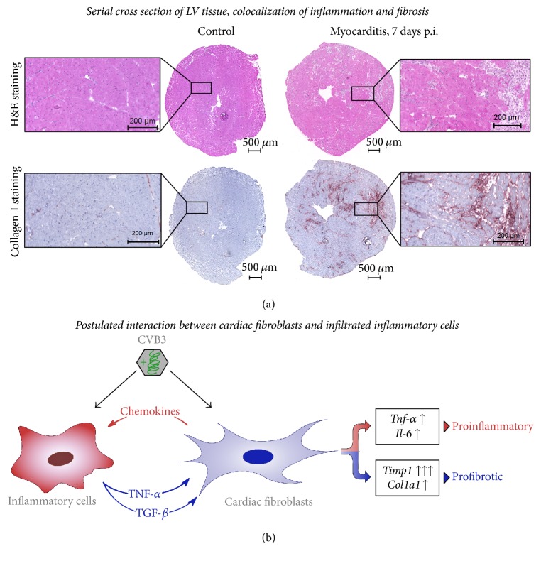
Background. Infection with Coxsackievirus B3 induces myocarditis. We aimed to compare the acute and chronic phases of viral myocarditis to identify the immediate effects of cardiac inflammation as well as the long-term effects after resolved inflammation on cardiac fibrosis and consequently on cardiac function. Material and Methods. We infected C57BL/6J mice with Coxsackievirus B3 and determined the hemodynamic function 7 as well as 28 days after infection. Subsequently, we analyzed viral burden and viral replication in the cardiac tissue as well as the expression of cytokines and matrix proteins. Furthermore, cardiac fibroblasts were infected with virus to investigate if viral infection alone induces profibrotic signaling. Results. Severe cardiac inflammation was determined and cardiac fibrosis was consistently colocalized with inflammation during the acute phase of myocarditis. Declined cardiac inflammation but no significantly improved hemodynamic function was observed 28 days after infection. Interestingly, cardiac fibrosis declined to basal levels as well. Both cardiac inflammation and fibrosis were reversible, whereas the hemodynamic function remains impaired after healed viral myocarditis in C57BL/6J mice.


Conflict of interest statement

The authors declare that there is no conflict of interests regarding the publication of this paper.

Figures

Figure 1
Figure 1
Cardiac inflammation and viral load in C57BL/6J mice 7 and 28 days after CVB3 infection. (a) Hematoxylin/eosin staining of cardiac tissue revealed higher numbers of invaded inflammatory cells 7 days after infection compared to control tissue. However, 28 days after infection inflammatory cells are hardly detectable and thus remarkably reduced compared to 7 days after infection. (b) Viral load in cardiac tissue of CVB3-infected C57BL/6J mice after 7 and 28 days. To determine the amount of progeny positive-strand RNA or the intermediate negative-strand RNA of CVB3, cDNA synthesis was performed with (+) or (−) strand-specific primers followed by quantification. More (+) RNA than (−) RNA was detected in cardiac tissue 7 as well as 28 days after infection. The amount of both strands was clearly reduced between 7 and 28 days demonstrating the elimination of CVB3 virus particles as well as the elimination of infected cells. However, the (+) RNA strand was detected in a higher extent compared to the (−) RNA strand; §significantly different compared to 7 days p.i.; §§§P < 0.001.
Figure 2
Figure 2
Immune cell infiltration: serial cross section of LVs from C57BL/6J mice without viral infection or 7 as well as 28 days after intraperitoneal CVB3 infection. Tissue sections were stained for various markers of inflammatory cells. Hematoxylin/eosin staining showed higher numbers of invaded cells to foci of inflammation 7 days after infection (indicated by arrows) compared to 28 days after infection. Increased numbers of CD80+, CD11b+, and CD68+ cells are detected within the identified foci of inflammation 7 days after infection.
Figure 3
Figure 3
Immune cell infiltration: gene expression and histological quantification of specific marker for various inflammatory cells in cardiac tissue of CVB3-infected mice 7 and 28 days after infection. (a) Gene expression analysis of specific marker for various immune cells. The gene expression of Cd3e, as a marker for T cells in general, was highly increased 7 days after infection. Subsequent analysis of Cd4 and Cd8a expression revealed that CD8+ cytotoxic T cell represents the predominant T cell population within the cardiac tissue during the acute phase of viral myocarditis. Furthermore, Cd19 expression, as a marker for B cells, was not increased 7 and 28 days after infection. (b) Detailed images of murine cardiac tissue stained for different cell surface markers of immune cells. Immune cells were quantified and plotted as positive cells per mm2. Tissue sections stained for CD11b+, CD68+, and CD80+ cells revealed significantly increased numbers 7 days after CVB3 infection compared to healthy controls. 28 days after infection no increased inflammation was detectable. The white bar represents healthy C57BL/6J mice, and grey bars represent mice infected with CVB3 for 7 days (light grey) or for 28 days (dark-grey). Data are presented as box plots; significantly different compared to noninfected mice (control); ∗∗P < 0.01; ∗∗∗∗P < 0.0001; nsnot  significant.
Figure 4
Figure 4
Cytokine expression in cardiac tissue and serum of CVB3-infected C57BL/6J mice 7 and 28 days after infection. (a) Cardiac tissue of CVB3-infected C57BL/6J mice was used for TaqMan based gene expression analysis of various chemotactic chemokines and cytokines. The gene expression of chemokines (Ccl2, Ccl5, Ccl7, Cxcl10, and Cxcl13) as well as the expression of Il-6 and Tnf-α was highly increased 7 days p.i. compared to healthy controls and returned to basal levels 28 days after infection. Gene expression of Il-23a expression was not altered in cardiac tissue after CVB3 infection. Data are presented as absolute mRNA expression (x-fold to the house keeping gene 18S) in box plots as well as in fold change to control animals as mean ± SEM above the corresponding bar using the formula 2−ΔΔCt. (b) Protein expression in cardiac tissue was determined using Bioplex. The protein expression of MCP-1 and RANTES was increased 7 days after infection and dropped down 28 days after infection. (c) Protein expression in serum was detected using protein profiler arrays. The chemokines MCP-1, RANTES, IP-10, and CXCL13 were increased in serum samples of CVB3 infected mice 7 days after infection. Expression levels from noninfected control mice are shown as white bars, from CVB3-infected animals 7 days p.i. as light grey bars and from CVB3-infected animals 28 days p.i. as dark-grey bars; significantly different compared to noninfected mice (control); P < 0.5; ∗∗P < 0.01; ∗∗∗P < 0.001; ∗∗∗∗P < 0.0001; §significantly different compared to 7 days p.i.; §P < 0.05; §§P < 0.01; nsnot  significant.
Figure 5
Figure 5
Remodeling: serial cross section of LVs from C57BL/6J mice without viral infection or 7 as well as 28 days after intraperitoneal CVB3 infection. Cardiac tissue of healthy or CVB3-infected C57BL/6J mice were stained for COL-I and COL-III. Hematoxylin/eosin staining revealed infiltrated cells. The foci of inflammation are clearly shown 7 days after infection indicated by arrows. Histological staining of COL-I showed a clear increase in fibrosis after CVB3 infection within the foci of inflammation. Immunohistochemistry of COL-III reveals no distinct effect of CVB3 infection.
Figure 6
Figure 6
Expression analysis of remodeling processes in cardiac tissue of CVB3-infected C57BL/6J mice 7 and 28 days after infection. (a) Gene expression analysis in cardiac tissue of healthy (white bar) and CVB3-infected C57BL/6J mice 7 days p.i. (light grey bar) as well as 28 days p.i. (dark-grey bar) was determined using TaqMan analysis. The mRNA expression of profibrotic genes Col1a1, Col3a1, and Ctgf was increased in mice 7 days after CVB3 infection and dropped down to normal expression levels 28 days after infection. Furthermore, expression of genes involved in the regulation of ECM degradation was increased within 7 days after infection. Timp1, the endogenous inhibitors for MMPs, as well as the collagenase Mmp13 was significantly upregulated in cardiac tissue 7 days after infection. Since the Timp1 expression raised more than the Mmp13 expression the ratio Timp1/Mmp13 revealed an increased Mmp13 inhibition. Data are presented as absolute mRNA expression (x-fold to the house keeping gene 18S) in box plots as well as in fold change to control animals as mean ± SEM above the corresponding bar using the formula 2−ΔΔCt. (b) Detailed histological stainings were performed to quantify cardiac remodeling and plotted as area fraction of healthy (white bar) and infected C57BL/6J mice 7 days p.i. (light grey bar) and 28 days p.i. (dark-grey bar). Tissue sections were stained for COL-I as well as COL-III. COL-I staining of cardiac tissue yielded in a significant increase of fibrosis in CVB3-infected mice 7 days after infection compared to healthy controls. Analyses of COL-III did not show significant changes in cardiac tissue after CVB3 infection; significantly different compared to noninfected mice (control); P < 0.05; ∗∗P < 0.01; ∗∗∗P < 0.001; ∗∗∗∗P < 0.0001; §significantly different compared to 7 days p.i.; §P < 0.05; §§P < 0.01; §§§P < 0.001;  nsnot  significant.
Figure 7
Figure 7
Gene expression analysis of murine cardiac fibroblasts after infection with CVB3 or stimulation with TGF-β. Altered gene expression of profibrotic genes was detected in cardiac fibroblasts in response to stimulation with 5 ng/mL TGF-β for 24 hours but not after infection with 0.5 MOI CVB3 for 24 hours. The mRNA expression of Col1a1, Ctgf, and Timp1 was significantly upregulated after TGF-β stimulation but remains unchanged for Col3a1 and Mmp13. Except for a slightly increased expression of Ctgf the viral infection with CVB3 did not result in a profibrotic gene expression within 24 hours after infection. Data are presented as relative mRNA expression (x-fold to untreated control cells) in box plots using the formula 2−ΔΔCt; significantly different compared to untreated control cells; ∗∗P < 0.01; ∗∗∗P < 0.001; ∗∗∗∗P < 0.0001; nsnot  significant.
Figure 8
Figure 8
Proinflammatory and profibrotic effects of CVB3 infection in murine heart tissue. (a) Serial cross sections of the left ventricle from healthy or CVB3-infected C57BL/6J mice were stained with hematoxylin/eosin or for COL-I. During acute myocarditis (7 days p.i.) infiltrated cells appear in the cardiac tissue clearly seen as foci of inflammation which are consistently colocalized with fibrosis, shown by COL-I staining. (b) Suggested interaction scheme of cardiac fibroblasts and inflammatory cells in CVB3-induced viral myocarditis. CVB3 infection of cardiac fibroblast alone did not result in profibrotic signaling but increased chemokine and cytokine expression was detected. Elevated chemokine levels might be one regulator to induce the recruitment of inflammatory cells into the infected cardiac tissue. In turn, the induced expression of TGF-β in CVB3-infected macrophages might be one key player to induce profibrotic gene expression in cardiac fibroblasts.

Similar articles

Cited by

References

    1. Taqueti V. R., Mitchell R. N., Lichtman A. H. Protecting the pump: controlling myocardial inflammatory responses. Annual Review of Physiology. 2006;68:67–95. doi: 10.1146/annurev.physiol.68.040104.124611. - DOI - PubMed
    1. Woodruff J. F. Viral myocarditis. A review. American Journal of Pathology. 1980;101(2):425–484. - PMC - PubMed
    1. Caforio A. L. P., Pankuweit S., Arbustini E., et al. Current state of knowledge on aetiology, diagnosis, management, and therapy of myocarditis: a position statement of the European Society of Cardiology Working Group on Myocardial and Pericardial Diseases. European Heart Journal. 2013;34(33):2636–2648. doi: 10.1093/eurheartj/eht210. - DOI - PubMed
    1. Drory Y., Turetz Y., Hiss Y., et al. Sudden unexpected death in persons <40 years of age. The American Journal of Cardiology. 1991;68(13):1388–1392. doi: 10.1016/0002-9149(91)90251-f. - DOI - PubMed
    1. Rose N. R. Myocarditis: infection versus autoimmunity. Journal of Clinical Immunology. 2009;29(6):730–737. doi: 10.1007/s10875-009-9339-z. - DOI - PubMed

LinkOut - more resources